View PDF - Australasian Plant Pathology Society
View PDF - Australasian Plant Pathology Society
View PDF - Australasian Plant Pathology Society

Create successful ePaper yourself
Turn your PDF publications into a flip-book with our unique Google optimized e-Paper software.
APPS 2009<br />
<strong>Plant</strong> Health Management:<br />
An Integrated Approach<br />
29 September – 1 October 2009<br />
Newcastle City Hall<br />
ISBN 978‐0‐646‐52919‐6<br />
Contents<br />
Welcome........................................................................................... 2<br />
Conference Organising Committee................................................... 2<br />
Sponsors ........................................................................................... 3<br />
Exhibitors.......................................................................................... 4<br />
Conference information ................................................................... 5<br />
General information ......................................................................... 5<br />
Social program.................................................................................. 6<br />
The McAlpine lecture........................................................................ 8<br />
Keynote biographies......................................................................... 9<br />
Venue map...................................................................................... 10<br />
Program .......................................................................................... 12<br />
Oral abstracts.................................................................................. 19<br />
Poster abstracts ............................................................................ 123<br />
List of posters ............................................................................... 124
Welcome<br />
On behalf of the Local Organising Committee welcome to<br />
Newcastle and the 17th <strong>Australasian</strong> <strong>Plant</strong> <strong>Pathology</strong> <strong>Society</strong><br />
Conference, an event that marks the 40th (or Ruby) anniversary<br />
of the <strong>Australasian</strong> <strong>Plant</strong> <strong>Pathology</strong> <strong>Society</strong>. It provides us with a<br />
good opportunity to reflect on the achievements of our<br />
profession over four decades of unprecedented discovery about<br />
the nature and management of plant disease. It is also a time to<br />
ponder the directions of our profession amidst the challenges<br />
posed by emerging and persistent plant diseases, food security,<br />
climate change, water shortages, rising atmospheric carbon<br />
dioxide levels, bioterrorism, consumer safety and preferences,<br />
and the opportunities presented to agriculture and horticulture<br />
by biofuels, phytomedicines and leisure activities.<br />
The conference theme ‘<strong>Plant</strong> Health Management: an<br />
integrated approach’ addresses these challenges from three<br />
angles—fundamental discovery, the application of these<br />
discoveries to practical problems and the adoption of research.<br />
Local and international keynote speakers have been invited to<br />
challenge you with their perspectives on the big questions in<br />
plant pathology. Many of you will have already been challenged<br />
by, and enjoyed, the supporting program of workshops and field<br />
trips.<br />
Newcastle is a bustling, historic, post‐industrial seaside city<br />
boasting exciting cultural activities, superb beaches, and other<br />
nearby attractions including the Hunter Valley, Barrington Tops<br />
National Park and more superb coastal scenery. Please take<br />
time to enjoy the location, catch up with friends and colleagues,<br />
meet new ones, and return home invigorated, wiser and happy.<br />
Conference Organising<br />
Committee<br />
• David Guest, Convenor<br />
• Rosalie Daniel<br />
• Robert Park<br />
• Peter Magee<br />
• Nerida Donovan<br />
• Len Tesoriero<br />
• Angus Carnegie<br />
• Chris Steel<br />
• Gavin Ash<br />
Workshop Convenors<br />
Microbial ecology—concepts and techniques for disease control<br />
—Kerry Everett<br />
Tree <strong>Pathology</strong> Workshop<br />
—André Drenth and Angus Carnegie<br />
Magical Mystery Vegetable Tour<br />
—Len Tesoriero and Nerida Donovan<br />
Biology and management of organisms associated with bunch<br />
rot diseases of grapes—Chris Steel<br />
David Guest<br />
Conference Convenor, APPS 2009<br />
Conference Secretariat<br />
Conference Logistics*<br />
PO Box 6150<br />
Kingston ACT 2604<br />
02 6281 6624 [ph]<br />
02 6285 1336 [fx]<br />
0448 576 105 [mobile]<br />
conference@conlog.com.au<br />
www.apps2009.org.au<br />
*acting as agent for APPS<br />
2 PLANT HEALTH MANAGEMENT: AN INTEGRATED APPROACH | APPS 2009
Sponsors<br />
The Local Organising Committee gratefully acknowledges the support of our sponsors:<br />
Conference sponsors<br />
Grains Research and Development<br />
Corporation<br />
HAL<br />
Gold sponsor<br />
APPS<br />
Silver sponsor<br />
Nufarm Australia and BASF<br />
Welcome Reception<br />
Cooperative Research Centre for<br />
National <strong>Plant</strong> Biosecurity<br />
International speaker and<br />
post‐conference tour sponsor<br />
Grape and Wine Research and<br />
Development Corporation<br />
Keynote speaker<br />
Forest and Wood Products Australia<br />
Limited<br />
Lunch, Day 2<br />
Agrichem<br />
Supporters<br />
<strong>Plant</strong> Health<br />
Australia<br />
Lomb Scientific<br />
AusVeg<br />
The Crawford Fund<br />
Mars<br />
The University of<br />
Sydney<br />
APPS 2009 | PLANT HEALTH MANAGEMENT: AN INTEGRATED APPROACH 3
Exhibitors<br />
APPS<br />
Nufarm<br />
The <strong>Australasian</strong> <strong>Plant</strong> <strong>Pathology</strong> <strong>Society</strong> is dedicated to the<br />
advancement and dissemination of knowledge of plant pathology and its<br />
practice in Australasia. Australasia is interpreted in the broadest sense to<br />
include not only Australia, New Zealand and Papua New Guinea, but also<br />
the Indian, Pacific and Asian regions. Although the <strong>Society</strong>’s activities are<br />
mainly focused on the <strong>Australasian</strong> region, many of the activities of our<br />
members are of international importance and significance.<br />
The <strong>Society</strong> was founded in 1969. Our members represent a broad range<br />
of scientific interests, including research scientists, teachers, students,<br />
extension professionals, administrators, industry and pest management<br />
personnel.<br />
FOR MORE INFORMATION:<br />
Dr Peter Williamson<br />
Business Manager<br />
APPS Inc<br />
Telephone (07) 4632 0467<br />
Facsimile (07) 46378326<br />
www.appsnet.org<br />
Nufarm Australia Limited and the link with BASF Australia Limited.<br />
Nufarm has become a successful crop protection company based in<br />
Australia but now with global activities that place it at number eight in<br />
the global ranking of agrochemical companies. The Nufarm head office is<br />
based at Laverton North in Victoria.<br />
In 2004 Nufarm entered into an agreement with BASF Australia Limited<br />
for Nufarm to market and develop BASF products within Australia. BASF<br />
has an excellent record for developing new horticultural products ,<br />
especially the discovery of new fungicides.<br />
For further information on the Nufarm/BASF range of products contact:<br />
doug.wilson@au.nufarm.com<br />
FOR MORE INFORMATION:<br />
Doug Wilson<br />
R&D Projects Co‐ordinator<br />
Nufarm Australia Limited<br />
Telephone (03) 9282 1427<br />
Facsimile (03) 9282 1022<br />
Mobile 0427 806 386<br />
e‐mail: doug.wilson@au.nufarm.com<br />
Leica Microsystems<br />
Leica Microsystems is a leading global designer and producer of<br />
innovative high‐tech precision optics systems for the analysis of<br />
microstructures.<br />
It comprises 11 manufacturing facilities in eight countries, sales and<br />
service companies in 20 countries and an international network of<br />
dealers; the company is also represented in over 100 countries and the<br />
international headquarters are based in Wetzlar, Germany.<br />
Leica Microsystems is one of the market leaders in each of the fields of<br />
microscopy, confocal laser scanning microscopy, microscope software,<br />
specimen preparation and medical equipment. The company<br />
manufactures a broad range of products for numerous applications<br />
requiring microscopic imaging, measurement and analysis. It also offers<br />
system solutions in the areas of life science, including biotechnology and<br />
medicine, as well as the science of raw materials and industrial quality<br />
assurance.<br />
Specific to this conference, we will be displaying automated compound<br />
and stereoscopic microscopes highlighting our imaging systems using<br />
Montage, multifocus 3D imaging and Web Module allowing viewing and<br />
analysis of images from a remote station via internet.<br />
FOR MORE INFORMATION:<br />
1800 625 286 [ph]<br />
www.leica‐microsystems.com<br />
Leica Microsystems Pty Ltd<br />
Unit 3, 112‐118 Talavera Road<br />
NORTH RYDE NSW 2113<br />
4 PLANT HEALTH MANAGEMENT: AN INTEGRATED APPROACH | APPS 2009
Conference information<br />
Registration desk<br />
The registration desk is located in the Concert Hall Foyer of City<br />
Hall. Please direct any questions you may have regarding<br />
registration, attendance, accommodation or social functions to<br />
the staff at this desk. The registration desk will be open during<br />
the following hours:<br />
Monday 28 September 1730–1900 (Newcastle Art Gallery)<br />
Tuesday 29 September 0800–1900<br />
Wednesday 30 September 0800–1730<br />
Thursday 1 October 0800–1730<br />
The registration desk can be contacted during these hours on<br />
0448 576 105.<br />
Name badges<br />
Your name badge is your entry to all sessions, exhibition, lunches<br />
and morning and afternoon teas. Please wear it at all times.<br />
Catering<br />
Morning and afternoon teas and lunches will be held in the<br />
Banquet Room, which is located on the ground floor of City Hall.<br />
Lunches will be served as an informal stand‐up buffet. We have<br />
arranged for special meals to be prepared for those delegates<br />
who have pre‐registered their special requirements. These meals<br />
will be available from the designated buffet stations during meal<br />
breaks. Please see a member of the banquet staff for assistance.<br />
Program changes<br />
The conference organisers cannot be held responsible for any<br />
program changes due to external or unforeseen circumstances.<br />
Please check the program board located outside the Concert Hall<br />
for any changes to sessions.<br />
Speakers preparation area<br />
A speaker preparation area is located in the Concert Hall Foyer<br />
of City Hall and will be open during the following hours:<br />
Monday 28 September 1730–1900 (Newcastle Art Gallery)<br />
Tuesday 29 September 0800–1900<br />
Wednesday 30 September 0800–1730<br />
Thursday 1 October 0800–1600<br />
All speakers must take their presentation to the speaker<br />
preparation area a minimum of four hours prior to their<br />
presentation, or the day before if presenting at a morning<br />
session. Speakers are also requested to assemble in their session<br />
room 15 minutes before the commencement of the session, to<br />
meet with their session chair and to familiarise themselves with<br />
the room and the audiovisual equipment.<br />
Noticeboard<br />
A noticeboard will be maintained adjacent to the registration<br />
desk showing program changes, messages and other<br />
information. Please check the board regularly for updates.<br />
Mobile phones<br />
As a courtesy to speakers and other delegates, please ensure<br />
that all mobile phones are switched off during sessions.<br />
Participant list<br />
The participant list has been included in the conference satchel.<br />
Those delegates who have indicated on their registration form<br />
that they do not wish to have their name and organisation<br />
appear on the participant list have not been included.<br />
General information<br />
Useful telephone numbers<br />
TAXIS<br />
Newcastle Taxis 13 33 00<br />
HOTELS<br />
Crowne Plaza Newcastle 4907 5065<br />
Travelodge Newcastle 4926 3777<br />
Ibis Newcastle 4925 2266<br />
PUBLIC TRANSPORT<br />
Buses 13 15 00<br />
www.newcastlebuses.info/timetables.htm<br />
AIRLINES<br />
Qantas 13 13 13<br />
Virgin Blue 13 67 89<br />
Jetstar 13 15 38<br />
Brindabella Airlines 1300 66 88 24<br />
Eating out in Newcastle<br />
Newcastle has a great food scene, with eateries to suit all<br />
budgets. There are four main dining precincts to explore in the<br />
inner city:<br />
• Darby Street in Cooks Hill (5–10 min walk from City Hall). A<br />
diverse, friendly, relaxed bohemian precinct. Darby Street<br />
has a vibrant cafe culture, and a good selection of<br />
restaurants, pubs and take away outlets.<br />
• Honeysuckle and the Harbour waterfront (5–10 min walk<br />
from City Hall). Down at the waterfront you will find cafes,<br />
bars and restaurants, with wonderful views across the<br />
wharves. The foreshore promenade offers a great way to<br />
walk off dessert!<br />
• Beaumont Street in Hamilton (10 min drive from City Hall).<br />
There is a strong Mediterranean focus along Beaumont<br />
Street, with many sidewalk cafes and a thriving pub‐scene.<br />
• The Junction (25 min walk / 5 min drive from City Hall). An<br />
upmarket shopping precinct with a smattering of first‐class<br />
restaurants and cafes to relax in.<br />
Dining options closest to City Hall are:<br />
• Civic Precinct, which has a few coffee shops and sandwich<br />
bars<br />
• Honeysuckle and Derby Street, which both have a great<br />
selection of sit‐down cafes, bars and restaurants.<br />
For further information on places to eat in Newcastle please visit<br />
www.eatlocal.com.au/.<br />
APPS 2009 | PLANT HEALTH MANAGEMENT: AN INTEGRATED APPROACH 5
Things to do in Newcastle<br />
CAROLE FRAZERS WALKS AND TALKS<br />
Discover what makes Newcastle unique and discover<br />
Newcastle’s best hidden treasures with Carole Frazer’s Walks<br />
and Talks. You may be surprised that Newcastle has many<br />
fascinating walks in and around Newcastle city. Included are the<br />
spectacular Bogey Hole, Leadlight Tower and historic houses, Art<br />
Gallery and cultural buildings. All walks include commentary on<br />
many city topics and especially of local Newcastle history. There<br />
are a number of different types of walks you can do catering for<br />
a diverse range of areas and interesting locations. Prices start at<br />
$10 per person for a 1 hour tour. For more information, log onto<br />
www.walks‐talks.com.au or phone Carole on 02 4952 1537<br />
BLACKBUTT RESERVE<br />
Blackbutt Reserve provides nature trails, wildlife exhibits,<br />
children’s playgrounds and recreational facilities. It is the perfect<br />
place for a relaxing family picnic or to explore the wonders of<br />
nature. Wildlife Exhibits open 9.00 am to 5.00 pm every day of<br />
the year. Picnic and recreation facilities open from 7.00 am to<br />
5.00 pm and entry is free. For more information log onto<br />
www.ncc.nsw.gov.au/discover_newcastle/blackbutt_reserve<br />
NEWCASTLES FAMOUS TRAM<br />
Everything about Newcastle’s Famous Tram is unique. Built from<br />
scratch in 1994, the tram is a genuine replica of the original<br />
Newcastle working tram, which was in service in 1923.<br />
Newcastle’s Famous Tram is a very novel and nostalgic way to<br />
visit the historical city of Newcastle. The Newcastle tour is a 45<br />
minute tour of our city, beaches and historical sites. A full<br />
commentary is provided. This service in the heart of Newcastle<br />
reveals to its passengers the beauty of the city and beach areas<br />
as well as an astonishing blend of history and current changes to<br />
the city lifestyle. Detailed information is provided about many<br />
historic sites. The tour is great value at $12 an adult and $6 and<br />
operates weekday tours from Newcastle’s Railway Station and<br />
the Crown Plaza in Wharf Road. The Tram operates at 11.00 am<br />
and 1.00 pm, with a special pick up at the Brewery Wharf Road<br />
at 12.55 pm, and during school holidays between 10.00 am<br />
11.00 am 12 noon and 1.00 pm, but please ring to confirm<br />
operating times. No service on weekends or public holidays. For<br />
more information log onto www.famous‐tram.com.au<br />
DARBY ST PRECINCT<br />
Conveniently situated and only 5 minutes from Newcastle<br />
Harbour and Foreshore, Darby St Precinct offers a diverse,<br />
friendly, and relaxed cosmopolitan destination. Consisting of<br />
over 20 cafes, outdoor dining and cosy retreats, including some<br />
award winning restaurants that boast the fine cuisine with<br />
friendly prices. Shoppers look out for unique fashion boutiques,<br />
art and gift galleries. You also have photography studios, homewares,<br />
everyday living, music and professional services. For more<br />
information log onto www.darbystreet.com.au<br />
Social program<br />
Welcome Reception<br />
Monday 28 September 2009<br />
5.30 pm – 7.00 pm<br />
Venue: Level 1, Newcastle Region Art Gallery, 1 Laman Street,<br />
Newcastle (opposite City Hall)<br />
Dress: Conference attire/neat casual<br />
Marking the opening of the conference, drinks and canapés will<br />
be served in the Newcastle Region Art Gallery. The welcome<br />
reception will give you the opportunity to register early and<br />
catch up with friends.<br />
Poster, Wine and Cheese Night<br />
Tuesday 29 September 2009<br />
6.00 pm – 7.00 pm<br />
Venue: Banquet Room and Concert Hall, Newcastle City Hall<br />
Dress: Conference attire/neat casual<br />
Cost: Included in full conference registration. $25 for extra<br />
attendees or other registration categories. If you wish to attend,<br />
please check with the registration desk staff if there are still<br />
tickets avaible.<br />
Conference Dinner<br />
Wednesday 30 September 2009<br />
7.00 pm (for 7.30 pm start) until late<br />
Venue: Auditorium 1, Newcastle Panthers Club, corner King and<br />
Union Streets, Newcastle (5 minutes walk from City Hall)<br />
Theme: Ruby—celebrating the 40th Anniversary of the APPS<br />
Dress: Smart casual (wear something ruby)<br />
Cost: Included in full conference registration. $110 for extra<br />
attendees or other registration categories. If you have not<br />
indicated on your registration form that you would like to<br />
attend, please see the registration desk staff to find our if there<br />
are still places or tickets available for purchase.<br />
It is not every day we turn 40. Come and help us celebrate our<br />
Ruby Anniversary at the Newcastle Panthers Club. You are<br />
assured of a night of great food, great wines, fun dancing and<br />
excellent company. Let’s paint Newcastle Ruby.<br />
Beach Party<br />
Thursday 1 October 2009<br />
6.30 pm – 10.30 pm<br />
Venue: Newcastle Surf Life Saving Club<br />
Dress: Casual<br />
Cost: The cost of the Beach Party is not included in registration<br />
fees. Cost to all delegates and guests is $55.. If you would like to<br />
attend, please check with the registration desk staff if there are<br />
still tickets avaible for purchase.<br />
Coach transfer: Departs from the front of City Hall, King Street at<br />
5.30 pm sharp and will return at 10.30 pm. Please be waiting at<br />
the front of City Hall at least 5 minutes before the scheduled<br />
departure time.<br />
6 PLANT HEALTH MANAGEMENT: AN INTEGRATED APPROACH | APPS 2009
APPS 2009 | PLANT HEALTH MANAGEMENT: AN INTEGRATED APPROACH 7
The McAlpine lecture<br />
The invitation to present the McAlpine lecture to the biennial<br />
conference of the <strong>Australasian</strong> <strong>Plant</strong> <strong>Pathology</strong> <strong>Society</strong> is<br />
extended to an eminent scientist in recognition of their<br />
significant contribution to <strong>Australasian</strong> plant pathology. The<br />
lecture is named after Daniel McAlpine, considered to be the<br />
father of plant pathology in the <strong>Australasian</strong> region. His most<br />
notable contributions were to study wheat rust following the<br />
1889 epidemic, to classify and describe Australian smuts, and to<br />
recognise Ophiobolus graminis (now Gaeumannomyces<br />
graminis) as the cause of wheat take‐all. He also collaborated<br />
with Farrer on resistance to rust in wheat (John Randles 1994,<br />
Stanislaus Fish 1976). Daniel McAlpine also contributed<br />
extensively to vegetable pathology. It is therefore fitting that a<br />
plant pathologist with extensive experience and passion such as<br />
Phil Keane be asked to deliver the McAlpine lecture in 2009.<br />
1976 Dr Lilian Fraser, Department of Agriculture, NSW<br />
Disease of citrus trees in Australia—the first hundred years<br />
1978 Dr David Griffin, Australian National University, ACT<br />
Looking ahead<br />
1980 Mr John Walker, Department of Agriculture, NSW<br />
Taxonomy, specimens and plant disease<br />
1982 Professor Richard Matthews, The University of Auckland, NZ<br />
Relationships between plant pathology and molecular biology<br />
1984 Professor Bob McIntosh, University of Sydney, NSW, and<br />
Dr Colin Wellings, Department of Agriculture, NSW<br />
Wheat rust resistance: the continuing challenge<br />
1986 Dr Allen Kerr, Waite Agricultural Research Institute, SA<br />
Agrobacterium: pathogen, genetic engineer and biological<br />
control agent<br />
1989 Dr Albert Rovira, CSIRO Division of Soils, SA<br />
Ecology, epidemiology and control of take‐all, rhizotomies bare<br />
patch and cereal cyst nematode in wheat<br />
1991 Mr John Walker, Department of Agriculture, NSW<br />
<strong>Plant</strong>s, diseases and pathologists in Australasia—a personal<br />
view<br />
1993 Dr John Randles (University of Adelaide, SA<br />
<strong>Plant</strong> viruses, viroids and virologists of Australasia<br />
1995 Dr Ron Close, Lincoln University, NZ<br />
The ever changing challenges of plant pathology<br />
1997 Professor John Irwin, CRC Tropical <strong>Plant</strong> <strong>Pathology</strong>, Qld<br />
Biology and management of Phytophthora spp. attacking field<br />
crops in Australia<br />
1999 Dr Dorothy Shaw, Department of Primary Industries, Qld<br />
Bees and fungi with special reference to certain plant pathogens<br />
2001 Dr Alan Dube, South Australian Research and Development<br />
Institute, SA<br />
Long‐term careers in plant pathology<br />
2003 Dr Mike Wingfield, University of Pretoria, South Africa<br />
Increasing threat of disease to exotic plantation forests in the<br />
southern hemisphere<br />
2007 Dr Graham Stirling, Biological Crop Protection, Qld<br />
The impact of farming systems on soil biology and soil‐borne<br />
diseases: examples from the Australian sugar and vegetable<br />
industries, the case for better integration of sugarcane and<br />
vegetable production and implications for future research<br />
2009 Assoc Prof Phil Keane, La Trobe University, Vic<br />
Lessons from the tropics—the unfolding mystery of vascularstreak<br />
dieback of cocoa, the importance of genetic diversity,<br />
horizontal resistance, and the plight of farmers<br />
McAlpine lecturer 2009: Philip Keane<br />
Philip grew up in the wheat/sheep belt<br />
of rural South Australia and gained his<br />
Bachelor of Agricultural Science (Hons)<br />
at the Waite Agricultural Research<br />
Institute, University of Adelaide in<br />
1968.<br />
He was awarded a PhD at the<br />
University of Papua New Guinea in<br />
1972 for his studies of vascular‐streak<br />
dieback, a serious epidemic disease of<br />
cocoa. He described and named the<br />
pathogen, Oncobasidium theobromae,<br />
and remains the world authority on what is a particularly<br />
unusual vascular wilt disease. Not only was the pathogen a new<br />
species, but also a new genus within the Basidiomycetes.<br />
Philip taught at UPNG before taking up a lectureship at La Trobe<br />
University in 1975. His time at La Trobe has been supplemented<br />
with sabbatical periods in the USA and Central America, as well<br />
as extensive project‐related travel through PNG and Indonesia.<br />
Since returning to Australia in 1975 Philip maintained his interest<br />
in diseases of cocoa in South East Asia and Papua New Guinea,<br />
and in agricultural development and education in tropical<br />
countries. His approach is focussed on the farmer—from<br />
listening to farmers, evaluating their ideas, then translating his<br />
research to be used by the farmers. Philip also initiated research<br />
into fungal diseases of crop plants and eucalypts, and co‐edited<br />
the standard monograph on Eucalypt Pathogens and Diseases.<br />
He is involved in research on a range of big questions in plant<br />
pathology, including the nature of resistance to crop diseases,<br />
especially cereal rusts, plant disease epidemiology, the diversity<br />
of macrofungi and broad questions in plant ecology.<br />
Philip is an enthusiastic undergraduate teacher and has trained<br />
many local and international PhD students, many of whom will<br />
be attending this conference. He has made a special and unique<br />
contribution to plant pathology in Australia and neighbouring<br />
countries, and it is a great honour that he has accepted our<br />
invitation to present the McAlpine Lecture.<br />
2005 Dr Gretna Weste, University of Melbourne, Vic<br />
A long and varied fungal foray<br />
8 PLANT HEALTH MANAGEMENT: AN INTEGRATED APPROACH | APPS 2009
Keynote biographies<br />
Barbara Christ<br />
Professor Barbara Christ, the current President of the American<br />
Phytopathological <strong>Society</strong>, is Senior Associate Dean in the College of<br />
Agricultural Sciences and Professor of <strong>Plant</strong> <strong>Pathology</strong> at<br />
Pennsylvania State University in the United States. Her research is<br />
focused on potato breeding and disease management, including<br />
basic research into understanding the inheritance of disease<br />
resistance as well as extension. Her research includes developing<br />
and releasing new varieties adapted for Pennsylvania growing<br />
conditions, developing disease‐resistant potato germplasm.<br />
examining the genetic variability and biology of potato pathogen<br />
populations, developing methods to detect and forecast potato<br />
diseases, developing integrated pest management strategies for<br />
potatoes in Pennsylvania, and evaluating new fungicides for efficacy<br />
against potato diseases.<br />
André Drenth<br />
Dr André Drenth is a Principal <strong>Plant</strong> Pathologist from the University<br />
of Queensland, and founder and Leader of the Tree <strong>Pathology</strong><br />
Centre which is a joint initiative between the University of<br />
Queensland and Queensland Primary Industries and Fisheries. André<br />
studied <strong>Plant</strong> Breeding and <strong>Pathology</strong> at Wageningen University and<br />
Cornell University, USA. André was Research Program Leader in the<br />
CRC for Tropical <strong>Plant</strong> Protection dealing with a large number of<br />
Tropical diseases. His ability to deliver practical outcomes from basic<br />
research in plant pathology is well recognised internationally. André<br />
has been involved in research on plant pathogens for nearly 20 years<br />
and has published widely on a range of plant diseases with a special<br />
focus on Phytophthora.<br />
Adrienne Hardham<br />
Professor Adrienne Hardham works in the Research School of<br />
Biology at the Australian National University. The main focus of her<br />
research is on cellular and molecular mechanisms responsible for<br />
the infection of plants by Phytophthora and rust fungi and the<br />
plant’s defence response to pathogen invasion.<br />
Greg Johnson<br />
Dr Greg Johnson is President of the <strong>Australasian</strong> <strong>Plant</strong> <strong>Pathology</strong><br />
<strong>Society</strong> (APPS) 2007–2009 and Secretary General of the International<br />
<strong>Society</strong> for <strong>Plant</strong> <strong>Pathology</strong> (ISPP) 2006–2013. Greg has had over 20<br />
years’ experience in development assistance in tropical horticulture<br />
and postharvest R&D collaboration with developing countries in Asia<br />
and the Pacific, and over 30 years’ experience in plant pathology<br />
practice, diagnostic advice and publishing on tropical and temperate<br />
plants and crops. His especial interest is postharvest diseases of<br />
mangoes. Greg currently operates a Canberra‐based consultancy,<br />
Horticulture 4 Development, that builds upon Greg’s background in<br />
managing a portfolio of projects and activities in postharvest<br />
technology, horticulture and crop protection with the Australian<br />
Centre for International Agricultural Research (ACIAR) in Asia and<br />
the Pacific. His recent activities have included an overview of the<br />
vegetable sector in tropical Asia and reviewing issues and priorities<br />
for postharvest disease management in mangoes.<br />
Eun Woo Park<br />
Professor Eun Woo Park is Dean of the College of Agriculture and<br />
Life Sciences in Seoul National University, Korea. Major research<br />
areas are epidemiology of airborne diseases with special emphasis<br />
on modeling and forecasting disease development, and applications<br />
of various information technologies to implement disease<br />
management strategies.<br />
Dov Prusky<br />
Professor Dov Prusky is Deputy Director Research and Development<br />
with the Agricultural Research Organization, Israel. He is also active<br />
in research in the Department of Postharvest Science of Fresh<br />
Produce of the ARO Technology and Storage of Agricultural Products<br />
Institute. Dov is currently Chair of the ISPP Postharvest Diseases<br />
Subject Matter committee. Dov’s research Interests include:<br />
• understanding the basic processes underlying the interactions<br />
between fruits and pathogenic fungi<br />
• studying biochemical and molecular mechanisms that are<br />
controlled by fungal virulence and fruit resistance factors<br />
• using transformation‐mediated gene disruption to create<br />
strains of pathogenic fungi that are specifically mutated in their<br />
ability to make cell‐wall degrading enzymes and other<br />
pathogenicity factors. These mutants are tested for their ability<br />
to cause disease and to elicit defense responses<br />
• studying the biochemical basis for modulation of pathogenicity<br />
factor affecting the transcription expression of nitrogen<br />
metabolism, ammonia secretion and the effect on the<br />
modulation of local pH<br />
• reduction of postharvest losses in deciduous and subtropical<br />
fruits.<br />
Robert Seem<br />
Robert C Seem has spent his 34‐year career as professor of plant<br />
pathology at Cornell University’s Agricultural Experiment Station in<br />
Geneva, New York. He specialises in the epidemiology of fruit and<br />
vegetable diseases. Robert also served in the station administration<br />
for 14 years. During this time he was instrumental in the<br />
development of the Cornell Agriculture and Food Technology Park<br />
Corporation, where he continues to serve a president of the board.<br />
Mike Wingfield<br />
Professor Michael Wingfield was born in South Africa. He graduated<br />
with BSc Hons (Natal) and MSc (Stellenbosch) degrees then<br />
completed his PhD in <strong>Plant</strong> <strong>Pathology</strong> (University of Minnesota),<br />
specialising in forest pathology and forest entomology. He returned<br />
to South Africa to establish the Tree Protection Co‐operative<br />
Programme (TPCP) at the University of the Free State, and in 1998<br />
established the Forestry and Agricultural Biotechnology Institute<br />
(FABI) at the University of Pretoria. FABI is now the Centre of<br />
Excellence in Tree Health Biotechnology. He is also an alumnus of<br />
the Harvard Business School Advanced Management Programme.<br />
Celeste Linde<br />
Celeste Linde investigates the population genetics of, for example,<br />
cereal pathogens, the influence of wild or weedy hosts on pathogen<br />
populations and their evolution of virulence. Her main focus has<br />
been with Rhynchosporium secalis, causing barley scald.<br />
APPS 2009 | PLANT HEALTH MANAGEMENT: AN INTEGRATED APPROACH 9
Venue map<br />
Level 1<br />
Level 2<br />
10 PLANT HEALTH MANAGEMENT: AN INTEGRATED APPROACH | APPS 2009
Level 3<br />
APPS 2009 | PLANT HEALTH MANAGEMENT: AN INTEGRATED APPROACH 11
Program<br />
Monday 28 September<br />
1400–<br />
1700<br />
1730–<br />
1800<br />
Registration open<br />
WELCOME RECEPTION<br />
Ground Floor Foyer, Newcastle Region Art Gallery<br />
Newcastle Region Art Gallery<br />
Tuesday 29 September<br />
0800–<br />
1900<br />
Registration open<br />
Concert Hall Foyer<br />
0800 ARRIVAL TEA AND COFFEE Concert Hall Foyer<br />
0830 Conference opening Concert Hall<br />
Prof David Guest, University of Sydney<br />
0845 Presidential address—‘Shield the young harvest from devouring blight’—Charles Darwin, Joseph Banks, Concert Hall<br />
Thomas Knight and wheat rust: discovery, adventure, and ‘getting the message out’<br />
Dr Greg Johnson, Horticulture 4 Development, ACT<br />
0930 Keynote address—The relevance of plant pathology in food production Concert Hall<br />
Dr André Drenth, Tree <strong>Pathology</strong> Centre, The University of Queensland and Primary Industries and Fisheries<br />
1030 MORNING TEA Banquet Room<br />
Session 1A<br />
Disease management<br />
Room: Concert Hall<br />
Chair: Andrew Miles<br />
1100 An integrated approach to<br />
husk spot management in<br />
macadamia<br />
Dr Olufemi Akinsanmi, The<br />
University of Queensland and<br />
Primary Industries and<br />
Fisheries, Qld<br />
1120 Application methods of<br />
phosphonate to control<br />
Phytophthora pod rot and<br />
stem canker on cocoa<br />
Dr Peter McMahon, La Trobe<br />
University, Vic<br />
1140 Botrytis bunch rot control<br />
strategies in cool climate<br />
viticultural regions of Australia<br />
and New Zealand<br />
Dr Jacqueline Edwards,<br />
Department of Primary<br />
Industries, Vic<br />
Session 1B<br />
Disease surveys<br />
Room: Cummings Room<br />
Chair: Eileen Scott<br />
Why Australia needs a<br />
coordinated national<br />
diagnostic system<br />
Ms Jane Moran, Department<br />
of Primary Industries, Vic<br />
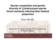
Development of a soil DNA<br />
extraction and quantitative<br />
PCR method for detecting two<br />
Cylindrocarpon species in soil<br />
Ms Chantal Probst, Lincoln<br />
University, NZ<br />
Bananas in Carnarvon—good<br />
news for growers in survey for<br />
quarantine plant pests and<br />
pathogens<br />
Dr Sarah Collins, Department<br />
of Agriculture and Food WA<br />
Session 1C<br />
Soilborne diseases<br />
Room: Hunter Room<br />
Chair: Nerida Donovan<br />
Can investment in building up<br />
soil organic carbon lead to<br />
disease suppression in<br />
vegetable crops?<br />
Dr Ian Porter, Department of<br />
Primary Industries, Vic<br />
Evaluation of soil health<br />
indicators in the vegetable<br />
industry of temperate<br />
Australia<br />
Ms Robyn Brett, Department<br />
of Primary Industries, Vic<br />
Rhizoctonia AG2.1 and AG3 in<br />
soil—competition or<br />
synergism?<br />
Dr Tonya Wiechel,<br />
Department of Primary<br />
Industries, Vic<br />
Session 1D<br />
Virology<br />
Room: Newcastle Room<br />
Chair: John Randles<br />
Towards universal detection of<br />
Luteoviridae<br />
Miss Anastasija Chomic,<br />
Lincoln University, NZ<br />
Massive parallel sequencing of<br />
small RNAs to identify plant<br />
viruses and virus‐induced small<br />
RNAs<br />
Dr Robin MacDiarmid, The<br />
New Zealand Institute for <strong>Plant</strong><br />
and Food Research Ltd, NZ<br />
Chickpea chlorotic stunt virus,<br />
an important virus of coolseason<br />
food legumes in Asia<br />
and North Africa and<br />
potentially in Australia<br />
Dr Safaa Kumari, International<br />
Center for Agricultural<br />
Research in the Dry Areas,<br />
Syria<br />
1200 LUNCH Banquet Room<br />
Editor’s meeting (1200–1400)<br />
Waratah Room<br />
Student mentor lunch (1200–1320)<br />
Mulumbinba Room<br />
1320 Keynote address—Emerging frontiers in forest pathology Concert Hall<br />
Prof Mike Wingfield, Forestry and Agricultural Biotechnology Institute, University of Pretoria, South Africa<br />
12 PLANT HEALTH MANAGEMENT: AN INTEGRATED APPROACH | APPS 2009
Session 2A<br />
Forest pathology/native<br />
Room: Concert Hall<br />
Chair: Angus Carnegie<br />
1410 Variability in pathogenicity of<br />
Quambalaria pitereka on<br />
spotted gums<br />
Mr Geoffrey Pegg, The<br />
University of Queensland/<br />
Primary Industries and<br />
Fisheries, Qld<br />
1430 Movement of pathogens<br />
between horticultural crops<br />
and endemic trees in the<br />
Kimberleys<br />
Ms Monique Sakalidis,<br />
Murdoch University, WA<br />
1450 Pathogenicity of Phytophthora<br />
multivora to Eucalyptus<br />
gomphocephala and<br />
E. marginata<br />
Dr Treena Burgess, Murdoch<br />
University, WA<br />
1510 Microscopy of progressive<br />
decay of fungi isolated from<br />
Meranti tree canker<br />
Dr Erwin Erwin, University of<br />
Mulawarman, Indonesia<br />
Session 2B<br />
Soilborne disease<br />
Room: Cummings Room<br />
Chair: Peter McGee<br />
Optimising conditions to<br />
investigate gene expression in<br />
pathogenic Streptomyces using<br />
RT‐qPCR<br />
Dr Tonya Wiechel,<br />
Department of Primary<br />
Industries, Vic<br />
Fusarium oxysporum and<br />
Pythium associated with<br />
vascular wilt and root rots of<br />
greenhouse cucumbers<br />
Mr Len Tesoriero, NSW<br />
Department of Primary<br />
Industries<br />
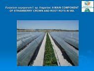
Fusarium oxysporum f. sp.<br />
fragariae: a main component<br />
of strawberry crown and root<br />
rots in Western Australia<br />
Dr Hossein Golzar,<br />
Department of Agriculture and<br />
Food WA<br />
Evaluation of resistant<br />
rootstocks for control of<br />
Fusarium wilt of watermelon in<br />
Nghe An Province, Vietnam.<br />
Prof Lester Burgess, The<br />
University of Sydney, NSW<br />
Session 2C<br />
Epidemiology<br />
Room: Hunter Room<br />
Chair: Chris Steel<br />
Bunch rot diseases and their<br />
management<br />
Prof Turner Sutton, NC State<br />
University, USA<br />
Inoculum and climatic factors<br />
driving epidemics of Botrytis<br />
cinerea in New Zealand and<br />
Australian vineyards<br />
Dr Rob Beresford, The New<br />
Zealand Institute for <strong>Plant</strong> and<br />
Food Research Limited, NZ<br />
Infection of apples by<br />
Colletotrichum acutatum in<br />
New Zealand is limited by<br />
temperature<br />
Dr Kerry Everett, The New<br />
Zealand Institute for <strong>Plant</strong> and<br />
Food Research Limited, NZ<br />
Epidemiology of walnut blight,<br />
caused by Xanthomonas<br />
arboricola pv. juglandis, in<br />
Tasmania, Australia<br />
Dr Katherine Evans, University<br />
of Tasmania<br />
Session 2D<br />
Disease management<br />
Room: Newcastle Room<br />
Chair: Robert Magarey<br />
Sugarcane smut—disease<br />
development and mechanism<br />
of resistance<br />
Dr Shamsul Bhuiyan, BSES<br />
Limited, Qld<br />
Dissemination of biological<br />
and chemical fungicides by<br />
bees onto Rubus and Ribes<br />
flowers<br />
Dr Monika Walter, The New<br />
Zealand Institute for <strong>Plant</strong> and<br />
Food Research Limited, NZ<br />
Current studies on divergence<br />
and management of pepper<br />
yellow leaf curl disease<br />
Indonesia<br />
Dr Sri Hidayat, Bogor<br />
Agricultural University,<br />
Indonesia<br />
Fungicide resistance in cucurbit<br />
powdery mildew<br />
Dr Chrys Akem, Primary<br />
Industries and Fisheries, Qld<br />
1530 AFTERNOON TEA Banquet Room<br />
1600 Keynote address—Population genetic analyses of plant pathogens: new challenges and opportunities Concert Hall<br />
Dr Celeste Linde, Research School of Biology, College of Medicine, Biology and Environment, Australian National University<br />
Session 3A<br />
Population genetics<br />
Room: Concert Hall<br />
Chair: Andre Drenth<br />
1640 Genetic diversity of Botryosphaeria parva<br />
(Neofusicoccum parvum) in New Zealand<br />
vineyards<br />
Mr Jeyaseelan Baskarathevan, Lincoln<br />
University, NZ<br />
1700 Anthracnose disease of chili pepper—<br />
genetic diversity, pathogenicity and<br />
breeding for resistance<br />
A/Prof Paul Taylor, The University of<br />
Melbourne, Vic<br />
1720 The diversity of Colletotrichum infecting<br />
lychee in Australia<br />
Ms Jay Anderson, Primary Industries and<br />
Fisheries, Qld and University of<br />
Queensland<br />
1740 Variation in Phytophthora palmivora on<br />
cocoa in Papua New Guinea<br />
Ms Josephine Saul Maora, PNG Cocoa<br />
Coconut Institute<br />
Session 3B<br />
Modelling and crop loss assessment<br />
Room: Cummings Room<br />
Chair: Ian Porter<br />
Spore traps for early warning of smut<br />
infestations in Australian sugarcane crops<br />
Dr Rob Magarey, BSES Limited, Qld<br />
Software‐assisted gap estimation (SAGE)<br />
for measuring grapevine leaf canopy<br />
density<br />
Mr Gareth Hill, The New Zealand Institute<br />
for <strong>Plant</strong> and Food Research Limited, NZ<br />
Evaluation of the efficacy of Brassica spot<br />
TM<br />
models for control of white blister in<br />
Chinese cabbage<br />
Mr Desmond Auer, Department of<br />
Primary Industries, Vic<br />
Evaluating an infection model of prune<br />
rust to improve the management of<br />
disease for almond and prune growers<br />
Mr Peter Magarey, South Australian<br />
Research and Development Institute<br />
Session 3C<br />
Disease management<br />
Room: Hunter Room<br />
Chair: Shane Hetherington<br />
Management of white blister on vegetable<br />
brassicas with irrigation and varieties<br />
Dr Elizabeth Minchinton, Department of<br />
Primary Industries, Vic<br />
Alternative screening methods for<br />
sugarcane smut using natural infection<br />
and tissue staining<br />
Dr Shamsul Bhuiyan, BSES Limited, Qld<br />
Interruption of cool chain and strawberry<br />
fruit rot by leak‐causing fungi Rhizopus<br />
species<br />
Dr Monika Walter, The New Zealand<br />
Institute for <strong>Plant</strong> and Food Research<br />
Limited, NZ<br />
Enhancing Papua New Guinea smallholder<br />
cocoa production through greater<br />
adoption of integrated pest and disease<br />
management<br />
Mr Yak Namaliu, PNG Cocoa Coconut<br />
Institute<br />
1800 DRINKS AND POSTERS Banquet Room and Concert Hall<br />
1830–<br />
2030<br />
Council of <strong>Society</strong> meeting<br />
Waratah Room<br />
APPS 2009 | PLANT HEALTH MANAGEMENT: AN INTEGRATED APPROACH 13
Wednesday 30 September<br />
0800–<br />
1730<br />
Registration open<br />
Concert Hall Foyer<br />
0800 ARRIVAL TEA AND COFFEE Concert Hall Foyer<br />
0830 Keynote address—Molecular cytology of Phytophthora‐plant interactions Concert Hall<br />
Prof Adrienne Hardham, <strong>Plant</strong> Cell Biology Group, Research School of Biology, The Australian National University<br />
Session 4A<br />
<strong>Plant</strong> pathogen interactions<br />
Room: Concert Hall<br />
Chair: David Guest<br />
0910 Gene expression changes<br />
during host‐pathogen<br />
interaction between<br />
Arabidopsis thaliana and<br />
Plasmodiophora brassicae<br />
Mrs Arati Agarwal,<br />
Department of Primary<br />
Industries, Vic<br />
0930 Hairpin RNA derived from viral<br />
NIa gene confers immunity to<br />
wheat streak mosaic virus<br />
infection in transgenic wheat<br />
plants<br />
Mr Muhammad Fahim, CSIRO<br />
<strong>Plant</strong> Industry, and Australian<br />
National University, ACT<br />
0950 Characterising inositol<br />
signalling pathways in<br />
Phytophthora spp. for future<br />
development of selective<br />
antibiotics<br />
Mr Dean Phillips, Deakin<br />
University, Vic<br />
1010 Systemic acquired resistance—<br />
a new addition to the IPM<br />
clubroot toolbox?<br />
Dr Caroline Donald,<br />
Department of Primary<br />
Industries, Vic<br />
Session 4B<br />
Disease surveys<br />
Room: Cummings Room<br />
Chair: Sandra Savocchia<br />
Prevalence and pathogenicity<br />
of Botryosphaeria lutea<br />
isolated from grapevine<br />
nursery materials in New<br />
Zealand<br />
Ms Regina Billones, Lincoln<br />
University, NZ<br />
Infection and disease<br />
progression of Neofusicoccum<br />
luteum in grapevine plants<br />
Mr Nicholas Amponsah,<br />
Lincoln University, NZ<br />
Carbohydrate stress increases<br />
susceptibility of grapevines to<br />
Cylindrocarpon black foot<br />
disease<br />
Miss Dalin Dore, Lincoln<br />
University, NZ<br />
Botryosphaeria spp. associated<br />
with bunch rot of grapevines in<br />
south‐eastern Australia<br />
Ms Nicola Wunderlich, Charles<br />
Sturt University, NSW<br />
Session 4C<br />
Epidemiology<br />
Room: Hunter Room<br />
Chair: Greg Johnson<br />
Honey bees— do they aid the<br />
dispersal of Alternaria radicina<br />
in carrot seed crops?<br />
Mr Rajan Trivedi, Lincoln<br />
University, NZ<br />
Translating research into the<br />
field: meta‐analysis of field pea<br />
blackspot severity and yield<br />
loss to extend model<br />
application for disease<br />
management in Western<br />
Australia<br />
Dr Moin Salam, Department of<br />
Agriculture and Food WA<br />
Development of a model to<br />
predict spread of exotic wind<br />
and rain borne fungal pests<br />
Dr Moin Salam, Department of<br />
Agriculture and Food WA<br />
Psyllid transmission of<br />
Huanglongbing from naturally<br />
infected Shogun mandarin to<br />
orange jasmine<br />
Dr Rantana Sdoodee, Prince of<br />
Songkla University, Thailand<br />
Session 4D<br />
Prokaryotic pathogens<br />
Room: Newcastle Room<br />
Chair: Lucy Tran‐Nguyen<br />
Transmission of 'Candidatus<br />
Phytoplasma australiense' to<br />
Cordyline and Coprosma<br />
Dr Ross Beever, Landcare<br />
Research, NZ<br />
Australian grapevine yellows<br />
phytoplasma found in<br />
symptomless shoot tips after a<br />
heat wave in South Australia<br />
Mr Peter Magarey, South<br />
Australian Research and<br />
Development Institute, SA<br />
Association of Phytoplasmas<br />
with papaya crown yellows<br />
(PCY) disease—a new disease<br />
of papaya in Northern<br />
Mindanao, Philippines<br />
Ms Regina Billones, Del Monte<br />
Phils Inc, Philippines<br />
Phytoplasma diseases in citrus<br />
orchards of Pakistan<br />
Dr Shazia Mannan, COMSATS<br />
Institute of Information<br />
Technology, Pakistan<br />
1030 MORNING TEA Banquet Room<br />
1100 Keynote address—Mechanisms modulating fungal attack in postharvest pathogen interactions and Concert Hall<br />
their modulation for improved disease control<br />
Prof Dov Prusky, Department of Postharvest Science of Fresh Produce, Agricultural Research Organization, Israel<br />
Session 5A<br />
<strong>Plant</strong> pathogen interactions<br />
Room: Concert Hall<br />
Chair: Rosalie Daniel<br />
1140 ABA‐dependant signalling of PR genes and<br />
potential involvement in the defence of<br />
lentil to Ascochyta lentis<br />
Dr Rebecca Ford, The University of<br />
Melbourne, Vic<br />
1200 Fundamental components of resistance to<br />
Phytophthora cinnamomi: using model<br />
system approaches<br />
Prof David Cahill, Deakin University, Vic<br />
1220 Genes involved in hypersensitive cell death<br />
responses during Fusarium crown rot<br />
infection in wheat<br />
Dr Jill Petrisko, University of Southern<br />
Queensland, Qld<br />
Session 5B<br />
Disease surveys<br />
Room: Cummings Room<br />
Chair: Aaron Maxwell<br />
Fishing For Phytophthora across Western<br />
Australia’s water bodies<br />
Dr Daniel Hüberli, Murdoch University,<br />
WA<br />
Incidence of fungi isolated from grape<br />
trunks in New Zealand vineyards<br />
Mr Dion Mundy, The New Zealand<br />
Institute for <strong>Plant</strong> and Food Research<br />
Limited, NZ<br />
Isolation and characterisation of strains of<br />
Pseudomonas syringae from waterways of<br />
the Central North Island of New Zealand<br />
Dr Joel Vanneste, The New Zealand<br />
Institute for <strong>Plant</strong> and Food Research<br />
Limited, NZ<br />
Session 5C<br />
Chemical control<br />
Room: Hunter Room<br />
Chair: Len Tesoriero<br />
Evaluation of fungicides to manage<br />
brassica stem canker<br />
Ms Lynette Deland, South Australian<br />
Research and Development Institute, SA<br />
Evaluation of spray programs for powdery<br />
mildew management in greenhouse<br />
cucumbers<br />
Dr Kaye Ferguson, South Australian<br />
Research and Development Institute, SA<br />
The incidence of copper resistant bacteria<br />
in Australian pome and stone fruit<br />
orchards<br />
Dr Chin Gouk, Department of Primary<br />
Industries, Vic<br />
14 PLANT HEALTH MANAGEMENT: AN INTEGRATED APPROACH | APPS 2009
1240 AGRICHEM LUNCH Banquet Room<br />
1340 Poster session Banquet Room and Concert Hall<br />
1430 AFTERNOON TEA Banquet Room<br />
1500 McAlpine lecture—Lessons from the tropics—the unfolding mystery of vascular‐streak dieback Concert Hall<br />
of cocoa, the importance of genetic diversity, horizontal resistance, and the plight of farmers<br />
Assoc Prof Phil Keane, Department of Botany, La Trobe University, Vic<br />
1600 AGM Concert Hall<br />
1730 Close of day<br />
1900 CONFERENCE DINNER Newcastle Panthers Club<br />
APPS 2009 | PLANT HEALTH MANAGEMENT: AN INTEGRATED APPROACH 15
Thursday 1 October<br />
0700 Regional Councillor’s meeting Waratah Room<br />
0700 CHAIRMAN’S BREAKFAST Mulumbinba Room<br />
0800–<br />
1730<br />
Registration open<br />
Concert Hall Foyer<br />
0800 ARRIVAL TEA AND COFFEE Concert Hall Foyer<br />
0830 Keynote address—Translating research into the field: how it started, how it is practised and Concert Hall<br />
how we carry out grape powdery mildew research<br />
Dr Bob Seem, Cornell University, USA<br />
0910 GRDC book launch: Mr James Clarke, Grains Research and Development Corporation Concert Hall<br />
Session 6A<br />
Cereal pathology 1<br />
Room: Concert Hall<br />
Chair: Mark Sutherland<br />
0925 Stem rust race Ug99: international<br />
perspectives and implications for Australia<br />
Dr Colin Wellings, The University of<br />
Sydney, NSW<br />
0945 Mitigating crop losses due to stripe rust in<br />
Australia: integrating pathogen<br />
population dynamics with research and<br />
extension programs<br />
Dr Colin Wellings, The University of<br />
Sydney, NSW<br />
1005 Impact of sowing date on crown rot losses<br />
Dr Steven Simpfendorfer, Department of<br />
Primary Industries, NSW<br />
1025 Symptom development and pathogen<br />
spread in wheat genotypes with varying<br />
levels of crown rot resistance<br />
Dr Cassandra Malligan, Queensland<br />
Primary Industries and Fisheries<br />
Session 6B<br />
Quarantine and exotic pathogens<br />
Room: Cummings Room<br />
Chair: Suzy Perry<br />
Development of an eradication strategy<br />
For exotic grapevine pathogens<br />
Dr Mark Sosnowski, South Australian<br />
Research and Development Institute, SA<br />
Green grassy shoot disease of sugarcane,<br />
a major disease in Nghe An Province,<br />
Vietnam<br />
Dr Rob Magarey, BSES Limited, Qld<br />
Molecular detection of Mycosphaerella<br />
fijiensis in the leaf trash of ‘Cavendish’<br />
banana<br />
Dr Seona Casonato, The New Zealand<br />
Institute for <strong>Plant</strong> and Food Research<br />
Limited, NZ<br />
Optimising responses to incursions of<br />
exotic plant pathogens<br />
Dr Mike Hodda, CSIRO Entomology, ACT<br />
Session 6C<br />
Alternatives to chemical control<br />
Room: Hunter Room<br />
Chair: Carolyn Blomley<br />
The influence of soil biotic factors on the<br />
ecology of Trichoderma biological control<br />
agents<br />
Prof Alison Stewart, Lincoln University, NZ<br />
Understanding Trichoderma bioinoculants<br />
in the root system of Pinus<br />
radiata<br />
Mr Pierre Hohmann, Lincoln University,<br />
NZ<br />
A bioassay to screen Trichoderma isolates<br />
for their ability to promote root growth in<br />
willow<br />
Mr Mark Braithwaite, Lincoln University,<br />
NZ<br />
Biofumigation for reducing Cylindrocarpon<br />
spp. in New Zealand vineyard and nursery<br />
soil<br />
Ms Carolyn Bleach, Lincoln University, NZ<br />
1045 MORNING TEA Banquet Room<br />
Session 7A<br />
Cereal pathology 2<br />
Room: Concert Hall<br />
Chair: Colin Wellings<br />
1100 Crown rot of winter cereals: integrating<br />
molecuar studies and germplasm<br />
improvement<br />
Prof Mark Sutherland, University of<br />
Southern Queensland, Qld<br />
1120 Infection of wheat tissues by Fusarium<br />
pseudograminearum<br />
Mr Noel Knight, University of Southern<br />
Queensland, Qld<br />
1140 Monitoring sensitivity to Strobilurin<br />
fungicides in Blumeria graminis on wheat<br />
and barley in Canterbury, New Zealand<br />
Dr Suvi Viljanen‐Rollinson, The New<br />
Zealand Institute for <strong>Plant</strong> and Food<br />
Research Limited, NZ<br />
1200 Cross inoculation of crown rot and<br />
Fusarium head blight isolates of wheat<br />
Mr Philip Davies, University of Sydney,<br />
NSW<br />
Session 7B<br />
Quarantine and exotic pathogens<br />
Room: Cummings Room<br />
Chair: Nerida Donovan<br />
Twenty years of quarantine plant disease<br />
surveillance on the island of New Guinea:<br />
key discoveries for Australia and PNG<br />
Mr Richard Davis, Australian Quarantine<br />
and Inspection Service, Qld<br />
The importance of reporting suspect exotic<br />
or emergency plant pests to your State<br />
Department of Primary Industry<br />
Dr Sophie Peterson, <strong>Plant</strong> Health<br />
Australia, ACT<br />
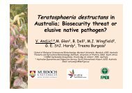
The use of sentinel plantings in forest<br />
biosecurity; results from mixed eucalypt<br />
species trails in South‐East Asia and<br />
Australia<br />
Dr Treena Burgess, Murdoch University,<br />
WA<br />
Methyl bromide alternatives for<br />
quarantine and pre‐shipment and other<br />
purposes—future perspectives<br />
Ms Janice Oliver, Office of the Chief <strong>Plant</strong><br />
Protection Officer, ACT<br />
Session 7C<br />
Alternatives to chemical control<br />
Room: Hunter Room<br />
Chair: Alison Stewart<br />
Fruit extracts of Azadirachta indica<br />
induces systemic acquired resistance in<br />
tomato against Pseudomonas syringae pv<br />
tomato<br />
Dr Prabir Paul, Amity University, India<br />
Fungal foliar endophytes induce systemic<br />
protection in cacao seedlings against<br />
Phytophthora palmivora<br />
Ms Carolyn Blomley, The University of<br />
Sydney, NSW<br />
Effectiveness of the rust Puccinia<br />
myrsiphylli in reducing populations of the<br />
invasive plant bridal creeper in Australia<br />
Dr Louise Morin, CSIRO Entomology, ACT<br />
Evaluation of essential oils and other plant<br />
extracts for control of soilborne pathogens<br />
of vegetable crops<br />
Ms Cassie Scoble, Department of Primary<br />
Industries and La Trobe University, Vic<br />
1230 LUNCH Banquet Room<br />
16 PLANT HEALTH MANAGEMENT: AN INTEGRATED APPROACH | APPS 2009
1330 Keynote address—Use of grid weather forecast data to predict rice blast development in Korea Concert Hall<br />
Prof Eun Woo Park, College of Agriculture and Life Sciences, Seoul National University, Korea<br />
1410 Investigating the impact of climate change on plant diseases<br />
Dr Jo Luck, Department of Primary Industries, Vic<br />
1430 Impact of climate change in relation to blackleg on oilseed rape and blackspot on field pea in Western Australia<br />
Dr Moin Salam, Department of Agriculture and Food, WA<br />
1450 Approaches to training in plant pathology capacity building projects in developing countries<br />
Prof LW Burgess, University of Sydney, NSW<br />
1510 Increasing global regulations on fumigants stimulates new era for plant protection and biosecurity<br />
Dr Ian Porter, Department of Primary Industries, Vic<br />
1530 AFTERNOON TEA Banquet Room<br />
1600 Keynote address—A world of possibilities Concert Hall<br />
Dr Barbara Christ, The Pennsylvania State University, USA<br />
1630 Incoming presidential address<br />
Dr Caroline Mohammed, School of Agricultural Science, University of Tasmania<br />
1645 Awards<br />
1700 Close of day<br />
1730 Bus leaves Civic Centre for Beach Party<br />
1800 BEACH PARTY Newcastle Surf Life Saving Club<br />
APPS 2009 | PLANT HEALTH MANAGEMENT: AN INTEGRATED APPROACH 17
Oral abstracts<br />
APPS 2009 | PLANT HEALTH MANAGEMENT: AN INTEGRATED APPROACH 19
President’s address<br />
‘Shield the young harvest from devouring blight’—Charles Darwin, Joseph Banks,<br />
Thomas Knight and wheat rust: discovery, adventure, and ‘getting the message out’<br />
G.I. Johnson{ XE "Johnson, G.I." }<br />
Horticulture 4 Development, PO Box 412, Jamison ACT 2614 Australia Email: greg.johnson@velocitynet.com.au<br />
1969: The year of the first moon landing (20 July 1969), the<br />
Woodstock Festival in upstate New York (15–18 August 1969),<br />
and (coinciding the last day of Woodstock) the beginning of the<br />
<strong>Australasian</strong> <strong>Plant</strong> <strong>Pathology</strong> <strong>Society</strong> (first AGM at 41st ANZAAS<br />
Meeting, Adelaide (18 August 1969) (Purss 1994)). All had a<br />
lengthy gestation and challenges along the way. All have<br />
changed the world!<br />
In the 17th President’s Address to the <strong>Australasian</strong> <strong>Plant</strong><br />
<strong>Pathology</strong> <strong>Society</strong>, David Guest (2001), noted: ‘I became a plant<br />
pathologist because the mechanisms organisms use to<br />
communicate fascinate me’. Well, I became a plant pathologist<br />
because I am gardener at heart. But I have learned along the<br />
way that communication is a critical issue—not only the<br />
communication amongst and between microorganisms and<br />
plants, but also that between plant pathologists, farmers,<br />
politicians and communities. And, communication that is timely,<br />
inspiring and, (preferably) accurate, often yields the most<br />
favourable outcomes.<br />
In this paper, I will explore some of the early communication<br />
relating to plant disease, particularly wheat rusts. I refer to<br />
Erasmus and Charles Darwin, Joseph Banks, Thomas Knight, and<br />
some pioneering Australian researchers, and the roles of<br />
conferences, publications and newspapers, to highlight how<br />
‘getting our message out’ was as important in the 19th and early<br />
20th centuries as it is now. And, finally, I will consider how a<br />
scientific society in the 21st century still has relevance and the<br />
potential to change the world.<br />
20 PLANT HEALTH MANAGEMENT: AN INTEGRATED APPROACH | APPS 2009
The relevance of plant pathology in food production<br />
André Drenth{ XE "Drenth, A." }<br />
Tree <strong>Pathology</strong> Centre, The University of Queensland and Primary Industries and Fisheries, Indooroopilly, Queensland 4068 Australia<br />
DrenthA@dpi.qld.gov.au<br />
<strong>Plant</strong>s are our only true primary producers of food, fibre and fuel<br />
through the process of photosynthesis. The objective of plant<br />
science in general is to understand the principles and processes<br />
involved in plant growth and reproduction and the objective of<br />
crop science concerns the productivity of our crops.<br />
Keynote address<br />
Over a period of 35 years from 1960 to 1995 the world food<br />
production doubled while the world population more than<br />
doubled from 2.5 billion to 5.6 billion. The present world<br />
population is 6.7 billion and expected to grow to 9 billion by<br />
2050. Agricultural production needs to increase 2.3% a year just<br />
to meet global food demand. At present we increase it by 1.5% a<br />
year. Thus the challenge for Agriculture is to double the global<br />
food production over the next three decades. In addition to<br />
meeting the challenge for food production Agriculture is also<br />
expected to provide renewable fuel. It should be clear that<br />
society needs Agriculture now more than ever before.<br />
The objective of the discipline of plant pathology is to reduce the<br />
impact of diseases on the production of plants for food, fibre<br />
and fuel. <strong>Plant</strong> pathology is an important biological science and<br />
arose out of need during times of famine, poor food security and<br />
large scale crop losses. Despite clearly defined objectives and a<br />
proud history of achievements many plant pathologists would<br />
agree with the statement ‘<strong>Plant</strong> pathology in relation to its<br />
importance to humanity continues to be a grossly underfunded<br />
discipline’ (Strange and Scott, 2005, Annual Review of<br />
Phytopathology). In order to ascertain the significance of our<br />
relatively small discipline we must recognise and document past<br />
contributions, identify and understand future challenges, and be<br />
actively working on tomorrow’s problems. The aim of my<br />
presentation is to address the following three questions:<br />
• What have been the contributions and impacts of plant<br />
science and more specifically plant pathology on food<br />
production?<br />
• What are the key challenges with regards to plant<br />
production which will enable Agriculture to feed a growing<br />
world population in the future?<br />
• What role can our discipline of plant pathology play in<br />
feeding the world?<br />
APPS 2009 | PLANT HEALTH MANAGEMENT: AN INTEGRATED APPROACH 21
Session 1A—Disease management<br />
An integrated approach to husk spot management in macadamia<br />
O.A. Akinsanmi{ XE "Akinsanmi, O.A." } and A. Drenth<br />
Tree <strong>Pathology</strong> Centre, The University of Queensland and Primary Industries and Fisheries, Queensland, 80 Meiers Road Indooroopilly,<br />
4068 Qld, Australia<br />
INTRODUCTION<br />
Husk spot, caused by Pseudocercospora macadamiae is a major<br />
fungal disease of macadamia in Australia (3). Husk spot occurs<br />
only in eastern Australia, costing over $10 million in lost<br />
productivity if the disease is not adequately controlled.<br />
P. macadamiae infects macadamia husks on which it continually<br />
produces inoculum (4), the infection causes premature<br />
abscission of diseased fruit, thus, resulting in extensive yield<br />
losses and reduced kernel quality. Application of fungicide is<br />
currently the only effective method for controlling husk spot (1).<br />
However, several factors including the upsurge in organic<br />
farming, the need for sustainable management practices, and<br />
possible development of fungicide resistant fungal strains and<br />
lack of quantitative information on levels of disease resistance in<br />
varieties require development of integrated management<br />
strategies for controlling husk spot. Systematic studies were<br />
performed to improve husk spot control through the provision of<br />
alternative fungicide, biological control options, effective<br />
cultural practices and diagnostic characters for disease resistant<br />
cultivars.<br />
MATERIALS AND METHODS<br />
In order to evaluate the efficacy of different fungicides and<br />
biocontrol options against husk spot, both laboratory and field<br />
trials were established. Macadamia trees treated with different<br />
fungicide products at three field sites in south east Queensland<br />
and northern New South Wales were evaluated for husk spot<br />
incidence and severity, from onset of visual symptoms to kernel<br />
maturity (2). The area under disease progress curves of the<br />
treatments were compared against each other and with the<br />
untreated control using analysis of variance. The activities of five<br />
Trichoderma species against P. macadamiae were assessed<br />
either singly or in combination with each other and bacteria<br />
(Bacillus subtilis and Pseudomonas fluorescence) in laboratory<br />
experiments. Five characters of macadamia varieties with<br />
varying incidence of husk spot were evaluated as possible<br />
diagnostic characters for husk spot resistant varieties and<br />
discriminant analysis was performed using the identified<br />
diagnostic characters to partition 12 macadamia varieties to<br />
husk spot resistance.<br />
stomata per unit area and number of lesion per fruit classified<br />
macadamia varieties into various resistance groups.<br />
Nut harvested<br />
100%<br />
80%<br />
60%<br />
40%<br />
20%<br />
0%<br />
Poor Moderate Good Very good<br />
None Once Twice<br />
Number of spray applications<br />
Figure 1. Proportion of quality of total macadamia kernel produced from<br />
trees that received varying number of spray applications.<br />
ACKNOWLEDGEMENTS<br />
We acknowledge the University of Queensland, Primary<br />
Industries and Fisheries Queensland, Australian Macadamia<br />
<strong>Society</strong> Ltd., Horticulture Australia Ltd. and Nufarm Australia Pty<br />
Ltd for the funding for this project.<br />
REFERENCES<br />
1. Akinsanmi, O.A., Miles, A.K., and Drenth, A. 2007. Timing of<br />
fungicide application for control of husk spot caused by<br />
Pseudocercospora macadamiae in macadamia. <strong>Plant</strong> Dis. 91:1675–<br />
1681.<br />
2. Akinsanmi, O.A., Miles, A.K., and Drenth, A. 2008. Alternative<br />
fungicides for controlling husk spot caused by Pseudocercospora<br />
macadamiae in macadamia. Australas. <strong>Plant</strong> Pathol. 37:141–147.<br />
3. Beilharz, V., Mayers, P.E., and Pascoe, I.G. 2003. Pseudocercospora<br />
macadamiae sp. nov., the cause of husk spot of macadamia.<br />
Australas. <strong>Plant</strong> Pathol. 32 (2):279–282.<br />
4. Miles, A.K., Akinsanmi, O.A., Sutherland, P.W., Aitken, E.A.B., and<br />
Drenth, A. 2009. Infection, colonisation and sporulation by<br />
Pseudocercospora macadamiae on macadamia fruit. Australas.<br />
<strong>Plant</strong> Pathol. 38 (1):36–43.<br />
RESULTS AND DISCUSSION<br />
Results of field trials showed that pyraclostrobin conferred<br />
significantly (P < 0.05) better protection than trifloxystrobin and<br />
also had somewhat similar efficacy as a tank‐mixture of<br />
carbendazim and copper against husk spot incidence and<br />
severity. The use of pyraclostrobin in rotation with tank mixture<br />
of carbendazim and copper would play a key role in the<br />
management of fungicide resistance in the industry. The<br />
reduction of copper usage would also provide additional benefits<br />
to the macadamia industry. Frequency or number of fungicide<br />
spray applications influenced total kernel quality (Fig. 1). In vitro<br />
volatile inhibition trials showed that growth rate of<br />
P. macadamiae was inhibited by 8% in mixed cultures of T. viride<br />
and T. harzianum, and by 5% in the mixed culture of T. koningii<br />
and T. harzianum but no mycoparasitism was observed in the<br />
hyphal interaction experiments. Our results showed that<br />
significant differences exist in the reaction of macadamia<br />
varieties to husk spot. The discriminant analysis on the disease<br />
incidence and severity, prevalence of stick‐tights, number of<br />
22 PLANT HEALTH MANAGEMENT: AN INTEGRATED APPROACH | APPS 2009
Application methods of phosphonate to control Phytophthora pod rot and stem<br />
canker on cocoa<br />
A. Purwantara, A. Wahab, Y. Imron, V.K.M. Dewi, P.J. McMahon{ XE "McMahon, P.J." }, S. Lambert, P.J. Keane, D.I. Guest<br />
Presenting author: Department of Botany, La Trobe University, Bundoora, Victoria 3086 Email: peter.mcmahon@latrobe.edu.au<br />
INTRODUCTION<br />
Stem canker and Phytophthora pod rot (PPR) or black pod<br />
caused by Phytophthora palmivora are among the most serious<br />
diseases of cocoa in Sulawesi, Indonesia, causing high yield<br />
losses for farmers. Potassium phosphonate (phosphite) has<br />
previously been demonstrated to effectively control canker and<br />
PPR in Papua New Guinea (1). To test the effectiveness of<br />
phosphonate in Sulawesi against Phytophthora diseases and to<br />
compare methods of application of the chemical, two<br />
experimental trials were conducted on cocoa farms in Sulawesi,<br />
Indonesia.<br />
Session 1A—Disease management<br />
METHODS<br />
1 The effect of trunk‐injected phosphonate on stem canker and<br />
PPR in Ladongi, Southeast Sulawesi. Fifty 10 year‐old hybrid<br />
cocoa trees were injected annually with 16 g a.i. phosphonate<br />
(Agrifos 600; Agrichem), fifty with water and fifty left untreated.<br />
For 4.5 years following the initial injection, trees were scored<br />
each month for canker severity and monitored for PPR and CPB<br />
incidence (% ripe pods affected). Treatments were compared by<br />
one‐way ANOVA followed by either the Bonferroni or the<br />
Games‐Howell post‐hoc tests.<br />
2 The effect of phosphonate on stem canker applied by three<br />
differing methods. In a blocked trial with four replicates, 2‐yearold<br />
grafted cocoa trees naturally infected with Phytophthora<br />
stem canker were treated with phosphonate either by trunk<br />
injection, bark painting (combined with Pentrabark; Agrichem),<br />
or implants?, or left untreated and were then scored monthly (as<br />
in Experiment 1) for five months for canker lesion size.<br />
RESULTS AND DISCUSSION<br />
1 Trunk injection. Phosphonate injection cured stem canker<br />
within four months of the initial injection (Fig. 1). Over a 2.5 year<br />
period, phosphonate significantly decreased cumulative PPR<br />
incidence (Table 1), while PPR incidence did not differ between<br />
the untreated and water‐injected trees indicating that the<br />
injection procedure itself did not influence these results. In both<br />
the control and phosphonate‐treated trees, cycles of PPR<br />
infection occurred in the wet season with PPR incidence<br />
fluctuating from less than 30% to greater than 75% (data not<br />
shown). These might have been due to variations in rainfall, the<br />
regular removal of infected pods in the experiment or natural<br />
cycles of sporulation and infection. CPB incidence did not differ<br />
significantly between treatments (data not shown).<br />
Figure 1. Mean canker scores for phosphonate‐injected trees (solid line<br />
and squares) and water‐injected (broken line, open diamond symbols)<br />
assessed from 2002 to 2006. Only trees that initially had canker<br />
infections are included. Trees were injected in June 2002, December<br />
2002 and then every year until the end of 2006.<br />
Table 1. Mean cumulative incidences of PPR in ripe pods from April 2004<br />
to December 2006.<br />
Treatment Mean PPR (%)<br />
Untreated<br />
43.4 ± 1.9 a<br />
Water‐injected<br />
40.2 ± 2.1 a<br />
Phosphonate‐injected<br />
26.3 ± 1.8 b<br />
Mean incidences were calculated from the total number of ripe pods and<br />
infested/infected pods harvested from each tree in the 2.5 years. Each treatment<br />
had 50 trees (replicates). Means (± SE) within columns followed by the same letter<br />
are not significantly different (P
Session 1A—Disease management<br />
Botrytis bunch rot control strategies in cool climate viticultural regions of Australia<br />
and New Zealand<br />
J. Edwards{ XE "Edwards, J." } 1 , D. Riches 1 , K.J. Evans 2 , R.M. Beresford 3 , G.N. Hill 3 , P.N. Wood 4 and D.C. Mundy 5<br />
1 Department of Primary Industries, 621 Burwood Highway, Knoxfield, Victoria 3180.<br />
2 Tasmanian Institute of Agricultural Research, University of Tasmania, New Town Research Laboratories, 13 St Johns Avenue, New Town,<br />
Tasmania 7008.<br />
3 <strong>Plant</strong> and Food Research, Mt Albert Research Centre, Private Bag 92‐169 Auckland, NZ<br />
4 <strong>Plant</strong> and Food Research, Hawke’s Bay Research Centre, Private Bag 1401, Havelock North, Hastings 4157, NZ<br />
5 <strong>Plant</strong> and Food Research, Marlborough Wine Research Centre, P.O. Box 845, Blenheim 7240, NZ<br />
INTRODUCTION<br />
Botrytis bunch rot can cause major losses in wine grapes, while<br />
negligible damage may occur in some seasons, even in the<br />
absence of control measures. We evaluated the effectiveness of<br />
different disease control strategies in five cool climate viticulture<br />
regions in the context of determining disease risk during the<br />
season (1) and economic outcomes for the grower.<br />
MATERIALS AND METHODS<br />
Standard measures of yield, weather, canopy density, bunch<br />
exposure, latent Botrytis cinerea incidence at pre‐bunch closure<br />
(PBC), and botrytis development post‐veraison were recorded at<br />
23 sites in Southern Tasmania, Southern Victoria and New<br />
Zealand between 2006–08 (Table 1). Untreated plots and<br />
combinations of treatments including fungicide timing, canopy<br />
modification (leaf plucking and shoot thinning), and inoculum<br />
reduction by removing bunch trash, were used to generate a<br />
wide range of harvest botrytis severities.<br />
RESULTS<br />
The efficacy of early season (5%, 80% cap‐fall), mid season (peasize,<br />
PBC), late season (veraison, pre‐harvest) fungicide<br />
applications and combination treatments varied considerably<br />
across the different trial sites and seasons. Only 8 out of 23 trials<br />
exceeded 3% botrytis severity at harvest (a level at which many<br />
wineries impose price penalties) on unsprayed vines. Fungicide<br />
applications reduced disease below the 3% threshold in only four<br />
of them (Table 1). Leaf plucking was as effective as the fungicide<br />
treatments in six New Zealand and one Victorian trial, but not in<br />
two other trials. Leaf removal combined with fungicide<br />
applications was the most effective treatment, reducing botrytis<br />
severity to below the 3% threshold even in trials where severity<br />
was >8% in the untreated plots. The experimental inoculum<br />
reduction treatment (bunch trash removal) did not reduce<br />
botrytis severity in these trials.<br />
Tactical decisions about the need for individual fungicide sprays<br />
or canopy management actions depend on the season, canopy<br />
density and bunch exposure, and underlying risk for the region.<br />
The data collected in these trials is being used to develop a<br />
botrytis risk model (1) to assist growers in making decisions for<br />
botrytis management in future.<br />
Table 1. Treatment effects on botrytis severity at harvest for trials<br />
conducted 2006–08 in Australia and New Zealand.<br />
Year<br />
Mean botrytis severity at harvest (%)<br />
Location and<br />
Leaf<br />
Variety 1 Control Spray program 2 pluck<br />
Spray<br />
+leaf pluck<br />
06–07 NZ (A) ‐ SB 25.3 7.7 (mid) ‐ ‐<br />
“ NZ (HB) ‐ SB 4.5 2.1 2.6 0.3<br />
“ NZ (HB) ‐ CH 9.6 5.9 ns 2.2 1.7<br />
“ NZ (M) ‐ SB1 0.2 0.1 ns(early) ‐ ‐<br />
NZ (M) ‐ SB2 0.6 ‐ ‐ ‐<br />
“ Tas ‐ SB 2.5 0.9 (mid) ‐ ‐<br />
“ Tas ‐ R 0.6 0.8 ns ‐ ‐<br />
“ Vic ‐ CH1 0.0 0.0 ns ‐ ‐<br />
“ Vic ‐ SB 0.1 0.0 ns ‐ ‐<br />
“ Vic ‐ CH2 0.0 0.0 ns ‐ ‐<br />
07–08 NZ (A) ‐ SB 1.6 0.1 (early) 0.7 0.3<br />
“ NZ (HB) ‐ SB 8.4 3.5 3.4 0.4<br />
“ NZ (HB) ‐ CH 9.5 1.9 1.9 0.3<br />
“ NZ (M) ‐ SB1 2.1 1.5 (mid/early) 1.1 0.2<br />
NZ (M) ‐ SB2 0.8 ‐ ‐ ‐<br />
“ NZ (M) ‐ SB3 5.3 0.6 ‐ ‐<br />
“ Tas ‐ SB 2.7 1.6 ns (mid) 2.2 1.9<br />
“ Tas ‐ R1 6.2 2.6 (mid) ‐ ‐<br />
“ Tas ‐ R2 1.9 0.8 ns (mid) ‐ ‐<br />
“ Tas ‐ CH 3.6 2.8 (late) ‐ ‐<br />
“ Vic ‐ CH1 2.0 0.0 2.1 0.0<br />
“ Vic ‐ SB 2.3 0.1 0.4 0.6<br />
“ Vic ‐ CH2 0.8 0.0 0.6 0.0<br />
DISCUSSION<br />
Assuming that 3% botrytis severity is a threshold level at which<br />
growers may suffer a price penalty on their crop, many of our<br />
trial sites did not reach the 3% severity threshold even when left<br />
untreated. Botrytis severity at harvest is strongly correlated with<br />
the length of the ripening period (1) and Auckland and Hawke’s<br />
Bay were most prone to serious botrytis epidemics, followed by<br />
Marlborough and Southern Tasmania, with Victoria the lowest<br />
risk. These results highlight the need for benefit‐cost analyses, as<br />
the full cost of a spray program (fuel, pesticide, labour, water,<br />
etc) may not always be recouped, particularly in drier viticultural<br />
regions like Victoria.<br />
Leaf plucking was as effective as a complete fungicide program<br />
at sites with dense canopies. The combined effects of leaf<br />
removal and fungicide application gave the lowest levels of<br />
botrytis, but risk of sunburn and effects on fruit quality are also<br />
important when considering leaf plucking for botrytis control.<br />
1<br />
M=Marlborough, HB=Hawke’s Bay, A=Auckland, SB=Sauvignon Blanc, R=Riesling,<br />
CH=Chardonnay<br />
2 Full fungicide program or the most effective fungicide timing (shown in brackets).<br />
ns—fungicide treatments not significantly different to untreated at P ≤ 0.05<br />
ACKNOWLEDGEMENTS<br />
GWRDC, New Zealand Winegrowers and our respective agencies<br />
for funding this research.<br />
REFERENCES<br />
1. Beresford and Hill (2008) Predicting in‐season risk of botrytis bunch<br />
rot in Australian and New Zealand vineyards. ‘Breaking the mould:<br />
a pest and disease update’, Australian <strong>Society</strong> of Viticulture and<br />
Oenology Seminar Proceedings, Mildura 24 July 2008.<br />
24 PLANT HEALTH MANAGEMENT: AN INTEGRATED APPROACH | APPS 2009
Development of a soil DNA extraction and quantitative PCR method for detecting two<br />
Cylindrocarpon species in soil<br />
INTRODUCTION<br />
C.M. Probst{ XE "Probst, C.M." }, M.V. Jaspers, E.E. Jones and H.J. Ridgway<br />
Faculty of Agriculture and Life Sciences, Lincoln University, PO Box 84, Lincoln 8150, New Zealand<br />
Cylindrocarpon black foot disease has been identified worldwide<br />
as a common cause of vine death in nurseries and in young<br />
vineyards (1), especially in sites converted from orchards or<br />
replanted from grapes. This indicates that the existing soil<br />
conditions may have contributed to the disease, although very<br />
little information is available regarding the survival of the<br />
pathogens in soil. In New Zealand, the three Cylindrocarpon<br />
species equally responsible for black foot disease on grapevines<br />
are C. destructans, C. macrodidymum and C. liriodendri (1).<br />
Recently, quantitative PCR (qPCR) has begun to supersede soil<br />
dilution plating as a method for precise determination of soil<br />
inoculum levels (2). In this research program, qPCR was<br />
optimised to test large soil samples for presence of two<br />
Cylindrocarpon species.<br />
MATERIALS AND METHODS<br />
Fungal isolates. The Lincoln University Culture Collection<br />
provided isolates of the three Cylindrocarpon species, which had<br />
been obtained from symptomatic New Zealand grapevines.<br />
Single spore colonies of 10 randomly selected isolates per<br />
species were grown on potato dextrose agar (PDA) at 20°C in the<br />
dark.<br />
RESULTS<br />
The primer pairs were specific for each of the two species and no<br />
cross reactivity was observed. They produced a 300 bp and 197<br />
bp product for C. macrodidymum and C. liriodendri, respectively.<br />
They were also suitable for a range of genetically diverse<br />
isolates. Each of the primers was able to detect as little as 30 pg<br />
DNA in a standard PCR and 3 pg DNA in a nested PCR. The qPCR<br />
could detect pure DNA at the same level as the nested PCR. For<br />
spore suspensions, the qPCR was able to detect as little as 100<br />
spores in soil. The time course experiment showed that for each<br />
species, less than half of the nuclei remained 1 week after<br />
infesting the soil with conidia. However, at this time, the DNA<br />
could still be visualised on agarose gels of the qPCR products<br />
(Fig 1). After 2, 3 and 6 weeks, the DNA could not be detected.<br />
M ng Weeks<br />
30 3 0.3 0.03 0.003 0 0 1 2<br />
Session 1B—Disease surveys<br />
Soil Infestation. A mixed conidium suspension (10 6 conidia /mL)<br />
was obtained for each species using three isolates. They were<br />
used to infest 5 L pots of soil, three replicates per species, with a<br />
final concentration of 10 5 conidia per gram of soil. Controls were<br />
treated with a similar quantity of water. The 5 L pots were sunk<br />
into, and level with, the ground in the Lincoln University<br />
Vineyard. Two 15 g soil samples were taken from each pot for<br />
DNA extraction at 0, 1, 2, 3 and 6 weeks after set up. All three<br />
species were inoculated into each pot to ensure detection<br />
specificity<br />
DNA extraction. For pure culture extraction, a small plug of<br />
mycelium was transferred from a 5 d old PDA culture to potato<br />
dextrose broth (PDB) and incubated for 7 d at room<br />
temperature, in 12 h light/ dark. The mycelium was harvested<br />
and stored at ‐80°C until DNA extraction using a PureGene DNA<br />
extraction kit (Qiagen). Spectrophotometry was used to quantify<br />
the genomic DNA. DNA was also extracted from 10 6 conidia of<br />
each isolate in a similar way.<br />
For DNA extraction from soil samples, 10 g of soil was placed in a<br />
250 mL bottle with 45 mL water containing 0.01% agar. The<br />
bottles were shaken for 10 min and left to stand for 10 min. The<br />
supernatant was put into two 15 mL tubes, and centrifuged at<br />
4000 x g for 15 min. The pellets were combined and centrifuged<br />
again, with the final pellet being used for DNA extraction using a<br />
PowerSoil kit (MO BIO Laboratories Inc.). Genomic DNA quality<br />
was assessed by visualisation on a 1% agarose gel.<br />
PCR amplification. Species specific primers for the β− tubulin<br />
region of C. macrodidymum and C. liriodendri, donated by Dr<br />
Lizel Mostert (University of Stellenbosch, South Africa), were<br />
used in qPCR with SYBR green chemistry and a Rox internal<br />
standard on an ABI Prism 7700 sequence detector. After the<br />
qPCR, the products were separated by electrophoresis on a 1.5%<br />
agarose gel and visualised under UV light.<br />
Figure 1. 1.5% agarose gel of products amplified during qPCR with the<br />
primers specific for C. liriodendri.<br />
DISCUSSION<br />
The results show that the species specific primers used in a qPCR<br />
system could detect as little as 3 pg of pure DNA, which is<br />
equivalent to 30 Cylindrocarpon macroconidia. When spores<br />
were recovered from soil a sensitivity of 100 spores was<br />
achieved. Following soil inoculation, DNA of fewer than 50% of<br />
the conidium nuclei in the soil was detected after 1 week.<br />
Additional unpublished data has indicated that in the soil<br />
environment, the 4‐celled conidia were often converted to 2<br />
chlamydospores (uninucleate). It is possible that the >50%<br />
decrease observed is a reflection of that conversion, rather than<br />
the death of conidia. Research is continuing to investigate the<br />
population dynamics of these Cylindrocarpon species in soil.<br />
Future research will include the development of C. destructans<br />
specific primers.<br />
ACKNOWLEDGEMENTS<br />
We gratefully acknowledge Lizel Mostert for providing the<br />
primers sequences and Winegrowers New Zealand and TECNZ<br />
for funding this project.<br />
REFERENCES<br />
1. Halleen, F., Fourie, P.H. and Crous P.W. (2006). A review of black<br />
foot disease of grapevine. Phytopathologia Mediterranea 45: S55‐<br />
S67.<br />
2. Kernaghan, G., Reeleder, R.D. and Hoke, S.M.T. (2007).<br />
Quantification of Cylindrocarpon destructans f. sp. Panacis in soils<br />
by real‐time PCR. <strong>Plant</strong> <strong>Pathology</strong> 56: 508–516.<br />
APPS 2009 | PLANT HEALTH MANAGEMENT: AN INTEGRATED APPROACH 25
Session 1B—Disease surveys<br />
Why Australia needs a coordinated national diagnostic system<br />
J.R. Moran{ XE "Moran, J.R." } 1 , J.H. Cunningtonv, F.J. Macbeth 2 , C.C. Murdoch 1 , A.M. Kelly 3 , D. Hailstones 4 , S.A. Peterson 5 , B. Hall 6 , S.<br />
Perry 7 , M. Williams 8<br />
1 Department of Primary Industries, Victoria, Private Bag 15, Ferntree Gully DC, 3156, VIC<br />
2 Office of the Chief <strong>Plant</strong> Protection Officer, Department of Agriculture, Fisheries and Forestry, GPO Box 858, Canberra, 2601, ACT<br />
3 NSW Department of Primary Industries, PMB 8 Camden, 2570, NSW<br />
4 CRC Diagnostics Research Program, PMG 8, Camden, 2570, NSW<br />
5 <strong>Plant</strong> Health Australia, 5/4 Phipps Close, Deakin, 2600, ACT<br />
6 South Australian Research and Development Institute, GPO Box 397, Adelaide, 5001, SA<br />
7 Department of Employment, Economic Development and Innovation, GPO Box 46, Brisbane, 4001, Qld<br />
8 Department of Primary Industries and Water, 13 St John’s Ave, New Town, 7008, Tasmania<br />
INTRODUCTION<br />
The accurate and rapid diagnosis of plant pests and diseases<br />
underpins all management activities aimed at preventing the<br />
establishment of exotic pests and is a critical component of<br />
surveillance activities.<br />
<strong>Plant</strong> pest diagnostic services also provide the foundation of<br />
endemic pest management through the timely identification of<br />
plant pests, if left uncontrolled, can disrupt agricultural plant<br />
production and restrict market access for agricultural produce.<br />
Rapid identification also supports quarantine processes, such as<br />
maintaining pest free areas that allow access to domestic and<br />
international markets.<br />
Diagnostic services are delivered by a range of agencies across a<br />
dispersed geography and climate range. The majority of<br />
diagnostic services are provided by state agencies with some<br />
being delivered by the Australian Government, commercial<br />
diagnostic laboratories, CSIRO, and the Universities (1). Services<br />
are provided on an ad‐hoc, commercial and nationally<br />
coordinated basis as required. Diagnostic operations are often<br />
performed in conjunction with collaborative research activities.<br />
The development of a national diagnostic system for plant pests<br />
and diseases has been the subject of discussion for over 10<br />
years. A number of reviews have been conducted of the national<br />
capability (1, 2) and issues of standards and accreditation have<br />
been addressed by the Subcommittee on <strong>Plant</strong> Health Diagnostic<br />
Standards (SPHDS) (3). More recently SPHDS has been given the<br />
responsibility to develop a National Diagnostic Strategy for plant<br />
health. This strategy will complement the National <strong>Plant</strong> Health<br />
Strategy that will be released this year.<br />
This paper reports on the activities of four diagnostic<br />
laboratories, their findings and implications for national<br />
biosecurity.<br />
METHODS AND RESULTS<br />
Data was collected from state government diagnostic<br />
laboratories in Tasmania, South Australia, Victoria and NSW.<br />
Data was also provided by the Office of the Chief <strong>Plant</strong><br />
Protection Officer (OCCPO).<br />
Between January 2006 and May 2009, 45 new pest or diseases<br />
have been brought to the attention of the OCPPO and have<br />
warranted action by the Consultative Committee on Exotic <strong>Plant</strong><br />
Pests. Of these 16 were fungi, 13 were invertebrates, 11 were<br />
virus or phytoplasmas and 5 were bacteria.<br />
interest is that 43% of these investigations arose from ad hoc<br />
samples submitted to the laboratory from industry. Most of<br />
these were ornamentals. The diagnostic investigations revealed<br />
15 new hosts of pathogens already present in Australia, three<br />
new pathogen records for Victoria, seven new pathogen records<br />
for Australia and one new undescribed species.<br />
Data from the other states proved difficult to obtain and to<br />
analyse. Diagnostic activity was not centrally coordinated and<br />
details were not easily accessed due to a lack of readily<br />
searchable databases. South Australian laboratories conducted<br />
in excess of 90 investigations into suspect emergency plant pest,<br />
NSW 88, Queensland 63 and Tasmania 20.<br />
The numbers of investigations conducted by DPI Vic have<br />
increased from 16 in 2006 to 64 in 2008. Similarly in NSW<br />
numbers increased from 19 in 2006 to 25 in the first five months<br />
of 2009. This is in part due to a better understanding by the<br />
diagnostic laboratory of reporting obligations under the<br />
Emergency <strong>Plant</strong> Pest Response Deed (4).<br />
DISCUSSION<br />
The data presented here demonstrates the increasing level of<br />
activity and importance of diagnostic activity nationally. It also<br />
highlights the benefits of a central coordination of diagnostic<br />
services within an agency. Those agencies without a centrally<br />
coordinated diagnostic service find it difficult to ascertain the<br />
level of diagnostic activity that underpins their states<br />
biosecurity. This shortcoming will be overcome as Australia<br />
works towards developing a National Diagnostic System.<br />
ACKNOWLEDGEMENTS<br />
To all the diagnosticians in Australia who have contributed to the<br />
data presented here.<br />
REFERENCES<br />
1. Moran JR and Muirhead IF, 2002, Assessment of the current status<br />
of the human resources involved in diagnostics for plant insect and<br />
diseases pests. A report prepared for <strong>Plant</strong> Health Australia.<br />
2. Anon, 2003, Developing a world class plant pathology diagnostic<br />
network. <strong>Plant</strong> Health Australia Workshop Proceedings<br />
3. Williams, MA, Hall, BH, Plazinski, J, Gray, P, Moran, JR, Stevens, P,<br />
Perry, S. (2009) Subcommittee on <strong>Plant</strong> Health Diagnostic<br />
Standards. APPS 2009.<br />
4. Peterson, SA, Macbeth, FJ, 2009, The importance of reporting<br />
suspect exotic emergency plant pests to your state department of<br />
primary industry, APPS 2009.<br />
In the same time the Victorian DPI diagnostic laboratory<br />
conducted investigations into 133 suspect emergency plant<br />
pests. Some of these investigations arose from detections<br />
interstate and consequent national surveillance activities. Of<br />
26 PLANT HEALTH MANAGEMENT: AN INTEGRATED APPROACH | APPS 2009
Bananas in Carnarvon—good news for growers in survey for quarantine plant pests<br />
and pathogens<br />
S.J. Collins{ XE "Collins, S.J." } A , A.E. Mackie A , J.H. Botha A , V.A.Vanstone A , J.M. Nobbs B and V.M. Lanoiselet A<br />
A<br />
Department of Agriculture and Food Western Australia, South Perth, 6151, Western Australia<br />
B<br />
SARDI <strong>Plant</strong> and Soil Health, GPO Box 397, Adelaide, 5001, South Australia<br />
INTRODUCTION<br />
The banana industry in Carnarvon, Western Australia, is unique<br />
among other Australian banana growing areas. While bananas are<br />
typically grown in tropical to sub‐tropical regions where rainfall is<br />
abundant, Carnarvon has an arid‐desert climate and growers depend<br />
on year round irrigation. Under these unique conditions, plant pests<br />
and pathogens common to other Australian banana growing areas<br />
may not be successful. However, pathogens and pests more suited<br />
to local conditions could potentially thrive. A survey to identify<br />
potential quarantine risks and exotic viruses, bacteria, fungi, insects<br />
and nematodes was initiated by HortGuard® and the Carnarvon<br />
Banana Producers Committee.<br />
METHODS<br />
From the approx. 55 banana growing properties in the Carnarvon<br />
area, 15 were selected for assessment based on a range in<br />
production and management practices, yields and years in<br />
production. Survey activities comprised three main areas, each with<br />
specific sampling methods.<br />
Nematodes. From each property, soil and roots were collected from<br />
ten trees using methods adapted from Pattison et al. (1). Sample<br />
trees were chosen randomly within older blocks where nematode<br />
numbers were likely to be higher. Roots were examined for<br />
symptoms. Nematodes were extracted from roots and soil in a mist<br />
chamber over 5 days, then quantified and identified.<br />
Virus, bacteria and fungi. A minimum of 100 plants per property<br />
were inspected. Leaves, pseudostems, suckers and bunches were<br />
visually assessed for symptoms of disease, mechanical and<br />
physiological damage. Symptoms were described in terms of type,<br />
quantity and severity. The most severely infected leaves were<br />
incubated in moist trays under natural light for 2–7 days. For<br />
identification, fruiting bodies on leaves were examined<br />
microscopically, on half strength Potato Dextrose Agar and on Water<br />
Agar. As no symptoms were observed for viruses, further testing was<br />
not conducted.<br />
Invertebrates. At each property 2–4 blocks of different aged plants<br />
were assessed using sweep netting and direct observation. Sweep<br />
net samples were collected from approx. 200 sweeps of the main<br />
canopy and from 100x10 minute sweeps near the plantation floor.<br />
Direct observation (10x hand lens) focused on invertebrates found<br />
on leaves, fruit, flowers and the plantation floor.<br />
RESULTS AND DISCUSSION<br />
Nematodes. No nematodes of quarantine significance were<br />
identified. Root Knot Nematode (RKN, Meloidogyne sp.) and Spiral<br />
Nematode (Helicotylenchus multicinctus) were identified from both<br />
the roots and soil (Table 1). Spiral Nematode and RKN were present<br />
in all samples. Root Lesion Nematode (Pratylenchus sp.) was<br />
identified from the roots, but not the soil in only one sample (2.7/g<br />
dry root). Root symptoms of both RKN and Spiral Nematode were<br />
observed. Typical symptoms of Burrowing Nematode (Radopholus<br />
similis) were absent, and this species was not identified from any<br />
sample.<br />
These nematode levels would not be regarded as a production<br />
constraint in tropical areas. In Carnarvon, where plants did not have<br />
well developed root systems, it is possible that Spiral and Root Knot<br />
Nematodes may have a greater impact (T. Pattison, pers. comm.,<br />
2009). In tropical areas, Spiral Nematode is of secondary importance<br />
to Burrowing Nematode. However, in areas where temperature and<br />
rainfall conditions are limiting, R. similis is rare, and H. multicinctus is<br />
the major nematode problem which can cause severe damage and<br />
decline in bananas.<br />
Table 1. Spiral and Root Knot Nematode (RKN) densities extracted from<br />
roots and soil.<br />
Nematodes/g dry root Nematodes/200g dry soil<br />
Property Spiral RKN Spiral RKN<br />
1 473.0 25.9 533.1 58.2<br />
2 0 56.6 8.5 357.6<br />
3 162.2 67.4 183.7 164.4<br />
4 142.9 32.0 1418.9 54.2<br />
5 638.9 9.7 892.4 29.6<br />
6 530.4 83.1 236.5 19.4<br />
7 254.8 22.8 245.6 18.9<br />
8 149.5 39.0 571.6 89.1<br />
9 458.1 21.7 577.2 25.5<br />
10 344.5 22.1 1141.9 26.8<br />
11 38.4 279.9 59.4 80.2<br />
12 675.5 1.3 640.6 7.7<br />
13 415.0 188.3 404.7 11.4<br />
14 433.8 3.8 450.1 8.4<br />
15 12.5 200.9 0 167.3<br />
Virus, bacteria and fungi. No diseases of quarantine significance<br />
were identified. From the 20 samples collected, secondary fungal<br />
pathogens were identified from 14, including Alternaria sp.,<br />
Stemphyllium sp., Penicillium sp. and Aspergillus sp. Colletotrichum<br />
sp. was identified from leaf spots on one sample. Pleospora<br />
herbarum was identified from one sample of a leaf spot with dark<br />
margin and bleached centre. P. herbarum is a cosmopolitan fungus<br />
that is occasionally a weak parasite known to cause leaf spots.<br />
Invertebrates. No pests of quarantine significance were identified.<br />
Pest levels were low over the entire area. Seventy per cent of the<br />
surveyed properties use biocontrol agents, which may account for<br />
the low pest numbers. Large numbers of spiders, which are<br />
beneficial in reducing pest invertebrates, were found on all<br />
properties. These included typical wheel‐web spiders (Araneidae)<br />
such as Eriophora spp. as well as Sparassidae, Lycosidae and<br />
Salticidae.<br />
CONCLUSION<br />
No exotic pests or diseases were identified. Only low levels of fungal<br />
pathogens and invertebrate pests were detected. The unique<br />
environmental conditions of Carnarvon, as well as its isolation from<br />
other banana growing areas, may contribute to this finding.<br />
Although Burrowing Nematode (R. similis) was not detected, Spiral<br />
and Root Knot Nematodes may pose a potential production<br />
constraint to bananas in this area.<br />
ACKNOWLEDGEMENTS<br />
Thanks to the Carnarvon Banana HortGuard ® Committee and the<br />
APC Carnarvon Banana Producers Committee for supporting and<br />
funding this work. D. Parr and S. Lawson assisted with sample<br />
collection. H. Hunter, X. Zhang, L. De Brincat, C. Wang, M. You and N.<br />
Eyres processed, and identified samples in the laboratory.<br />
REFERENCES<br />
1. Pattison T, Stanton J, Treverrow N, Lindsay S, Campagnolo D (2000)<br />
Managing Banana Nematodes. 2nd ed. Qld Horticulture Institute,<br />
DPIQ.<br />
Session 1B—Disease surveys<br />
APPS 2009 | PLANT HEALTH MANAGEMENT: AN INTEGRATED APPROACH 27
Session 1C—Soilborne diseases<br />
Can investment in building up soil organic carbon lead to disease suppression in<br />
vegetable crops?<br />
INTRODUCTION<br />
I. Porter{ XE "Porter, I." } A , R. Brett A , S. Mattner A , C. Hall A , R. Gounder A , N. O’Halloran B , P. Fisher B and J. Edwards A<br />
A Biosciences Research Division, Knoxfield Centre, Private Bag 15, Ferntree Gully Delivery Centre, 3156<br />
B Future Farming Systems Division, Tatura Centre, 3616, DPI Victoria<br />
Over the past few years, there has been increasing recognition<br />
of soil as an important non‐renewable asset that needs to be<br />
managed well and looked after. Practices to improve soil health<br />
are increasingly being recommended such as building up soil<br />
organic carbon by the addition of green manures, animal<br />
manures, organic mulches, composts, etc.<br />
Soilborne diseases have traditionally been controlled with the<br />
use of chemicals such as fumigants and fungicides. These options<br />
are being withdrawn worldwide for human health and<br />
environmental reasons. Alternative methods of control such as<br />
the use of biofumigant crops and strategic nutrient application<br />
are now being used more widely by industries. However, what is<br />
the penalty cost of using these methods when conditions are<br />
unfavourable for disease? Can improvements in soil health such<br />
as building up organic carbon be used to create disease<br />
suppressive conditions on vegetable farms?<br />
MATERIALS AND METHODS<br />
Trials were set up to examine the impact of disease<br />
management practices and soil organic amendments on crop<br />
productivity and profitability at two vegetable farms in southern<br />
Victoria.<br />
At site 1, with a history of clubroot of brassica (Plasmodiophora<br />
brassicae), chemical fumigation, biofumigation, fungicides,<br />
nitrate and slow release ammonium fertilisers and organic soil<br />
conditioners (chicken manures, silage and composted green<br />
waste) were applied to broccoli grown during winter when<br />
conditions were unfavourable for disease.<br />
Long‐term trials were also established at both sites to compare<br />
the effect of organic amendments with the standard practice of<br />
metham fumigation on crop yield, profitability and soil health<br />
characteristics. Treatments wer applied in autumn and spring of<br />
2008. Crop rotations included lettuce, broccoli, endive, leeks and<br />
celery.<br />
Overhead irrigation, base fertiliser, insecticide and herbicides<br />
were applied as required, according to local grower practice.<br />
Trial designs were randomised complete blocks with treatments<br />
replicated 4 times. Yield data were analysed using ANOVA.<br />
Effects on soil biological, chemical and physical characteristics<br />
were measured but data analysis is still under way.<br />
RESULTS AND DISCUSSION<br />
Tailored site‐specific nutrient applications were able to provide<br />
equivalent yields (Figure 1) and profit as soil fumigation, with<br />
potentially better benefits to the environment. The<br />
biofumigants, Voom and Fumifert, and composted organic mulch<br />
gave yields equivalent to standard grower practice. Fluazinam, a<br />
pesticide registered for clubroot control, resulted in lower yields<br />
than standard grower practice due to its tendency to cause a<br />
delay in crop maturity.<br />
9<br />
8<br />
7<br />
6<br />
5<br />
4<br />
3<br />
2<br />
1<br />
0<br />
Fluaz.+GBA+CaNO3<br />
3rd harvest<br />
2nd harvest<br />
1st harvest<br />
Rustica fertilzer<br />
Perlka<br />
Fluazinam<br />
Broccoli Yields (kg/plot) Lamattina, 2008<br />
GBA<br />
Green Org. Compost<br />
Grower practice<br />
Voom<br />
Fumifert<br />
Alzon<br />
Metham 800 L/ha<br />
Metham 425 L/ha<br />
GBA+CaNO3<br />
Figure 1. Short term study: Marketable yield of broccoli (3 harvests)<br />
grown with different soil treatments in winter 2008 at Boneo, Victoria,<br />
LSD = 1.24.<br />
Broccoli yield (kg)<br />
10<br />
9<br />
8<br />
7<br />
6<br />
5<br />
4<br />
3<br />
2<br />
1<br />
0<br />
Standard grower - low<br />
Metham - low<br />
Average Total Broccoli Yield<br />
Standard grower - high<br />
Composted mulch - low<br />
Metham - high<br />
Silage - low<br />
Composted mulch - high<br />
Treatment<br />
Chicken Manure - low<br />
Chicken Manure - high<br />
Silage - high<br />
Yield cut 4<br />
Yield cut 3<br />
Yield cut 2<br />
Yield cut 1<br />
Figure 2. Long term study: Marketable yield of broccoli grown with<br />
different organic amendments during winter 2008.<br />
Organic amendments, although producing moderate yields<br />
(Figure 1), reduced grower profit in the short term study, but<br />
increased yields and maintained profit in the long term study<br />
(Figure 2). The trials also confirmed better water and nutrient<br />
use efficiency and biological indicators are showing an increase<br />
in biological activity which leads to better soil resilience and<br />
possible disease suppression. Current and future trials have been<br />
set up to evaluate the long term effect of repeated treatments,<br />
particularly with respect to disease suppression. A cost/benefit<br />
model is also being developed to help growers determine the<br />
benefits of soil health for different crop production systems.<br />
ACKNOWLEDGEMENTS<br />
Horticulture Australia Ltd, DPI Victoria and the Vegetable<br />
Industry funded this research.<br />
28 PLANT HEALTH MANAGEMENT: AN INTEGRATED APPROACH | APPS 2009
Evaluation of soil health indicators in the vegetable industry of temperate Australia<br />
INTRODUCTION<br />
R.W. Brett{ XE "Brett, R.W." }, R.K. Gounder, S.W. Mattner, C.R. Hall and and I.J. Porter<br />
Biosciences Research Division, DPI Victoria, Private Bag 15, Ferntree Gully Delivery Centre, 3156, Vic<br />
Soil health benchmarking trials commenced in the temperate<br />
vegetable industry in 2007 as part of a national program to<br />
better understand soil health and its impact on vegetable<br />
production efficiency and soilborne diseases.<br />
The aim of the benchmarking trials was to identify soil biological,<br />
physical and chemical tests that could be used to detect changes<br />
in soil caused by various farm management practices. Ultimately,<br />
the study aims to use these indicators to identify good soil<br />
health strategies that suppress soilborne pathogens.<br />
MATERIALS AND METHODS<br />
In 2007, 14 commercial production sites and three nonproduction<br />
(“control”) sites were selected at two vegetable<br />
farms on sandy soil at Cranbourne, Victoria.<br />
Olsen P (mg/kg)<br />
300<br />
250<br />
200<br />
150<br />
100<br />
50<br />
0<br />
optimal P<br />
TS3<br />
PS5<br />
TSNC<br />
PS4<br />
PS3B<br />
PS2<br />
PS3A<br />
PS1<br />
TS2<br />
TS1<br />
TS43b<br />
TS43a<br />
TS33a<br />
TS43d<br />
TS33b<br />
TS43c<br />
TS4<br />
Session 1C—Soilborne diseases<br />
Sites that were at different stages of production (table 1) were<br />
compared with each other and with non‐production sites. A<br />
suite of biological, chemical and physical tests were evaluated<br />
for their robustness as indicators of soil health under a range of<br />
farm practices (1).<br />
Table 1. Site selection for benchmarking studies<br />
Site name<br />
TS1<br />
TS2<br />
TS4<br />
PS1, PS3A<br />
PS2, PS3B<br />
PS4<br />
TS33a, TS43a,c<br />
TS33b, TS43b,d<br />
PS5, TS3, TSNC<br />
Crop stage at sampling<br />
6 week spinach<br />
Fallow<br />
Celery tansplant<br />
Leeks residue incorporated<br />
Leeks mature<br />
Kohl rabi residue incorporated<br />
Fumigated, lettuce transplant<br />
Lettuce transplant<br />
Non‐production<br />
RESULTS AND DISCUSSION<br />
Biological tests. Populations of free‐living nematodes were<br />
quantified as an indicator of a site’s previous history and<br />
potential resilience following disturbance. Populations and<br />
community structure of free‐living nematodes were affected by<br />
soil disturbances such as tillage, fumigation, or residue<br />
incorporation, but tended to recover over time. These data<br />
indicate that although tillage, fumigation and crop residue<br />
management can stimulate flushes of bacterial colonisation,<br />
populations of microbial organisms may stabilise during crop<br />
growth. This was more evident for crops of longer (leeks) rather<br />
than shorter (lettuce) duration. These observations are<br />
important when considering the resilience of soils and long term<br />
sustainability of vegetable production.<br />
Chemical tests. Available phosphorus was always higher in<br />
production compared with non‐production sites, irrespective of<br />
cropping history (Figure 1). Combined with wide‐ranging levels<br />
of potassium and sulphur measured in many cropped sites, these<br />
results suggest inappropriate fertiliser application may be<br />
common in vegetable production. The impact of this on control<br />
of soilborne diseases could be important and will be considered<br />
in future studies.<br />
Figure 1. Phosphorus levels were higher than required for vegetable<br />
production at cropped sites compared with non‐production sites,<br />
irrespective of cropping history.<br />
CONCLUSION<br />
On the sandy Cranbourne soils, nematode communities<br />
responded rapidly to changing soil conditions caused by farm<br />
practices and are a good indicator of past soil management, and<br />
potentially a good indicator of soil resilience and soil health.<br />
Some chemical measures such as available phosphorus,<br />
potassium and sulphur were useful in identifying poor fertiliser<br />
management and therefore poor soil health practices.<br />
Additional chemical and biological measures such as nitrogen,<br />
carbon and soil‐borne disease thresholds are being evaluated in<br />
current field trials to identify indicators that show how altered<br />
farm management can improve suppression of soilborne<br />
diseases.<br />
The indicators of soil health that prove to be robust predictors of<br />
the relationship between on‐farm practices, crop yield and soil<br />
health will be further developed so that growers can improve<br />
nutrient, water and disease management on farm. The<br />
information will become part of a national database which will<br />
enable growers to better manage farm inputs and improve the<br />
sustainability and soil health of the vegetable industry in<br />
temperate Australia.<br />
ACKNOWLEDGEMENTS<br />
This work was funded by Horticulture Australia Ltd, the Victorian<br />
Vegetable Industry and the Victorian DPI. We thank the<br />
vegetable farmers who provided land and resources to conduct<br />
these trials.<br />
REFERENCES<br />
1. Porter, I., Brett, R., and Mattner, S. (2007) Management of soil<br />
health for sustainable vegetable production. Horticulture Australia<br />
Ltd VG06090 Final report.<br />
APPS 2009 | PLANT HEALTH MANAGEMENT: AN INTEGRATED APPROACH 29
Session 1C—Soilborne diseases<br />
INTRODUCTION<br />
Rhizoctonia AG2.1 and AG3 in soil—competition or synergism?<br />
T.J. Wiechel{ XE "Wiechel, T.J." } and N.S. Crump<br />
Biosciences Research Division, DPI Victoria, Knoxfield Centre, Private Bag 15, Ferntree Gully Delivery Centre 3156<br />
Rhizoctonia solani causes stem and stolon canker, which delay<br />
and reduce emergence, as well as black scurf on tubers. AG2.1<br />
and AG3 are the most dominant anastomosis groups isolated<br />
from potato plants (1). The interactions between AG2.1 and 3<br />
have not been fully examined. Strains within the one species<br />
that co‐occur in a habitat (soil) and may have similar resource<br />
requirements may be expected to compete with each other.<br />
Competition between fungi has been categorised as either 1)<br />
colonisation of unoccupied habitat or 2) colonisation of habitat<br />
that is already occupied (2). This study used radish as a model<br />
system to investigate whether AG2.1 and AG3 compete with<br />
each other in soil, or act synergistically to produce disease.<br />
MATERIALS AND METHODS<br />
Soil inoculation. An isolate of AG2.1 and AG3 (both originally<br />
from potato) were inoculated into soil in combination at various<br />
rates: 1—1 plate fungal mycelium, 0.5—half plate fungal<br />
mycelium, 0.25 quarter plate fungal mycelium per 8 kg soil<br />
(Table 1). Nine radish seeds were planted per pot with 5<br />
replicate pots, and grown at 17°C in a growth room (cool) or<br />
27°C in the glasshouse (warm). Emergence and pruning of the<br />
radish seedlings was assessed weekly for 5 weeks. <strong>Plant</strong>s were<br />
harvested and assessed for yield, after 4 weeks (warm<br />
conditions), or 8 weeks (cool conditions).<br />
There was a good negative relationship between AG2.1 soil<br />
inoculum treatments and radish emergence (R 2 ‐0.9).<br />
Emergence at 7 days<br />
10<br />
9<br />
8<br />
7<br />
6<br />
5<br />
4<br />
3<br />
2<br />
1<br />
0<br />
lsd 0.5217 p
INTRODUCTION<br />
Towards universal detection of Luteoviridae<br />
A. Chomic{ XE "Chomic, A." } A , M. Pearson B and K. Armstrong A<br />
A<br />
Bio‐Protection Research Centre, Lincoln University, PO Box 84, Lincoln 7647, New Zealand<br />
B School of Biological Sciences, University of Auckland, PB 92019, Auckland, New Zealand<br />
Luteoviridae is a plant virus family of 26 species which causes<br />
yield losses in cereals, potatoes and other economically<br />
important crops worldwide. Accurate detection and fast<br />
identification are important components of controlling the<br />
spread of luteoviruses and reducing yield losses.<br />
3. C1F1/C2R3 detected ScYLV species.<br />
None of the primer combinations detected PEMV‐1 or CtRLV.<br />
Session 1D—Virology<br />
PCR based detection is often the method of choice for<br />
Luteoviridae as it is more sensitive than serological methods [1]<br />
which frequently fail to detect infection due to the low<br />
concentration of luteoviruses in plants. Since the currently<br />
available luteovirus primers are mostly species specific and work<br />
under different PCR conditions, universal primers are desirable.<br />
Following PCR amplification sequencing may be required to<br />
confirm virus identity, often resulting in several days delay. A<br />
possible alternative is the use of melt curve analysis (MCA)<br />
which requires less time than sequencing and can be preformed<br />
in most real‐time PCR equipment. MCA is already used for<br />
identification of some animal and plant viruses.<br />
We tested 7 primers designed to target the most conserved<br />
regions of the Luteoviridae genomes and which possess high<br />
homology to over 75% of Luteoviridae species. We also tested<br />
the suitability of MCA in identification of Luteoviridae species.<br />
MATERIALS AND METHODS<br />
Virus isolates. Thirty luteovirus isolates representing 15 species<br />
were obtained from New Zealand and overseas (either as<br />
infected freeze dried plant material or as cDNA). These included<br />
all 5 species from the Luteovirus genus, 8 of the 9 species from<br />
the Polerovirus genus (except CYDV‐RPS), PEMV‐1 (the only<br />
species from the Enamovirus genus) and CtRLV which is not<br />
assigned to a genus.<br />
PCR. Reactions were performed using three combinations of<br />
primers (7 primers in total):<br />
1. C1F1/C1F2/C1R1/C1R2 (129bp and 148bp amplicons)<br />
2. C2F1/C2F2/C2R3 (68bp amplicon)<br />
3. C1R1/C2R3 (75bp amplicon)<br />
All primers have a low level of degeneracy and amplify a<br />
fragment from the coat protein gene. Amplification products<br />
were sequenced to confirm their identity.<br />
Figure 1. Amplification of Luteoviridae with primers C2F1, C2F2 and<br />
C2R3. Lanes: 1 – BWYV, 2 – BMYV, 3 – BClV, 4 – BLRV, 5 – PLRV, 6 – TuYV,<br />
7 – CABYV, 8 – CYDV‐RPV, 9 – SbDV, 10 – Negative (water), M – 1kB<br />
Ladder (NEB).’<br />
The primers are homologous to 6 other Luteoviridae species<br />
(SPLSV, BYDV‐SGV, BYDV‐GPV, BYDV‐RMV, GRAV and TVDV) but<br />
amplification of those species is yet to be tested.<br />
Melt curve analysis showed that at least 5 Luteoviridae species<br />
can potentially be distinguished by different melt temperatures;<br />
but exact temperatures are yet to be determined.<br />
ACKNOWLEDGMENTS<br />
We acknowledge Dave Saul and Karin Farreyrol (Univ. Auckland)<br />
for the original primer design. We thank J.Fletcher, C.Delmiglio,<br />
O.LeMaire, M.Stevens, B.Wintermantel, S.Saumtally, T.van<br />
Antwerpen and S.Fuentes for kindly supplying virus isolates and<br />
MAF Bio‐Security New Zealand, in particular J.Tang, for technical<br />
assistance. This work was supported by MAF Biosecurity New<br />
Zealand and Better Border Biosecurity.<br />
REFERENCES<br />
1. D’Arcy, C.J., L.L. Domier, and L. Torrance, Detection and Diagnosis<br />
of Luteoviruses, in The Luteoviridae, H.G. Smith, and Barker, H.,<br />
Editor. 1999, CABI Publishing.<br />
Melt Curve Analysis. Real‐time PCR was performed using the<br />
same three primer combinations as above, SYBR GreenER I<br />
fluorescent dye and an ABI PRISM ® 7000 real‐time cycler. The<br />
melting of amplification products was performed at a rate of<br />
0.2ºC/min.<br />
RESULTS AND DISCUSSION<br />
Between them the three combinations of primers detected all 13<br />
of the the Luteovirus and Polerovirus species tested:<br />
1. C2F1/C2F2/C2R3 primers detected nine species (Fig.1).<br />
2. C1F1/C1F2/C1R1/C1R2 primers detected BYDV‐PAV, BYDV‐<br />
MAV and BYDV‐PAS.<br />
APPS 2009 | PLANT HEALTH MANAGEMENT: AN INTEGRATED APPROACH 31
Session 1D—Virology<br />
Massive parallel sequencing of small RNAs to identify plant viruses and virus‐induced<br />
small RNAs<br />
R.M. MacDiarmid{ XE "MacDiarmid, R.M." } A , D. Cohen A , A.G. Blouin A and L.J. Collins B<br />
A<br />
The New Zealand Institute for <strong>Plant</strong> and Food Research Ltd, 120 Mount Albert Road, Auckland, New Zealand<br />
B<br />
Allan Wilson Centre for Molecular Ecology and Evolution and Institute of Molecular BioSciences, Massey University, Palmerston North,<br />
Massey University, Palmerston North, New Zealand<br />
INTRODUCTION<br />
Small RNAs are short (20–25 nt) RNAs that can guide the<br />
degradation of target mRNA or alternatively inhibit their<br />
translation. The net result of either process is the lack of<br />
functionality of the target RNA. In plants, small RNAs are<br />
generated from two distinct sources: the plant genome or<br />
infecting viruses 1 . The intrinsic small RNAs encoded by a plant<br />
are mainly composed of microRNAs that are processed from<br />
longer RNA transcripts and target other plant‐encoded mRNAs.<br />
Small interfering (si) RNAs are derived from an infecting virus<br />
and target that same virus RNA for degradation, thus forming<br />
the basis of a highly malleable, sequence‐specific defence<br />
mechanism.<br />
Recent advances in sequencing technologies now permit the<br />
economic identification of millions of RNA sequences from a<br />
single sample of plant tissue. Knowledge of the siRNA sequences<br />
in a plant infected with an unknown virus provides a potential<br />
tool for identification of the virus from which the sequences are<br />
derived. This process is aimed at use at the border by Biosecurity<br />
New Zealand.<br />
Figure 1. Venn diagram illustrating the pools of small RNA sequences<br />
derived from uninfected and infected tissue.<br />
MATERIALS AND METHODS<br />
<strong>Plant</strong> and virus samples. Nicotiana occidentialis plants grown at<br />
20°C with 10 hours low light per day were either mockinoculated<br />
or inoculated from leaf samples infected with one of<br />
four known viruses or from six plants infected with unknown<br />
viruses. Small RNAs were isolated and sequenced using the<br />
Genome Analyser (Illumina®) at the Allan Wilson Centre<br />
Genome Sequencing Service.<br />
Bioinformatic programs. Data were matched to known targets<br />
using ELAND (Illumina®), or formed into contiguous sequences<br />
using Velvet 2 or Edena 3 before matching to virus sequences in<br />
publicly available sequence databases using Basic Local<br />
Alignment Search Tool (BLAST) 4 .<br />
RESULTS AND DISCUSSION<br />
We have sequenced small RNAs from both uninfected and virus<br />
infected samples, resulting in over 22 million sequences<br />
(~500,000 unique sequences). The unique sequences present in<br />
uninfected tissue were subtracted in silico from the infected<br />
sequence pool to identify the small RNAs specific to uninfected<br />
tissue and those common to both uninfected and virus‐infected<br />
tissues (Figure 1). The remaining sequences that were present<br />
only in the virus‐infected tissue were then used either to match<br />
to a known infecting virus (Figure 2) or to develop a method to<br />
identify unknown virus infectious agents also present in the<br />
tissue. To increase search specificity, contiguous sequences were<br />
generated before BLAST analyses.<br />
Subtraction in silico of the infecting virus siRNAs from the<br />
sequences found only in virus‐infected plants left a large pool of<br />
small RNAs (~240,000 unique sequences) that were<br />
(presumably) not of viral origin. The identity of these novel<br />
plant‐derived small RNA sequences present in response to virus<br />
infection is being investigated and will be discussed.<br />
Figure 2. Alignment of small interfering RNAs to the genome of a<br />
potexvirus. The potexvirus genome (five arrows representing open<br />
reading frames) is aligned with the ~4,000 unique, cognate siRNA<br />
sequences (dashes).<br />
ACKNOWLEDGEMENTS<br />
This research is funded by the New Zealand Foundation for<br />
Research, Science and Technology (C06X0710).<br />
REFERENCES<br />
1. Mlotshwa S, Pruss GJ, Vance V. (2008) Small RNAs in viral infection<br />
and host defense. Trends <strong>Plant</strong> Sci. 13(7): 375–82.<br />
2. Zerbino DR, Birney E. (2008) Velvet: algorithms for de novo short<br />
read assembly using de Bruijn graphs. Genome Res. 18(5):821–9.<br />
3. Hernandez D, Francois P, Farinelli L, Osteras M, Schrenzel J. (2008)<br />
De novo bacterial genome sequencing: millions of very short reads<br />
assembled on a desktop computer. Genome Res. 18:802–809.<br />
4. Altschul SF, Madden TL, Schäffer AA, Zhang J, Zhang Z, Miller W. &<br />
Lipman DJ. (1997) Gapped BLAST and PSI‐BLAST: a new generation<br />
of protein database search programs. Nucleic Acids Res. 25:3389–<br />
3402.<br />
32 PLANT HEALTH MANAGEMENT: AN INTEGRATED APPROACH | APPS 2009
Chickpea chlorotic stunt virus, an important virus of cool‐season food legumes in Asia<br />
and North Africa and potentially in Australia<br />
Safaa G. Kumari{ XE "Kumari, S.G." } A , Nouran Attar A , H. Josef Vetten B and Joop van Leur c<br />
A International Center for Agricultural Research in the Dry Areas (ICARDA), P.O. Box 5466, Aleppo, Syria<br />
B Julius Kuehn Institute, Federal Research Centre for Cultivated <strong>Plant</strong>s (JKI), Messeweg 11/12, 38104 Braunschweig, Germany<br />
C New South Wales Department of Primary Industries, Tamworth Agricultural Institute, 4 Marsden Park Road, Calala NSW 2340, Australia<br />
INTRODUCTION<br />
Chickpea chlorotic stunt virus (CpCSV), a proposed new member<br />
of the genus Polerovirus (family Luteoviridae) was first described<br />
in Ethiopia in 2006 (1) and has since been reported from Eritrea<br />
(4), Syria (3), Egypt, Morocco and Sudan (2). It naturally infects<br />
many legume crops (e.g., chickpea, lentil, field pea, faba bean) as<br />
well as some leguminous weeds and four wild non‐legume plant<br />
species (1, 2, 3, 4). Typical symptoms of CpCSV‐infected plants<br />
are leaf rolling, yellowing and stunting (Figure 1‐A). CpCSV is a<br />
phloem‐limited virus that is present in very low concentrations<br />
and transmitted only by aphids (Aphis craccivora Koch.) in a<br />
persistent manner (1, 3).<br />
This study reports the use of a few monoclonal antibodies to<br />
CpCSV (1, 2) for detecting different CpCSV isolates from 8<br />
countries in Asia and North Africa.<br />
MATERIALS AND METHODS<br />
Sample collections, diagnostic techniques and reagents used—A<br />
total of 3265 food legume samples showing yellowing/stunting<br />
symptoms were collected from 8 countries (Azerbaijan, China,<br />
Eritrea, Ethiopia, Lebanon, Syria, Tunisia and Yemen) and tested<br />
for the presence of CpCSV using the following three mixtures of<br />
CpCSV monoclonal antibodies (MAbs) in tissue‐blot<br />
immunoassay (TBIA): M‐I = 1‐1G5 + 1‐3H4 + 1‐4B12; M‐II = 5‐2B8<br />
+ 5‐3D5; and M‐III= 5‐5B8 (1, 2). In addition, over 500 TBIA blots<br />
from chickpea, faba bean and field peas collected in northern<br />
NSW, Australia, for different types of symptoms were processed<br />
with the CpCSV MAbs.<br />
RESULTS AND DISCUSSION<br />
Figure 1‐B shows how CpCSV is detected in infected plants by<br />
using TBIA.<br />
A<br />
B<br />
Figure 1. (A) Symptoms produced<br />
on a chickpea plant infected with<br />
chickpea chlorotic stunt virus<br />
(CpCSV); (B) TBIA detection of<br />
CpCSV in a stem blot from an<br />
infected chickpea plant (top) as<br />
compared to that from a noninfected<br />
plant (bottom).<br />
Table 1 summarises the reactions of the MAbs with the samples<br />
from 8 countries in Asia and North Africa. Results obtained<br />
placed the tested samples in four groups: group A comprised<br />
1218 samples (from Azerbaijan, Lebanon, Syria, Tunisia and<br />
Yemen) reacting with MAbs M‐II and M‐III; group B contained<br />
254 samples (from Azerbaijan, China, Eritrea, Ethiopia and<br />
Tunisia) reacting only with MAb M‐I; group C included 77<br />
samples (from Azerbaijan and Ethiopia) reacting with MAbs M‐I<br />
and M‐II; and group D consisted of 38 samples (from Ethiopia<br />
and Tunisia) reacting only with MAb M‐II. The presence of CpCSV<br />
was confirmed in a representative number of samples from each<br />
group and country by RT‐PCR using specific primer sets. This is<br />
the first record of CpCSV in Azerbaijan, China, Lebanon, Tunisia<br />
and Yemen.<br />
Testing of the Australian samples gave CpCSV‐positive reactions<br />
for several chickpea, faba bean and field pea samples, which<br />
tested negative to antisera specific for other luteoviruses. These<br />
findings require reconfirmation, but are an indication that CpCSV<br />
(or a closely related non‐described virus) may be present in<br />
Australia. Sequence analysis of RT‐PCR amplicons is in progress<br />
and will shed more light on the relatedness among the<br />
aforementioned groups and to the two major CpCSV strains<br />
proposed by Abraham et al. (2).<br />
Table 1. TBIA reactions of different luteovirus isolates with three groups<br />
of monoclonal antibodies raised against CpCSV<br />
No. of<br />
samples<br />
No. of TBIApositive<br />
TBIA reaction with<br />
different CpCSV MAbs*<br />
Country/Crop tested samples M‐I M‐II M‐III<br />
Eritrea<br />
Chickpea 211 32 + ‐ ‐<br />
Tunisia<br />
Chickpea 711 6 + ‐ ‐<br />
8 ‐ + ‐<br />
335 ‐ + +<br />
Faba bean 127 8 ‐ + +<br />
Azerbaijan<br />
Chickpea 320 11 ‐ + +<br />
2 + + ‐<br />
153 + ‐ ‐<br />
Lentil 86 1 + ‐ ‐<br />
Ethiopia<br />
Chickpea 129 70 + + ‐<br />
29 ‐ + ‐<br />
1 + ‐ ‐<br />
Lentil 9<br />
5 + + ‐<br />
1 ‐ + ‐<br />
Faba bean 190 4 + ‐ ‐<br />
Fenugreek 80 55 + ‐ ‐<br />
China<br />
Faba bean 15 2 + ‐ ‐<br />
Syria<br />
Chickpea 579 460 ‐ + +<br />
Faba bean 362 279 ‐ + +<br />
Lentil 78 46 ‐ + +<br />
Pea 21 12 ‐ + +<br />
Yemen<br />
Pea 35 19 ‐ + +<br />
Lebanon<br />
Chickpea 32 3 ‐ + +<br />
Faba bean 232 43 ‐ + +<br />
Pea 35 1 ‐ + +<br />
Lentil 13 1 ‐ + +<br />
* M‐I= 1‐1G5+1‐3H4+1‐4B12; M‐II= 5‐2B8+5‐3D5; M‐III= 5‐5B8 (1, 2)<br />
REFERENCES<br />
1. Abraham AD, Menzel W, Lesemann DE, Varrelmann M, Vetten HJ<br />
(2006) Chickpea chlorotic stunt virus: A new polerovirus infecting<br />
cool‐season food legumes in Ethiopia. Phytopathology 96, 437–<br />
446.<br />
2. Abraham, AD, Menzel W, Varrelmann M, Vetten HJ (2009)<br />
Molecular, serological and biological variation among chickpea<br />
chlorotic stunt virus isolates from five countries of North Africa and<br />
West Asia. Archives of Virology 154 (Published online, April 5, 2009)<br />
3. Asaad NY, Kumari SG, Kassem AH, Shalaby A, Al‐Chaabi S, Malhotra<br />
RS (2009) Detection and characterization of chickpea chlorotic<br />
stunt virus in Syria. Journal of Phytopathology 157 (Published<br />
online, April 15, 2009).<br />
4. Kumari SG, Makkouk KM, Loh M, Negassi K, Tsegay S, Kidane R,<br />
Kibret A, Tesfatsion Y (2008) Viral diseases affecting chickpea crop<br />
in Eritrea. Phytopathologia Mediterranea 47, 42–49.<br />
Session 1D—Virology<br />
APPS 2009 | PLANT HEALTH MANAGEMENT: AN INTEGRATED APPROACH 33
Keynote address<br />
INTRODUCTION<br />
Emerging frontiers in forest pathology<br />
Michael J. Wingfield{ XE "Wingfield, M.J." }<br />
Forestry and Agricultural Biotechnology Institute, University of Pretoria, Pretoria 0002, South Africa<br />
If one uses the first text book on the topic as starting point, the<br />
study of tree diseases is a little more than 100 years old. Thus,<br />
Robert Hartig’s fundamental text published in 1874 and<br />
translated into English in 1894 as “Textbook of the diseases of<br />
trees” provided the first firm foundation for forest pathology. It<br />
is interesting, that it was not long after the publication of<br />
Hartig’s forest pathology text book that chestnut blight caused<br />
by Cryphonectria parasitica was first found outside its native<br />
range in the United States in 1904. At the time, this might have<br />
been considered co‐incidental, but it was clearly linked to<br />
growing trade, particularly between northern hemisphere<br />
countries and particularly including wood and wood products.<br />
Thus, outbreaks of devastating diseases such as white pine<br />
blister rust, Dutch elm disease and others caused by non‐native<br />
pathogens first emerged more or less during the same period of<br />
time.<br />
The study of tree diseases and the field of forest pathology have<br />
grown firmly during the course of the last Century. The causal<br />
agents of diseases of unknown aetiology have been discovered,<br />
new diseases have been recorded and the biology of the biology<br />
of many important tree pathogens has been elucidated. A suite<br />
of outstanding text books have been produced both for the<br />
teaching environment as well as for diagnosticians. Furthermore,<br />
we have celebrated the careers of many outstanding forest<br />
pathologists, working in many different parts of the world and<br />
contributing substantially to our understanding of tree<br />
pathogens and the diseases that they cause.<br />
While forest pathology has become a well‐established field of<br />
study and outstanding forest pathologists practice in many<br />
countries, there are causes for concern. In this regard, new and<br />
in some cases very serious tree diseases continue to appear<br />
regularly, both in natural forests and in plantation environments.<br />
This implies that in many situations, we are failing to manage the<br />
global threats to forests and forestry due to diseases. Ironically,<br />
there are also indications, at least in some countries that<br />
positions for forest pathologists and educators in this field are<br />
decreasing, rather than increasing in number.<br />
NEW TREE PATOHGEN TRENDS<br />
The threats relating to new diseases emerging from the<br />
accidental movement of pathogens from one area to another is<br />
well‐recognised. This is also a category of disease that continues<br />
to increase, despite global efforts to manage the movement of<br />
plants and plant products. A worrying trend that appears to be<br />
increasing is the adaptation of pathogens to new hosts.<br />
Apparent host and vector shifts, at levels previously unexpected<br />
are occurring for host specific pathogens. Novel pathogens are<br />
also emerging through hybridisation. The processes leading to<br />
host/vector shifts and hybridisation leading to the evolution of<br />
so‐called “new pathogens” are pathogens” are poorly<br />
understood and they deserve increased attention.<br />
FOREST PATHOLOGY OPPORTUNITIES<br />
New tree diseases and new categories of tree disease are<br />
emerging and this is a trend that is likely to continue to grow.<br />
This will also bring new and exciting challenges for forest<br />
pathologists. New tools and technologies continue to become<br />
available for research and these will clearly also enhance the<br />
depth and breadth of tree pathology investigations.<br />
Identification of tree pathogens and disease diagnosis is a field<br />
that has changed markedly during the course of the last two<br />
decades. The emergence of DNA‐based tools for these purposes<br />
is now commonly used and this will be increasingly true in the<br />
future. Many tree pathogens that have been treated as single<br />
species are now known to represent suites of cryptic taxa and<br />
this is already having a substantial impact on for example<br />
quarantine regulations. DNA based techniques have likewise<br />
made it possible to recongnise hybrids between species and they<br />
have shifted our understanding of pathogen population biology<br />
to new and exciting levels. Furthermore, diagnostics have<br />
become much more rapid and also more accurate. These are all<br />
areas that will grow substantially in the foreseeable future.<br />
For many years, quarantine efforts to restrict the movement of<br />
pathogens, was based on lists of names of undesirable<br />
organisms. It is now widely recognised that such lists have many<br />
flaws, many that have also emerged from the more<br />
sophisticated taxonomy and population biology tools now<br />
available. The focus has clearly shifted to understanding<br />
pathways that allow pathogens to move globally. Many<br />
opportunities remain to be explored in this domain and exciting<br />
new strategies are likely to emerge in the future<br />
In order to understand the importance and impact of climate<br />
change on tree health, there will clearly need to be a greater<br />
focus on team research. The importance of integrating the<br />
efforts of scientists from different disciplines has long been<br />
recognised. Yet there are disappointingly few examples of tree<br />
health research conducted by formally constituted and<br />
supported multidisciplinary teams.<br />
CONCLUSION<br />
Forest pathology is more important today than it has ever been<br />
in the past. The threat of tree diseases in their many different<br />
manifestations has increased steadily ever since they were first<br />
formally recognised. This trend is likely to continue to grow. The<br />
challenge for forest pathologists will be to fight for a fair share of<br />
biological science budgets for tree health research. This funding<br />
can then be expended to capture the many new and exciting<br />
opportunities and technologies that can contribute substantially<br />
to improving the current and future health of the world’s<br />
forests.<br />
Climate change is one of the most important issues facing the<br />
world and it will clearly also impact on tree health. Very little<br />
focused research has been conducted on the likely impact of<br />
climate change on tree diseases. This is a complex area of study<br />
but it is also one that will require the attention of forest<br />
pathologists globally.<br />
34 PLANT HEALTH MANAGEMENT: AN INTEGRATED APPROACH | APPS 2009
INTRODUCTION<br />
Variability in pathogenicity of Quambalaria pitereka on spotted gums<br />
G.S. Pegg{ XE "Pegg, G.S." } A , A.J. Carnegie B , M.J. Wingfield C , A. Drenth A<br />
A<br />
Tree <strong>Pathology</strong> Centre, The University of Queensland/Primary Industries and Fisheries, Queensland, Australia, 4068<br />
B<br />
Forest Resources Research, NSW Department of Primary Industries, New South Wales, Australia, 2119<br />
C<br />
Forestry and Agricultural Biotechnology Institute, University of Pretoria, South Africa, 0002<br />
Quambalaria shoot blight (QSB) is a serious disease affecting the<br />
expanding eucalypt plantation estate in subtropical and tropical<br />
eastern Australia (1,2). Quambalaria pitereka has been isolated<br />
from foliage, stems and woody tissue of species in the genera<br />
Corymbia, Blakella and Angophora in Australia (1,2,3).<br />
Quambalaria pitereka affects the new flush of Corymbia foliage<br />
causing spotting, necrosis and distortion of expanding leaves and<br />
green stems.<br />
The aim of this investigation was to identify if variation in<br />
pathogenicity levels existed between isolates of Q. pitereka<br />
collected from a range of species and regions in NSW and<br />
Queensland.<br />
MATERIALS AND METHODS<br />
Isolates of Q. pitereka were collected from spotted gum<br />
plantations and Corymbia species trials in north Queensland<br />
(NQ), south east Queensland (SEQ) and northern New South<br />
Wales (NSW). Isolates used were: Q107 from C. torelliana (NQ),<br />
Q147 from C. citriodora (NQ), Q151, Q152 from C. variegata<br />
(SEQ) and Q191, Q200 from C. variegata (NSW).<br />
Spotted gum seedlings (1/2 sibling Woondum provenance) and<br />
cuttings were grown in steam sterilised soil mix and fertilised<br />
with slow release Osmocote ® (Native Trees) as required and<br />
irrigated twice a day for 10 minutes each using overhead<br />
sprinklers. Glasshouse temperatures were maintained at 25–<br />
28°C during the day and 20–22°C overnight. Seedlings were<br />
grown for three months prior to inoculation with Q. pitereka.<br />
QSB Score (x/100)<br />
35<br />
30<br />
25<br />
20<br />
15<br />
10<br />
5<br />
0<br />
Q107 Q147 Q151 Q152 Q191 Q200<br />
Quambalaria pitereka isolate<br />
Figure 1. Comparison of pathogenicity of a range of isolates of<br />
Q. pitereka on spotted gum, Woondum provenance.<br />
DISCUSSION<br />
Variability in pathogenicity between isolates of Q. pitereka has<br />
not previously been studied. This preliminary study using isolates<br />
from a range of regions identifies only a small degree of<br />
variability. More extensive studies are needed to investigate the<br />
significance of pathogen virulence on disease development,<br />
especially in relation to selecting for disease resistance. Further<br />
studies are required to investigate Q. pitereka population<br />
genetics and potential variability in isolate virulence.<br />
ACKNOWLEDGEMENTS<br />
We thank Queensland Primary Industries Innovation and<br />
Biosecurity Program Investment, Forest <strong>Plant</strong>ations Queensland,<br />
Integrated Tree Cropping, Forest Enterprises Australia and<br />
Forests NSW for providing the necessary funding for this<br />
research.<br />
Session 2A—Forest pathology/native<br />
Isolates of Q. pitereka were obtained from single lesions and<br />
grown on Potato Dextrose Agar (PDA) for 2 to 3 weeks in the<br />
dark at 25°C. A spore suspension (1x10 6 spores/ml) was obtained<br />
by washing plates with sterile distilled water (SDW) to which two<br />
drops of Tween 20 had been added prior to inoculation.<br />
Seedlings were inoculated using a fine mist spray (2.9 kPa<br />
pressure) generated by a compressor driven spray gun (Iwata<br />
Studio series 1/6 hp; Gravity spray gun RG3, Portland, USA), to<br />
the upper and lower leaf surfaces of the seedlings until runoff<br />
was achieved. All seedling trees were covered with plastic bags<br />
immediately after inoculation to maintain high humidity levels<br />
and to increase the period of leaf wetness. Bags were removed<br />
after 48 hours and plants watered using overhead irrigation<br />
systems twice a day for a period of 10 minutes. Sub‐samples of<br />
the spore suspension applied to the trees were placed onto PDA<br />
and incubated at 25°C for 48 hours to ensure that the spores<br />
were viable.<br />
REFERENCES<br />
1. Carnegie AJ, 2007a. Forest health condition in New South Wales,<br />
Australia, 1996–2005. I. Fungi recorded from eucalypt plantations<br />
during forest health surveys. <strong>Australasian</strong> <strong>Plant</strong> <strong>Pathology</strong> 36, 213–<br />
24.<br />
2. Pegg GS, O’Dwyer C, Carnegie AJ, Wingfield MJ, Drenth A 2008.<br />
Quambalaria species associated with plantation and native<br />
eucalypts in Australia. <strong>Plant</strong> <strong>Pathology</strong> 57, 702–14.<br />
3. Simpson JA, 2000. Quambalaria, a new genus of eucalypt<br />
pathogens. <strong>Australasian</strong> Mycologist 19, 57–62.<br />
Each treatment was replicated six times with a single inoculation<br />
per tree and assessed for disease incidence and severity 14 days<br />
later. A QSB score was then calculated to compare levels of<br />
pathogenicity between isolates. A comparison between isolates<br />
was done using ANOVA (Stat<strong>View</strong>®).<br />
RESULTS<br />
Significant variability in pathogenicity was observed between<br />
isolates. Isolate Q152 was significantly more virulent than all<br />
other isolates.<br />
APPS 2009 | PLANT HEALTH MANAGEMENT: AN INTEGRATED APPROACH 35
Session 2A—Forest pathology/native<br />
Movement of pathogens between horticultural crops and endemic trees in the<br />
Kimberleys<br />
INTRODUCTION<br />
T.I. Burgess A , J.D. Ray B , M.L. Sakalidis{ XE "Sakalidis, M.L." } A , V. Lanoiselet C , G.E.StJ. Hardy A<br />
A<br />
School of Biological Sciences and Biotechnology, Murdoch University, Murdoch, 6150, Australia<br />
B<br />
Australian Quarantine and Inspection Service, NAQS/OSP, Marrara NT, 0812<br />
C Department of Agriculture and Food , Baron‐Hay Court , South Perth, 6151, WA<br />
Recently a survey of endophytes associated with boabs<br />
(Adansonia gregorrii) and associated tree species in the<br />
Kimberleys, Western Australia has resulted in the description of<br />
seven new species in the Botryosphaeriaceae (Pavlic et al. 2008).<br />
Additionally several common species of Lasiodiplodia,<br />
(L. theobromae, L. pseudoptheobromae and L. parva) were also<br />
isolated as endophytes of endemic tree species.<br />
Concurrently, surveys in the Ord River Irrigation Area (ORIA)<br />
have revealed Mangiferum indica trees showing symptoms of<br />
dieback and cankers. In this project we isolated, identified and<br />
determined the pathogenicity of fungi associated with these<br />
cankers.<br />
MATERIALS AND METHODS<br />
Isolation and identification. Fungi were isolated from cankers<br />
using standard techniques. Due to the similarity in morphological<br />
features among the Botryosphaeriaceae, molecular<br />
identification was conducted by extracting DNA, amplifying and<br />
sequencing the ITS and EF1‐α gene regions (Burgess et al. 2005)<br />
and conducting phylogenetic analysis to identify cryptic species.<br />
Pathogenicity trails. Trials were conducted using unripe but<br />
mature Kensington pride mangoes The mangoes were washed in<br />
water then submerged in 1.5% NaOH (Bleach) solution for 2<br />
minutes to disinfest. Mangoes were then allowed to air dry and<br />
were stored overnight at temp 28–33°C. Mangoes were<br />
inoculated with 11 fungal isolates (9 replicates per isolate), by<br />
wounding with the tip of a sterile pipette and immediately<br />
placing a colonised agar plug mycelia side down over the wound.<br />
After 6 days the lesions were measured.<br />
RESULTS AND DISCUSSION<br />
Seven species from the Botryosphaeriaceae were isolated from<br />
mangoes in the ORIA. Three of these species, L. theobromae,<br />
N. ribis and N. dimidiatum are known pathogens of mangoes<br />
causing either cankers or post‐harvest disease. Four species,<br />
P. adansoniae, N. novaehollandia, L. pseudotheobromae and<br />
L. parva have not been reported previously from mangoes, but<br />
are commonly found as tree endophytes in the surrounding<br />
region.<br />
Table 1. Species of Botrysphaeriaceae isolated as endophytes of endemic<br />
trees in the Kimberleys and causing disease in mangoes in Kununurra<br />
Species Mangoes Trees<br />
Pseudofusicoccum adansoniae #<br />
Pseudofusicoccum kimberleyense #<br />
Pseudofusicoccum ardesiacum #<br />
Lasiodiplodia theobromae<br />
Lasiodiplodia pseudotheobromae<br />
Lasiodiplodia parva<br />
Lasiodiplodia margaritacea #<br />
Lasiodiplodia crassispora<br />
Dothiorella longicollis #<br />
Fusicoccum ramosum #<br />
Neoscytalidium novaehollandia #<br />
Neoscytalidium dimidiatum<br />
Neofusicoccum ribis<br />
# denotes species newly described by Pavlic et al. 2008<br />
Common endophytes and latent pathogens of mangoes,<br />
Neofusicoccum mangiferae, N. parvum and Botryosphaeria<br />
dothidea, were not isolated in the ORIA. In addition N. ribis was<br />
isolated very rarely. The most commonly isolated pathogens<br />
were L. theobromae, P. adansoniae and the two Neoscytalidium<br />
spp. interestingly these fungi are also common in the<br />
surrounding endemic vegetation. It appears that many of the<br />
pathogens of mangoes in the ORIA have moved onto the trees<br />
from the surrounding environment rather than arriving with<br />
nursery stock. The differences in the species found in the ORIA<br />
compared with other mango growing regions could be due to<br />
the geographic isolation of the region.<br />
REFERENCES<br />
1. Burgess TI, Barber PA, Hardy GESJ, 2005. Botryosphaeria spp.<br />
associated with eucalypts in Western Australia including<br />
description of Fusicoccum macroclavatum sp. nov. <strong>Australasian</strong><br />
<strong>Plant</strong> <strong>Pathology</strong>. 34: 557–567.<br />
2. Pavlic D, Barber PA, Hardy GESJ, Slippers B, Wingfield MJ, Burgess<br />
TI, 2008. Seven new species of the Botryosphaeriaceae discovered<br />
on baobabs and other native trees in Western Australia. Mycologia.<br />
100: 851–866.<br />
All tested species were pathogenic to mangoes, with<br />
Lasiodiplodia theobromae causing the largest lesion followed by<br />
the Neoscytalidium spp. P. adansoniae caused the smallest<br />
lesions.<br />
36 PLANT HEALTH MANAGEMENT: AN INTEGRATED APPROACH | APPS 2009
Pathogenicity of Phytophthora multivora to Eucalyptus gomphocephala and<br />
E. marginata<br />
P.M. Scott 1 , P. Barber 1 , T. Jung 1 , B.L. Shearer 1,2 , G.E. Hardy, T.I. Burgess{ XE "Burgess, T.I." } 1<br />
1 Centre of Phytophthora Science and Management, School of Biological Sciences and Biotechnology, Murdoch University, Australia<br />
2 Science Division, Department of Environment and Conservation, Australia<br />
INTRODUCTION<br />
Since the early 1990s there has been a significant decline of<br />
E. gomphocephala, and more recently E. marginata, in the tuart<br />
forest in tuart woodland in Yalgorup National Park SW Western<br />
Australia, although no satisfactory aetiology has been<br />
established to explain the decline. Characteristics of the canopy<br />
dieback and decline distribution are reminiscent of other forest<br />
declines known to involve Phytophthora soil pathogens and<br />
indicate that a Phytophthora species may be involved in the<br />
decline. In 2007 isolates of Phytophthora multivora, recently<br />
described by (1), were recovered from rhizosphere soil of<br />
declining or dead trees of Eucalyptus gomphocephala and<br />
E. marginata. For E. gomphocephala and E. marginata, the<br />
pathogenicity of P. multivora was tested: ex situ on seedlings<br />
using a soil infestation method; and in situ on stems using an<br />
under bark infestation method.<br />
MATERIALS AND METHODS<br />
Ex situ soil inoculation trial: The roots of E. gomphocephala<br />
seedlings, grown in neutral coarse river sand, were infested with<br />
a vegetable juice—vermiculite medium colonised with five<br />
isolates of P. multivora isolated across the Yalgorup decline and<br />
two isolates of P. cinnamomi; while the roots of E. marginata<br />
seedlings were infested with one isolate of P. multivora as<br />
described (2). After one year the roots of infested seedlings were<br />
scanned and the lengths of roots of different diameters were<br />
calculated using the WINRHIZO Pro V 2007d software (Reagent<br />
Instruments, Québec, Canada). Isolates were recovered from the<br />
roots of all infested seedlings.<br />
In situ under bark inoculation trial: The stems of less than 1.5 m<br />
tall E. gomphocephala saplings, naturally growing on site in<br />
Yalgorup National Park, were under bark inoculated with five<br />
isolates of P. multivora; while saplings of E. marginata were<br />
inoculated with one isolate of P. multivora as described (3).After<br />
nine weeks saplings were harvested and lesion lengths<br />
calculated.<br />
RESULTS<br />
Ex situ soil inoculation trial: Preliminary results from the ex situ<br />
soil infestation trial indicate that E. gomphocephala seedlings<br />
treated with P. multivora isolate Pm1 and Pm2 and P. cinnamomi<br />
isolates Pc1 and Pc2, had significantly less roots between 0–2<br />
mm in diameter compared to the control (Figure 1). Eucalyptus<br />
gomphocephala seedlings infested with P. multivora isolates<br />
Pm3, Pm4 and Pm5 did not have significantly less roots<br />
compared to the control across any size class. Eucalyptus<br />
marginata seedlings infested with P. multivora isolate Pm1, did<br />
not have significantly less roots compared to the control across<br />
any size class.<br />
In situ under bark inoculation trial: Saplings of E. gomphocephala<br />
and E. marginata inoculated with all P. multivora isolates had<br />
significantly longer lesions compared to the control. When<br />
harvested the average lesion length on P. multivora inoculated<br />
E. gomphocephala and E. marginata seedlings was 13.6 mm and<br />
90.5 mm respectively.<br />
Root length (cm)<br />
20000<br />
16000<br />
12000<br />
8000<br />
4000<br />
0<br />
Con Pm1 Pm2 Pm3 Pm4 Pm5 Pc1 Pc2<br />
Isolates<br />
Figure 1. Length of live roots (mean ± SE) of E. gomphocephala seedlings<br />
for roots 0–2 mm in diameters Con, control; Pm 1–5, P. multivora<br />
isolates 1–5; Pc 1–2 P. cinnamomi isolates.<br />
DISCUSSION<br />
The significant reduction in root diameter in E. gomphocephala<br />
seedlings after infestation with isolates Pm1 and Pm1 indicates<br />
that P. multivora is a pathogen of E. gomphocephala under<br />
glasshouse conditions and may be a significant soil pathogen to<br />
E. gomphocephala in the field. The variation in pathogenicity of<br />
P. multivora isolates used in the soil infestation trial suggests<br />
that there is variation in the pathogenicity of P. multivora<br />
isolates within the field.<br />
The significant lesion lengths measured in E. gomphocephala and<br />
E. marginata sapling inoculated with P. multivora isolates<br />
confirms that P. multivora is a pathogen to both host species<br />
under conditions where P. multivora can colonise the vascular<br />
tissue in the field. The lesion lengths indicate that P. multivora<br />
can be especially E. marginata saplings.<br />
The variation in pathogenicity observed between isolates and<br />
species in both soil infestation and under bark inoculation trials<br />
suggests that P. multivora can be significantly aggressive to both<br />
E. gomphocephala and E. marginata; although further research<br />
is needed to understand the population dynamics of the<br />
pathogen and its impact within the tuart decline in Yalgorup<br />
National Park.<br />
REFERENCES<br />
1. Scott PM, Burgess TI, Barber PA, Shearer BL, Stukely MJC, Hardy<br />
GESJ, Jung T (2009) Phytophthora multivora sp. nov., a new species<br />
recovered from declining Eucalyptus, Banksia, Agonis and other<br />
plant species in Western Australia. Persoonia.<br />
2. Jung T, Blaschke H, Neumann P (1996) Isolation, identification and<br />
Pathogenicity of Phytophthora species from declining oak stands.<br />
European Journal of Forest <strong>Pathology</strong> 26, 253–272.<br />
3. Shearer BL, Michaelsen BJ, Somerford PJ (1988) Effects of isolate<br />
and time of inoculation invasion of secondary phloem of Eucalyptus<br />
spp. and Banksia grandis by Phytophthora spp. <strong>Plant</strong> Disease 72,<br />
121–126.<br />
Session 2A—Forest pathology/native<br />
APPS 2009 | PLANT HEALTH MANAGEMENT: AN INTEGRATED APPROACH 37
Session 2A—Forest pathology/native<br />
INTRODUCTION<br />
Microscopy of progressive decay of fungi isolated from meranti tree canker<br />
E. Erwin{ XE "Erwin, E." } A , Won‐Joung Hwang B , Shuhei Takemoto C and Yuji Imamura D<br />
A Forestry Faculty, University of Mulawarman, Jln. Ki Hajar Dewantara No.1, Samarinda 75123, Indonesia<br />
B Institute of Wood Technology, Akita Prefectural University, 11‐1 Kaieizaka, Noshiro, Akita 016‐0876, Japan<br />
C <strong>Plant</strong> <strong>Pathology</strong> Research Team, National Institute of Fruit Tree Science, Tsukuba, Ibaraki 305‐8605, Japan<br />
D Research Institute for Sustainable Humanosphere (RISH), Kyoto University, Uji, Kyoto 611–0011, Japan<br />
White rot basidiomycetes are especially important in the forest<br />
ecosystem because they are the only fungi capable of degrading<br />
all cell wall components (cellulose, lignin, hemicelluloses) of<br />
wood. Micromorphological aspects of two main types of white<br />
rot, selective delignification and simultaneous rot, have been<br />
distinguished [1]. Light red meranti (Shorea smithiana) and<br />
yellow meranti (Shorea gibossa) trees which growing in one area<br />
of natural dipterocaprs in Kalimantan, Indonesia inhabited by<br />
white‐rot fungi, Schizophyllum commune [2] and Phlebia<br />
brevispora [3], respectively. It is of interest that S. commune and<br />
P. brevispora in the decayed wood of standing S. smithiana and<br />
S. gibbosa and have been causing simultaneous decay.<br />
This study was performed to confirm their progressive decay<br />
patterns in artificial laboratory conditions (in vitro).<br />
MATERIALS AND METHODS<br />
Fungal strain and decay test procedure. The decay fungi isolated<br />
from decayed wood of S. smithiana and S. gibbosa cankerous<br />
trees, were genetically identified by their ITS sequence as<br />
Schizophyllum commune [2] and Phlebia brevispora [3],<br />
respectively. Twelve sound wood‐blocks (20 x 20 mm in crosssection<br />
x 10 mm in length) obtained from the uninfected<br />
portions of the stem disks were inoculated with the identified<br />
fungi, and incubated in accordance with the JIS K 1571 soil‐block<br />
test procedure [4].<br />
Microscopic observations. Various stages of the decay wood<br />
were examined using light and scanning electron microcopy. Six<br />
exposure times were analyzed: 2, 4, 6, 8, 10 and 12 weeks.<br />
RESULTS<br />
After 12 weeks of exposure in the laboratory decay test, wood<br />
blocks of S. smithiana that had been inoculated with S. commune<br />
fungus sustained an average weight loss of 1.82%, whereas P.<br />
breviospora decayed S. gibbosa wood more aggressively than S.<br />
commune (Table 1).<br />
Table 1. Weight loss in S. smithiana and S. gibbosa wood infected with S.<br />
commune and P. brevispora, respectively.<br />
Weight loss percentage<br />
Incubation period<br />
(Mean±SE)<br />
(weeks) S. smithiana S. gibbosa<br />
2 0.42 ± 0.32 0.91 ± 0.10<br />
4 0.50 ± 0.44 2.24 ± 0.60<br />
6 0.54 ± 0.50 5.02 ± 1.03<br />
8 0.60 ± 0.62 8.23 ± 1.22<br />
10 0.80 ± 0.40 11.80 ± 5.15<br />
12 1.82 ± 0.40 12.34 ± 2.76<br />
Figure 1. Decay wood caused by fungus for 12 weeks incubation. (A)<br />
Hyphal branches of S. commune fungus passed through pits in vessels of<br />
S. smithiana (arrows). Bar 5 μm; (B) Erosion channels in parenchyma<br />
cells adjacent to infected vessels of S. gibbosa (arrow). Bar 10 μm.<br />
DISCUSSION<br />
Slight erosion of wood cell walls in S. smithiana over 12 weeks’<br />
incubation was classified as the early stage of simultaneous<br />
decay, and showed a similar pattern to that observed in<br />
naturally decayed wood samples.<br />
P. brevispora reduced S. gibbosa wood weight by 0.91–12.34%<br />
and produced progressive simultaneous decay over 2–12 weeks’<br />
incubation in vitro. The first 6 weeks of incubation was classified<br />
as the early stages decay, in which pit erosion and slight erosion<br />
of cell walls facilitated hyphal between cells. Numerous and<br />
conspicuous holes as well as erosion troughs in cell walls, which<br />
were found at the end of 8 weeks’ incubation, showed that an<br />
intermediate stage of decay had occurred. Furthermore,<br />
complete degradation of wood cell components, termed the<br />
advanced stage of decay, was found in some areas of wood<br />
blocks after 12 week’s incubation.<br />
REFERENCES<br />
A<br />
1. Blanchette RA (1984) Screening wood decayed by white‐rot fungi<br />
for preferential lignin degradation. Appl Environ Microbiol 48:647–<br />
653<br />
2. Erwin, Takemoto S, Hwang WJ, Takeuchi M, Itoh T, Imamura Y<br />
(2008) Anatomical characterization of decayed wood in standing<br />
light red meranti and identification of the fungi isolated from the<br />
decayed area. Journal of Wood Science 5:233–241<br />
3. Erwin, Hwang WJ, Takemoto S and Imamura, Y (2007)<br />
Micromorphological changes of wood and decay fungi in yellow<br />
meranti (Shorea gibossa) stem canker. 57th Annual Meeting of the<br />
Japan Wood Research <strong>Society</strong>. Hiroshima, August 8–10.<br />
4. JIS K 1571 (2004) Test methods for determining the effectiveness of<br />
wood preservatives and their performance requirements. Japanese<br />
Industrial Standard (JIS), Japanese Standards Association, Tokyo.<br />
B<br />
38 PLANT HEALTH MANAGEMENT: AN INTEGRATED APPROACH | APPS 2009
Optimising conditions to investigate gene expression in pathogenic Streptomyces<br />
using RT‐qPCR<br />
INTRODUCTION<br />
T.J. Wiechel{ XE "Wiechel, T.J." }, R.C. Ates and N.S. Crump<br />
Biosciences Research Division, DPI Victoria, Knoxfield Centre, Private Bag 15, Ferntree Gully Delivery Centre 3156<br />
Pathogenic Streptomyces species cause common scab of potato.<br />
The pathogen produces a phytotoxin, thaxtomin A, that induces<br />
a host response that creates the corky symptoms of common<br />
scab that can be superficial, raised, netted or deep pitted. QPCR<br />
is being increasingly used as a method for mRNA quantification<br />
but only a relatively few studies have reported the use of RTqPCR<br />
for plant pathogens. This study aimed at optimising total<br />
RNA isolation from pathogenic Streptomyces spp. and using RTqPCR<br />
conditions to quantify thaxtomin A transcripts.<br />
Ct value<br />
Session 2B—Soilborne disease<br />
MATERIALS AND METHODS<br />
Pathogenic Streptomyces strain. Streptomyces scabies strain S29<br />
was grown on yeast malt extract agar (YME) at 27°C. After one<br />
week S29 was inoculated into 3 types of growth media (yeast<br />
malt extract broth YME, thaxtomin defined medium broth TDM,<br />
oatmeal broth OM) both with and without cellobiose (1). The<br />
inoculated media were incubated at 27°C on a rotating shaker<br />
for one week then filtered through cheese cloth to separate the<br />
Streptomyces cultures from the media. The cultures were used<br />
fresh or stored at ‐20°C until ready to extract RNA.<br />
RNA extraction. RNAqueous 4 PCR (Ambion) for isolation of<br />
DNA‐free RNA was used to extract total RNA from the<br />
Streptomyces cultures according to the manufacturers<br />
instructions.<br />
cDNA synthesis and quantification. Total RNA was treated with<br />
DNaseI and ABI High capacity cDNA reverse transcription kit was<br />
used to transcribe RNA into cDNA according to the<br />
manufacturers instructions. The resulting cDNA was quantified<br />
using Nanodrop ND‐1000 Spectrophotometer.<br />
cDNA dilutions and qPCR. The cDNA was diluted to the following<br />
concentrations 500, 250, 125 and 62.5 ng/5 µL and replicated 3<br />
times. QPCR was performed in 0.2 mL tubes in a Rotor Gene<br />
3000 machine (Corbett Research). Each 25 µL reaction consisted<br />
of 50 ng template cDNA, 1x SYBR qPCR Super Mix (Invitrogen)<br />
0.1 µM of primers TxTATq1 and TxTATq2 (2). The thermal cycle<br />
protocol was 50°C for 2 min, 95°C for 2 min and 45 cycles of 95°C<br />
for 15 sec and 60°C for 60 sec and Melt cycle of 60–95°C in 1°C<br />
intervals. Negative controls without cDNA template were run<br />
with every assay to rule out contaminations.<br />
RESULTS<br />
The RNA isolation method RNAqueous 4 PCR provided a high<br />
quantity of high integrity RNA. The RNA was transcribed into<br />
cDNA using the ABI High capacity cDNA reverse transcription kit<br />
and gave good quality cDNA. The transcript levels of thaxtomin A<br />
were quantified using SYBR qPCR with primers specific to<br />
thaxtomin A. A standard curve was constructed from the means<br />
of the serially diluted cDNA replicates (Figure 1). The small<br />
standard errors with these replicates demonstrated that the<br />
assay is reproducible and highly robust. The transcript levels of<br />
thaxtomin A were greater in media amended with cellobiose<br />
than without, with oatmeal broth having the highest transcript<br />
levels as expressed by Ct value (Figure 2).<br />
Concentration ng/ 5 µL<br />
Figure 2. Standard curve obtained from serially diluted cDNA. Ct values<br />
are the means of 3 replicates. Error bars represent standard error of the<br />
means.<br />
Fluorescence<br />
OM+<br />
YME-<br />
TDM+<br />
OM-<br />
+ve<br />
YME+<br />
TDM-<br />
Figure 3. Amplification curves for amended media. OM oatmeal, OM+<br />
oatmeal with cellobiose, TDM thaxtomin defined medium, TDM+<br />
thaxtomin defined medium with cellobiose, YME yeast malt extract,<br />
YME+ yeast malt extract with cellobiose.<br />
DISCUSSION<br />
This proof of concept study optimised the RNA extraction<br />
method from pathogenic Streptomyces and RT‐ qPCR conditions<br />
to quantify thaxtomin A transcripts. The levels of thaxtomin A<br />
varied depending on the growth media used, with oatmeal plus<br />
cellobiose giving the highest quantity. The next stage of this<br />
research will be to use these optimum conditions to study gene<br />
expression of pathogenic Streptomyces spp.<br />
ACKNOWLEDGEMENTS<br />
This project was facilitated by HAL in partnership with the<br />
processing potato industry, funded by the processing potato levy<br />
and voluntary contributions from industry partners. The<br />
Australian Government provided matched funding for HAL’s R&D<br />
activities. DPI Victoria contributed funding to this project.<br />
REFERENCES<br />
1. Johnson EG, Joshi MV, Gibson DM, Loria R (2007) Cellooligosaccharides<br />
released from host plants induce pathogencity in<br />
scab‐causing Streptomyces species. Physiological and Molecular<br />
<strong>Plant</strong> <strong>Pathology</strong> 71:18–25.<br />
2. Crump NS (2005) Prediction and molecular detection of soilborne<br />
pathogens of potato. Final Report HAL PT01019.<br />
-ve<br />
APPS 2009 | PLANT HEALTH MANAGEMENT: AN INTEGRATED APPROACH 39
Session 2B—Soilborne disease<br />
Fusarium oxysporum and Pythium species associated with vascular wilt and root rots<br />
of greenhouse cucumbers<br />
INTRODUCTION<br />
L.A. Tesoriero{ XE "Tesoriero, L.A." } A , L.M. Forsyth A and L.W. Burgess B<br />
A NSW Department of Primary Industries, EMAI, PMB 8, Camden, 2570, NSW<br />
B The University of Sydney, 2006, NSW<br />
Fusarium wilt of cucumber caused by F. oxysporum f.sp.<br />
cucumerinum (F.o.cuc) and root rots caused by Pythium species<br />
have a worldwide distribution. F. oxysporum f. sp. radiciscucumerinum<br />
(F.o.r‐cuc) has more recently been described in<br />
Europe and North America causing stem and root rots of<br />
greenhouse cucumbers (1, 2). F.o.r‐cuc was shown to be<br />
genetically distinct from F.o.cuc (3).<br />
RESULTS AND DISCUSSION<br />
Pythium isolates were assigned to nine taxonomic groups. Seven<br />
conformed to named species: P. aphanidermatum (Edson)<br />
Fitzpatrick, P. coloratum Vaartaja, P. irregulare Buisman, P.<br />
mamillatum Meurs, P. oligandrum Drechsler, P. spinosum<br />
Sawada, and P. ultimum Trow var. ultimum Trow. The remaining<br />
two taxa were placed in Pythium ‘Group T’ and the Pythium<br />
species ‘HS Group’ (4).<br />
Pythium species are commonly associated with roots from a<br />
wide botanical host range (4). Member species and isolates can<br />
be aggressive pathogens, weakly pathogenic, secondary<br />
invaders, saprophytes or hyperparasites.<br />
Except for the one previous study of Fusarium wilt in Australian<br />
greenhouse cucumbers (5), the causes and severity of wilt, stem<br />
and root rots are unknown.<br />
Preliminary surveys of greenhouse cucumbers in crops grown in<br />
the peri‐urban districts of Sydney revealed severe vascular wilt,<br />
stem and root rots associated with heavy crop losses. The<br />
purpose of this study was to identify potential agents<br />
responsible for these diseases and to determine their<br />
significance across major Australian production areas.<br />
MATERIALS AND METHODS<br />
Greenhouse cucumber crops were surveyed across major<br />
Australian production areas in NSW, Qld., S.A. and W.A. between<br />
2001 and 2006. Cucumber plants were assessed in‐situ for<br />
stunting, wilting, discolouration of hypocotyl tissue, and pinkorange<br />
sporulation on stem lesions. Samples of diseased plants<br />
that were not permanently wilted were collected, transported to<br />
a laboratory and processed within 24 hours where possible.<br />
Growers freighted further samples directly to the laboratory.<br />
Sub‐samples of symptomatic roots and stems were washed<br />
clean of soil or media, surface‐sterilised in a hypochlorite<br />
solution and rinsed in sterile‐distilled water.<br />
Pythium species were isolated from roots and crown tissue that<br />
had been plated to potato carrot agar amended with 5 ppm<br />
pimaricin and 10 ppm rifampicin. Fusarium species were<br />
similarly isolated from roots and stem vascular tissue plated to<br />
¼‐strength potato dextrose agar amended with 100 ppm<br />
novobiocin. Plates were incubated at 25°C and examined for the<br />
presence of typical features of these genera under a compound<br />
light microscope. Pythium species were sub‐cultured and<br />
identified (4). Fusarium isolates were single‐spored and<br />
identified (6).<br />
Representative isolates of Pythium taxa were further<br />
characterised by sequence analysis of ITS rDNA regions and<br />
compared with published sequences (7).<br />
Fusarium isolates were compared with F.o.cuc and F.o.r‐cuc<br />
reference isolates from overseas by determining their vegetative<br />
compatibility groupings (VCGs), repPCR profiles and sequences<br />
of their β−tubulin, calmodulin and α−elongation factor genes.<br />
One unique VCG of F. oxysporum dominated in all production<br />
areas. It also had a unique repPCR profile while gene sequences<br />
aligned closely with F.o.cuc. A number of other isolates each had<br />
unique VCGs while they shared similar repPCR profiles. A subset<br />
of this group clustered with gene sequences of F.o.r‐cuc. Further<br />
VCG studies will be required for F.o.cuc isolates since the<br />
publication of four new groups from China (8).<br />
This study confirmed that F.o.cuc is the major subspecies of<br />
Fusarium oxysporum associated with greenhouse cucumber<br />
vascular wilt disease in Australia. Isolates consistent with F.o.rcuc<br />
were also identified as a minor population. Pathogenicity<br />
and host range studies completed the characterisation of<br />
representative Fusarium and Pythium isolates. Results of these<br />
studies will be reported elsewhere.<br />
ACKNOWLEDGEMENTS<br />
NSW DPI, the Australian Government and HAL Ltd funded this<br />
study. L. Gunn, S. Azzopardi, F. Lidbetter, C. Howard and S.<br />
Peterson assisted with molecular studies. Thanks also to B.<br />
Summerell and E. Liew for guidance and use of the laboratory at<br />
the RBG, Sydney.<br />
REFERENCES<br />
1. Vakalounakis, D.J. 1996. Root and stem rot of cucumber caused by<br />
Fusarium oxysporum f. sp. Nov., <strong>Plant</strong> Disease 80:313–316.<br />
2. Punja, Z.K. and Parker, P. 2000. Development of Fusarium root and<br />
stem rot, a new disease on greenhouse cucumber in British<br />
Columbia, caused by Fusarium oxysporum f.sp. radiciscucumerinum,<br />
Canadian Journal of <strong>Plant</strong> <strong>Pathology</strong> 22:349–363.<br />
3. Vakalounakis, D.J. and Fragkiadakis, G.A. 1999. Genetic diversity of<br />
Fusarium oxysporum isolates from cucumber: differentiation by<br />
pathogenicity, vegetative compatibility, and RAPD fingerprinting,<br />
Phytopathology 89:161–168.<br />
4. Plaats‐Niterink, A.J. van der. 1981. Monograph of the genus<br />
Pythium. Studies in Mycology 21, 242pp.<br />
5. Wicks, T.J., Volle, D. and Baker, B.T. 1978. The effect of soil<br />
fumigation and fowl manure on populations of Fusarium<br />
oxysporum f.sp. cucumerinum in glasshouse soil and on the<br />
incidence of cucumber wilt. Agricultural Record 5:5pp.<br />
6. Burgess L.W., Summerell B.A., Bullock S, Gott K.P. and Backhouse D<br />
1994. Laboratory Manual for Fusarium Research (3rd Edition) The<br />
University of Sydney, 133pp.<br />
7. Lévesque, C.A. and De Cock, A. 2004. Molecular phylogeny and<br />
taxonomy of the genus Pythium. Mycological Research 108:1363–<br />
1383.<br />
8. Vakalounakis, D.J, Wang, Z., Fragkiadakis, G.A., Skaracis, G. and Li,<br />
D‐B. 2004. Characterisation of Fusarium oxysporum isolates<br />
obtained from cucumber in China by pathogenicity, VCG and RAPD.<br />
<strong>Plant</strong> Disease 88:645–649.<br />
40 PLANT HEALTH MANAGEMENT: AN INTEGRATED APPROACH | APPS 2009
Fusarium oxysporum f. sp. fragariae: a main component of strawberry crown and root<br />
rots in Western Australia<br />
INTRODUCTION<br />
H. Golzar{ XE "Golzar, H." } and D. Phillips<br />
Department of Agriculture and Food Western Australia, 3 Baron‐Hay Court South Perth WA 6151<br />
Root and crown rots are important diseases of strawberry crops<br />
worldwide. The fungi Phytophthora spp., Verticillium spp.,<br />
Fusarium spp., Gnomonia fragariae and Colletotrichum spp. have<br />
been reported as causal agents of strawberry crown and root<br />
rot, and they caused considerable yield reduction. In many cases<br />
the causes of root rot are referred to several pathogens of which<br />
Pythium spp., Rhizoctonia spp., Cylindrocarpon spp., Phoma spp.,<br />
Coniothyrium spp. and Fusarium spp. have been the most<br />
common in the root rot complex (1, 3). Fusarium oxysporum f.<br />
sp. fragariae has been reported in Queensland, Western<br />
Australia and Japan as an important pathogen of strawberry (2,<br />
3, 4).<br />
MATERIALS AND METHODS<br />
Field survey. During the surveys conducted a high incidence of<br />
strawberry death was observed in coastal districts up to 50 km<br />
north of Perth areas in 2005 and 2006. A total of 50 partially<br />
diseased and asymptomatic plants were randomly collected<br />
from ten major strawberry fields. Roots were carefully washed<br />
under running tap water and the crown of each plant was<br />
dissected lengthwise. Vascular discolouration of the crown and<br />
root was evident on some of the samples collected.<br />
Isolation method. Crowns and roots of diseased and<br />
asymptomatic plants were surface‐sterilised by immersion in a<br />
1.25% aqueous solution of sodium hypochlorite for 1 min, rinsed<br />
in sterile water and dried in a laminar flow cabinet. Out of 50<br />
samples 500 root and crown specimens with equal numbers of<br />
10 pieces per sample were selected randomly. Specimens were<br />
separately placed on potato dextrose agar (PDA), water agar and<br />
selective media (P10VPH and P10VP) and then incubated at 22 ±<br />
3°C. Emerged fungal colonies were sub‐cultured on carnation<br />
leaf agar, PDA and V‐8 juice agar and incubated at 25°C with a<br />
12 h dark and light cycle. Growth rate, colony morphology and<br />
morphological characteristics of the isolated fungi were<br />
determined.<br />
Pathogenicity test. The pathogenicity of 10 Fusarium isolates<br />
was tested on Fragaria × ananassa cv. Camarosa, Lycopersicon<br />
lycopersicum cv. Petula and Cucumis sativus (Lebanese<br />
cucumber) in a glasshouse experiment. Strawberry runners and<br />
4‐week‐old seedlings of tomato and cucumber were inoculated<br />
by dipping the roots in a spore suspension (10 5 spores/mL)<br />
before planting. Controls were dipped in tap water.<br />
RESULTS AND DISCUSSION<br />
During the surveys, a high incidence of decline and death of<br />
strawberries was observed. Mortality of Camarosa and Gaviota<br />
varieties of strawberry (Fragaria × ananassa) was between 0 and<br />
60% in some strawberry fields.<br />
recovery of the fungi (Table1) was indicated that Fusarium spp.<br />
(74%) was predominant while Phytophthora spp. (21%), Pythium<br />
spp. (23.5%), Phoma spp. (3%), Rhizoctonia spp. (9%),<br />
Colletotrichum spp. (1.5%) and Macrophomina spp. (16%) were<br />
minor components of root and crown rots of strawberries. In<br />
most cases combination of fungi were recovered from both root<br />
and crown strawberry samples tested. A culture of F. oxysporum<br />
f. sp. fragariae has been deposited in the culture collection of<br />
Department of Agriculture and Food Western Australia as WAC<br />
12708.<br />
Table 1. Average percentage of fungi associated with root and crown<br />
rots of Camarosa cultivar in 2005 and 2006<br />
2005 and 2006<br />
Total recovery %<br />
Recovered Fungi Diseased Healthy*<br />
Fusarium oxysporum f. sp. fragariae 74.0 10.0<br />
Phytophthora spp. 21.0 1.5<br />
Pythium spp. 23.5 9.0<br />
Phoma spp. 3.3 0.0<br />
Rhizoctonia spp. 9.3 2.0<br />
Colletotrichum spp. 1.5 0.0<br />
Macrophomina spp. 16.0 1.0<br />
Others 9.0 1.0<br />
* Asymptomatic plant<br />
Strawberry root and crown rots seem to be caused by diverse<br />
fungi. Determining the specific fungus or fungi causing crown<br />
and root rots is complicated, because the fungi isolated from<br />
diseased tissue may only be capable of causing disease under<br />
specific conditions or only in specific associations with other<br />
organisms.<br />
ACKNOWLEDGEMENT<br />
The authors would like to thank the Western Australia<br />
strawberry industry for financial support.<br />
REFERENCES<br />
1 D’Ercole N, Nipoti P, Manzali D (1989) Research on the root rot<br />
complex of strawberry plants. Acta Horticulturae 265, 497–506.<br />
2 Golzar, H, Phillips, D. and Mack S. 2007. Occurrence of strawberry<br />
root and crown rot in Western Australia. <strong>Australasian</strong> <strong>Plant</strong><br />
Disease Notes. 2, P. 145–147<br />
3 Maas JL (1998) ‘Compendium of strawberry diseases.’ 2nd edn.<br />
(APS Press: St Paul, MN)<br />
4 Winks BL, Williams YN (1965) A wilt of strawberry caused by a new<br />
form of Fusarium oxysporum. Queensland Journal of Agricultural<br />
and Animal Sciences 22, 475–479.<br />
Session 2B—Soilborne disease<br />
Of the 500 root and crown specimens, Fusarium spp. was<br />
consistently isolated from diseased plants. Fusarium isolates<br />
used were pathogenic on the strawberry runners but nonpathogenic<br />
on the tomato and cucumber plants tested. On the<br />
basis of morphological characteristics and pathogenicity tests, F.<br />
oxysporum f. sp. fragariae was identified as a main component<br />
of root and crown rots of strawberries. Average percentage<br />
APPS 2009 | PLANT HEALTH MANAGEMENT: AN INTEGRATED APPROACH 41
Session 2B—Soilborne disease<br />
Evaluation of resistant rootstocks for control of Fusarium wilt of watermelon in Nghe<br />
An Province, Vietnam<br />
V.T. Dau A , N.V. Dang B , D.H. Nguyen A , L.T. Pham A , T.T.M. Le A , H.T. Phan C , L.W. Burgess{ XE "Burgess. L.W." } D<br />
A Nghe An <strong>Plant</strong> Protection Sub‐Department, 19 Tran Phu St., Vinh, Nghe An, Vietnam<br />
B<br />
Syngenta, 16 3A St., Bien Hoa 2 Industrial Area, Dong Nai, Vietnam<br />
C National Institute of Medicinal Materials, 3B Quang Trung St, Hanoi, Vietnam<br />
D Faculty of Agriculture, Food and Natural Resources, The University of Sydney, Sydney, 2006, New South Wales, Australia<br />
INTRODUCTION<br />
Fusarium wilt of watermelon, caused by Fusarium oxysporum<br />
f.sp. niveum, has caused serious losses in watermelon crops in<br />
Nghia Dan District, Nghe An Province, Vietnam over the past two<br />
years, and is threatening the viability of the industry (1). We<br />
demonstrated that the local watermelon cultivar could be<br />
grafted successfully onto a resistant rootstock in 2008. In this<br />
paper we report on the evaluation of a range of potential<br />
resistant rootstocks in relation to graft compatibility and<br />
watermelon production and quality, and the assessment of three<br />
rootstock‐scion combinations under commercial conditions.<br />
MATERIALS, METHODS AND RESULTS<br />
We evaluated the local cultivar Bau trang (Lagenaria sinceraria)<br />
together with five hybrid cucurbits for use as resistant<br />
rootstocks. The hybrids, provided by Syngenta, Thailand, were<br />
Kazako, Carnivar and Bulrojangsaeng (S. sinceraria), and<br />
Emphasis and Argentaria (pumpkin). The simple grafting<br />
technique as refined by Syngenta, is efficient and can be taught<br />
quickly to farmers. The three basic tools used by our cooperating<br />
farmers are a razor blade, and home‐made scalpel and stylet<br />
(Figure 1).<br />
Figure 2. Successful graft of watermelon on resistant rootstock (A) and<br />
graft junction on mature plant (B).<br />
The Bulrojangsaeng hybrid was the best rootstock on all criteria<br />
but the farmers consider it too expensive. Consequently the Bau<br />
trang cultivar was used as a resistant rootstock in an initial 0.1ha<br />
field trial in soil with a history of severe watermelon wilt, in<br />
spring 2009. Two watermelon cultivars from Syngenta, Thuy Loi<br />
and Phu Dong, were used as scions. The grafted plants were not<br />
affected by Fusarium wilt and produced marketable fruit.<br />
Staff from Nghe An <strong>Plant</strong> Protection Sub‐Department and<br />
Syngenta held farmer field schools on Fusarium wilt and the<br />
grafting technique in March, 2009. A cooperating farmer planted<br />
an eight‐hectare summer crop of grafted seedlings in late April,<br />
2009. He used the Bau trang rootstock and two watermelon<br />
cultivars in his trial, Hac My Nhan and Dat Viet (Nong Viet<br />
Company), selections based on cost of seed. The results of this<br />
planting will be reported.<br />
Figure 1. Basic tools used for grafting watermelon onto Fusarium wilt<br />
resistant rootstock. Home made scalpel (top), stylet (middle) and razor<br />
blade (bottom)<br />
The grafting process involves excising the stem of the rootstock<br />
seedling immediately above the cotyledons, at the first true leaf<br />
stage. A narrow cone shaped slot is then made in the stem with<br />
the stylet. The upper part of the stem of a watermelon seedling<br />
(0.8 to 1.0cm long), at the cotyledon stage, is then removed with<br />
a razor blade, making an oblique transfer cut. The scion is then<br />
inserted in the slot in the rootstock stem. The grafting process<br />
takes about 10 to 15 seconds. The success rate is approximately<br />
80% or greater depending on experience. A successful graft is<br />
shown in Figure 2.<br />
DISCUSSION<br />
The use of watermelon grafted onto resistant rootstocks has<br />
been shown to be a successful approach for the prevention of<br />
Fusarium wilt in soils with high inoculum levels. The cost of seed<br />
in relation to yield, fruit quality and market acceptance in Hanoi<br />
will determine the preferred combinations of scions and<br />
rootstocks. The widespread use of tomatoes grafted onto<br />
resistant rootstocks for control of bacterial wilt in Vietnam (2)<br />
indicates the potential for using resistant rootstocks to prevent<br />
losses from a range of diseases caused by soil‐borne pathogens.<br />
ACKNOWLEDGEMENTS<br />
Financial support from the Australian Centre for International<br />
Agricultural Research (ACIAR CP/2002/115) is gratefully<br />
acknowledged.<br />
REFERENCES<br />
1. Dau VT, Burgess LW, Pham LT, Phan HT, Nguyen HD, Le TV, Nguyen<br />
DH (2009) First report of Fusarium wilt in watermelon in Vietnam.<br />
<strong>Australasian</strong> <strong>Plant</strong> Disease Notes 4, 1–3.<br />
42 PLANT HEALTH MANAGEMENT: AN INTEGRATED APPROACH | APPS 2009
INTRODUCTION<br />
Bunch rot diseases and their management<br />
T.B. Sutton{ XE "Sutton, T.B." }, J. Miranda Longland, K. Whitten Buxton, O. Anas<br />
Department of <strong>Plant</strong> <strong>Pathology</strong>, NC State University, Raleigh, NC 27695 USA<br />
Bunch rot diseases are of concern in all regions of the world<br />
where grapes are grown but nowhere are they more<br />
problematic than in moist and temperate growing regions.<br />
Among the most important bunch rot diseases in the<br />
southeastern United States are black rot, phomopsis, botrytis,<br />
bitter rot, ripe rot, and sour rot. This paper summarises a series<br />
of studies designed to gain a better understanding of the biology<br />
and epidemiology of the pathogens that cause bitter rot<br />
[Greeneria uvicola (Berk. & Curtis) Punith.] and ripe rot<br />
[homothallic and heterothallic strains of Colletotrichum<br />
gloeosporioides (Penz.) Penz. & Sacc. (teleomorph Glomerella<br />
cingulata (Stonem.) Spauld. & Schrenk) and C. acutatum J.H.<br />
Simmonds (teleomorph G. acutata Guerber & Correll)] and how<br />
to manage them.<br />
BIOLOGY AND EPIDEMIOLOGY OF G. UVICOLA AND<br />
COLLETOTRICHUM SPP.<br />
Fruit susceptibility to G. uvicola and Colletotrichum spp.<br />
through the growing season. From 2003–2007 a series of<br />
studies were conducted to determine the period during the<br />
growing season that fruit were susceptible to infection by G.<br />
uvicola and homothallic isolates of C. gloeosporioides. Fruit of<br />
Chardonnay, Merlot, and Cabernet Franc were susceptible to<br />
infection by G. uvicola from bloom until harvest but were most<br />
susceptible just prior to and during véraison. Although fruit were<br />
susceptible to C. gloeosporioides from bloom to harvest, the<br />
period of peak susceptibility was not as obvious. Chardonnay<br />
was most susceptible at bloom and véraison, Seyval blanc during<br />
bloom, post‐bloom, and véraison, and Cabernet Franc at postbloom,<br />
closing, véraison, and preharvest.<br />
Influence of temperature and leaf wetness on infection of fruit<br />
by G. uvicola. Detached fruit of V. vinifera (cv Chardonnay,<br />
Cabernet Franc, and Cabernet Sauvignon) were atomised with a<br />
conidial suspension of G. uvicola (10 5 spores/ml), placed in moist<br />
chambers and subjected to 14, 18, 22, 26, and 30°C for 6, 12, 18,<br />
or 24 h of wetting. Optimum conditions for infection were 6 or<br />
12 h of wetting and temperatures ranging from 22.4 to 24.6°C<br />
(mean=23.3°C) (1).<br />
Colletotrichum species in the diverse geographical regions of<br />
North Carolina and to examining differences based on host<br />
genera. The relative proportion of Colletotrichum spp. in a<br />
vineyard varied with the cultivar/species present and location<br />
(2).<br />
MANAGEMENT<br />
Effect of cane pruning. An experiment was conducted from<br />
2004–2006 on the cultivars Chardonnay, Merlot, and Cabernet<br />
Sauvignon in a 12‐year‐old vineyard in the Piedmont of North<br />
Carolina designed to evaluate the effect of cane pruning vs spur<br />
pruning on the incidence and severity of bitter rot and ripe rot.<br />
Cordons 1, 2, and 3‐years‐old were established during the course<br />
of the experiment and the incidence and severity of bitter rot,<br />
ripe rot and sour rot was compared to the 12‐year‐old spur<br />
pruned vines There was a significant reduction in the incidence<br />
and severity of bitter rot and ripe rot in the first year which<br />
carried over during the 3 years of the study. Cane pruning did<br />
not reduce the incidence of sour rot, but did reduce its severity.<br />
The disease management program for ripe rot and bitter rot in<br />
the southeastern US. The backbone of the disease management<br />
program in the southeastern US is cultural practices which are<br />
designed to reduce the initial inoculum and create an<br />
environment within the vine canopy less favorable for disease<br />
development. However, a fungicide program beginning at bloom<br />
and continuing until harvest is necessary to effectively manage<br />
summer bunch rot diseases. Captan, applied on a 10–14 day<br />
interval is the principle fungicide used from closing to harvest.<br />
REFERENCES<br />
1. Miranda, J.G. and Sutton, T.B. 2008. Factors affecting the infection<br />
of fruit of Vitis vinifera by the bitter rot pathogen, Greeneria<br />
uvicola. Phytopathology 98: 580–584.<br />
2. Whitten Buxton, K.R. and Sutton, T.B. 2008. Biology and<br />
epidemiology of Colletotrichum species associated with ripe rot of<br />
grapes. Phytopathology 98:S170.<br />
Session 2C—Epidemiology<br />
Relative susceptibility of cultivars to G. uvicola and<br />
Colletotrichum spp. Fruit of 38 grape cultivars or selections were<br />
evaluated for their susceptibility to G. uvicola while 35 cultivars<br />
or selections were tested for their susceptibility to a homothallic<br />
isolate of C. gloeosporioides in a laboratory assay<br />
There was a wide variation in the susceptibility of fruit to G.<br />
uvicola. Fruit of V. vinifera were more susceptible than the<br />
French American hybrids. V. aestavalis Cynthiana was the most<br />
resistant to G. uvicola (1). There was also a wide range in the<br />
susceptibility of cultivars and selections to C. gloeosporioides;<br />
however; there was not a clear difference between the relative<br />
susceptibility of V. vinifera and hybrids to C. gloeosporioides.<br />
Early harvest Cynthiana, Chardonnay, Merlot, Petit Syrah, and<br />
Pride were among the most susceptible cultivars to C.<br />
gloeosporioides.<br />
Population structure of Colletotrichum spp.associated with ripe<br />
rot of grapes. This study was conducted from 2004–2007 with<br />
the objectives of determining the population structure of<br />
APPS 2009 | PLANT HEALTH MANAGEMENT: AN INTEGRATED APPROACH 43
Session 2C—Epidemiology<br />
Inoculum and climatic factors driving epidemics of Botrytis cinerea in New Zealand<br />
and Australian vineyards<br />
R.M. Beresford{ XE "Beresford, R.M." } A , G.N. Hill A , K.J. Evans B , J. Edwards C , D. Riches C , P.N. Wood D and D.C. Mundy E<br />
A The New Zealand Institute for <strong>Plant</strong> & Food Research Limited (<strong>Plant</strong> & Food Research), Private Bag 92 169, Auckland 1142, New Zealand<br />
B Tasmanian Institute of Agr. Research, University of Tasmania, New Town Res. Laboratories, 13 St Johns Avenue, New Town, Tasmania<br />
7008<br />
C Department of Primary Industries, 621 Burwood Highway, Knoxfield, Victoria 3180<br />
D <strong>Plant</strong> & Food Research, Hawke’s Bay Research Centre, Private Bag 1401, Havelock North, Hastings 4157, New Zealand<br />
E <strong>Plant</strong> & Food Research, Marlborough Wine Research Centre, P.O. Box 845, Blenheim 7240, New Zealand<br />
INTRODUCTION<br />
Botrytis bunch rot (botrytis) reduces grape yield and wine quality<br />
in seasons when wet weather occurs during grape ripening.<br />
Grape growers need to be able to predict when there is a high<br />
risk of botrytis so that fungicide applications, vine canopy<br />
management and harvesting operations can be more effectively<br />
planned. This study investigated climatic predictors of harvest<br />
botrytis severity that were measured in non‐fungicide treated<br />
plots between key vine growth stages: 1) early season (flowering<br />
to pre‐bunch closure (PBC)), 2) mid season (PBC to beginning of<br />
ripening (veraison)), 3) late season (veraison to harvest).<br />
MATERIALS AND METHODS<br />
Weather data came from weather stations within 1–5 km of<br />
each vineyard trial site (1). Relationships between harvest<br />
botrytis severity (2) and rainfall, daily mean, minimum and<br />
maximum temperature and surface wetness duration were<br />
investigated. Surface wetness duration and temperature during<br />
wetness were summarised using the “Bacchus” model (3) from<br />
Hortplus TM (www.hortplus.com).<br />
RESULTS AND DISCUSSION<br />
There were strong regional associations between harvest<br />
botrytis severity and some climatic variables during some growth<br />
stage intervals (Figures 1–3). The figures show the 3% severity<br />
threshold at which wineries may impose price penalties for<br />
botrytis‐affected grapes. Amount of rainfall, even late in the<br />
growing season, was, surprisingly, a poor predictor of harvest<br />
botrytis severity. Further research is using the relationships<br />
found in this study, together with fungicide and vine<br />
management factors, to develop a botrytis risk prediction model.<br />
ACKNOWLEDGEMENTS<br />
This study was funded by New Zealand Winegrowers, the<br />
Australian Grape and Wine Research and Development<br />
Corporation, the N.Z. Foundation for Research, Science and<br />
Technology (C06X0810) and <strong>Plant</strong> and Food Research.<br />
REFERENCES<br />
1. Beresford RM, Hill GN 2008. Predicting in‐season risk of botrytis<br />
bunch rot in Australian and New Zealand vineyards. In: Breaking<br />
the mould—a pest and disease update. Proceedings of the ASVO<br />
seminar, Mildura, Victoria. Australian <strong>Society</strong> of Viticulture and<br />
Oenology Inc.: Adelaide, South Australia: 24–28.<br />
2 Beresford RM, Evans KJ, Wood PN, Mundy DC 2006. Disease<br />
assessment and epidemic monitoring methodology for bunch rot<br />
(Botrytis cinerea) in grapevines. New Zealand <strong>Plant</strong> Protection 59:<br />
355–360.<br />
3. Kim KS, Beresford RM, Henshall WR 2007. Prediction of disease risk<br />
using site‐specific estimates of weather variables. New Zealand<br />
<strong>Plant</strong> Protection 60: 128–132.<br />
Harvest botrytis severity (%)<br />
20<br />
15<br />
10<br />
5<br />
Auckland<br />
Hawke’s Bay<br />
Marlborough<br />
Tasmania<br />
Yarra Valley<br />
Bacchus index at<br />
3% severity= 0.57<br />
y = 38.287x - 19.009<br />
R 2 = 0.74<br />
0<br />
0.3 0.5 0.7 0.9<br />
Mean Bacchus index, early-season<br />
Figure 1. “Bacchus” index (surface wetness duration and temperature)<br />
was the most useful early‐season indicator of harvest botrytis severity.<br />
Harvest botrytis severity (%)<br />
20<br />
15<br />
10<br />
5<br />
y = 7E+07e -0.7139x<br />
R 2 = 0.81<br />
Auckland<br />
Hawke’s Bay<br />
Marlborough<br />
Tasmania<br />
Yarra Valley<br />
Mean max.temp. at<br />
3% severity= 23.8 o C<br />
0<br />
20 22 24 26<br />
Mean max. temp., early-season<br />
Figure 2. Mean daily maximum air temperature in the early part of the<br />
season was strongly inversely related to harvest botrytis severity.<br />
Harvest botrytis severity (%)<br />
20<br />
15<br />
10<br />
5<br />
Auckland<br />
Hawke’s Bay<br />
Marlborough<br />
Tasmania<br />
Yarra Valley<br />
Mean interval at<br />
3% severity= 40.8 days<br />
y = 0.005e 0.1567x<br />
R 2 = 0.87<br />
0<br />
0 20 40 60 80<br />
Mean late-season interval (days)<br />
Figure 3. A longer ripening period (late‐season interval) was associated<br />
with greater harvest botrytis severity. This regional trend is often<br />
reflected in individual vineyards, where a delay in harvest date to<br />
achieve higher grape sugar content is accompanied by increased risk of<br />
botrytis.<br />
44 PLANT HEALTH MANAGEMENT: AN INTEGRATED APPROACH | APPS 2009
Infection of apples by Colletotrichum acutatum in New Zealand is limited by<br />
temperature<br />
K.R. Everett{ XE "Everett, K.R." }, O.E. Timudo‐Torrevilla, I.P.S. Pushparajah, R.W.A. Scheper, P.W. Shaw, T.M. Spiers, A. Ah Chee, J.T.<br />
Taylor, P. Wood, D.R. Wallis, M.A. Manning<br />
The New Zealand Institute for <strong>Plant</strong> and Food Research Limited, P.B. 92169, Mt Albert, Auckland, New Zealand<br />
INTRODUCTION<br />
Colletotrichum acutatum infects the surface of apples in New<br />
Zealand to cause small, 1–2 mm diameter dark spots, usually on<br />
the side of the fruit exposed to the sun, which can enlarge to<br />
cover the entire fruit surface with one or several orange,<br />
sporulating lesions. Infection eventually results in fruit drop.<br />
Symptoms express in summer. Little is known of the<br />
epidemiology of C. acutatum infecting apples, either in New<br />
Zealand or elsewhere (1). A study was conducted to investigate<br />
the epidemiology of this fungus on apples in New Zealand, to<br />
facilitate the design of strategies to achieve control with no<br />
residues.<br />
MATERIALS AND METHODS<br />
Laboratory inoculations. ‘Royal Gala’ apples were harvested<br />
monthly starting immediately after fruit set in November 2005<br />
until harvest in 2006. At each time, apples were woundinoculated<br />
with 10 6 spores/ml C. acutatum and placed at 5, 10,<br />
15, 17.5, 20, 25 or 30°C in humid conditions for 7 days. Lesion<br />
diameter was then measured.<br />
Field inoculations. ‘Royal Gala’ apples in two orchards in each of<br />
three major apple growing regions in New Zealand, viz. Waikato,<br />
Hawke’s Bay and Nelson, were wound‐inoculated monthly with<br />
10 6 spores/ml C. acutatum beginning in November 2005 until<br />
harvest in February 2006. Lesion diameter was then measured.<br />
Lesion diameter (mm)<br />
30<br />
25<br />
20<br />
15<br />
10<br />
5<br />
0<br />
November<br />
December<br />
January<br />
February<br />
5 10 15 20 25 30<br />
Temperature o (C)<br />
Figure 1. Mean lesion diameter (mm) of detached apple fruit inoculated<br />
at harvest with 10 6 spores/ml Colletotrichum acutatum. A Boltzmann<br />
plot was fitted.<br />
RESULTS<br />
Detached ‘Royal Gala’ apples were susceptible to infection by C.<br />
acutatum when a temperature of c. 15°C was exceeded,<br />
regardless of maturity (Fig. 1). A wetness period of 72 hours was<br />
required for infections without wounding (results not shown). In<br />
the field ‘Royal Gala’ apples were infected by C. acutatum after a<br />
temperature of 15.2°C was exceeded (Fig. 2).<br />
mean number of infected fruit (max. = 5)<br />
5<br />
4<br />
3<br />
2<br />
1<br />
0<br />
Y = 3.1 + (0.02 - 3.1)/(1+exp ((x-15.2/0.01))<br />
R 2 = 73%<br />
12 13 14 15 16 17 18 19 20 21<br />
mean daily temperature ( o C)<br />
Figure 2. The mean number of infected apple fruit out of five inoculated<br />
with 10 6 spores/ml of Colletotrichum acutatum plotted against the mean<br />
daily temperature for 72 hours after inoculation. A Boltzmann plot was<br />
fitted. The x0 value (inflection point) = 15.2°C.<br />
DISCUSSION<br />
In New Zealand mean daily temperatures of 15°C are exceeded<br />
during December, January and February. Effective control<br />
without residues may be able to be achieved by reducing<br />
inoculum early in the season before temperatures are above<br />
mean daily termperatures of c. 15°C, as was achieved for control<br />
of B. dothidea (2). A biological control agent or a benign<br />
chemical such as calcium chloride (3) could be used to protect<br />
the fruit from infection during summer when temperature is not<br />
limiting infections.<br />
ACKNOWLEDGEMENTS<br />
This work was funded by MAF Sustainable Farming Fund, Pipfruit<br />
New Zealand, Waikato Fruitgrowers’ Association and Nelson<br />
Group 8.<br />
REFERENCES<br />
1. Peres NA, Timmer LW, Adaskaveg JE, Correll JC (2005) Lifestyles of<br />
Colletotrichum acutatum. <strong>Plant</strong> Disease 89, 784–96.<br />
2. Everett KR, Timudo‐Torrevilla OE, Taylor JT and Yu J (2007)<br />
Fungicide timing for control of summer rots of apples. New Zealand<br />
<strong>Plant</strong> Protection 60, 15–20.<br />
3. Boyd‐Wilson KSH and Walter M (2007) Effect of nutrients on the<br />
biocontrol activity of yeasts on apple pathogens. IN: Conference<br />
Handbook, 16th Biennial Conference of <strong>Australasian</strong> <strong>Plant</strong><br />
<strong>Pathology</strong> <strong>Society</strong>. P. 43.<br />
Session 2C—Epidemiology<br />
APPS 2009 | PLANT HEALTH MANAGEMENT: AN INTEGRATED APPROACH 45
Session 2C—Epidemiology<br />
Epidemiology of walnut blight, caused by Xanthomonas arboricola pv. juglandis, in<br />
Tasmania, Australia<br />
M.D. Lang A , K.J. Evans{ XE "Evans, K.J." } B and S.J. Pethybridge C<br />
A<br />
Tasmanian Institute of Agricultural Research (TIAR), University of Tasmania (UTAS), P.O. Box 3523, Burnie, 7320, TAS, Australia<br />
B<br />
TIAR, UTAS, 13 St Johns Ave, New Town, 7008, TAS, Australia<br />
C<br />
Botanical Resources Australia—Agricultural Services Pty Ltd., 44–46 Industrial Drive, Ulverstone, 7315, TAS, Australia<br />
INTRODUCTION<br />
Walnut blight is an important bacterial disease of walnuts worldwide,<br />
and can cause the premature drop of almost all fruit in<br />
some Australian orchards (1). In glasshouse trials, only five<br />
minutes of continuous wetness on the fruit surface is necessary<br />
for infection by Xanthomonas arboricola pv. juglandis (2), and<br />
extensive wetness periods may explain polycyclic disease<br />
epidemics in California (3). Objectives of this study were to<br />
characterise the temporal progression of disease incidence and<br />
severity in walnut fruit in Tasmania, Australia, and the<br />
relationship between disease severity and yield of marketable<br />
nuts.<br />
Table 1. Linear models of the temporal progression of walnut blight<br />
incidence in Vina and Franquette in Tasmania.<br />
Year Model R 2 A Intercept Slope Day 50% B<br />
Vina<br />
2004-05 Monomolecular 0.9677 -0.3203 0.0121 84<br />
2005-06 Logistic 0.9922 -10.0603 0.1926 52<br />
2006-07 Monomolecular 0.9886 -0.0655 0.0018 -<br />
Franquette<br />
2005-06 Gompertz 0.9926 -3.5383 0.0771 51<br />
2006-07 Monomolecular 0.9946 -0.1298 0.0042 -<br />
A Back transformed R 2 ; B Days (predicted) to 50% disease incidence.<br />
MATERIALS AND METHODS<br />
Randomised, complete block trials were conducted on single<br />
tree plots (n = 6) of Vina and Franquette over three and two<br />
growing seasons, respectively, in northern Tasmania. On nontreated<br />
trees, disease incidence and severity was assessed on<br />
the same 100 fruits from fruit set to harvest, or until a fruit fell.<br />
Severity was estimated visually as the perc ent area of the fruit<br />
covered with blight symptoms. Incidence was derived from the<br />
severity data as the percentage of fruit with symptoms. Daily<br />
cumulative disease incidence was analyzed as a function of time<br />
with linear, monomolecular, exponential, logistic and Gompertz<br />
models. Crop yield in non‐treated plots was defined as the<br />
percentage of the 100 fruit present at fruit set that produced<br />
marketable nuts at harvest.<br />
RESULTS AND DISCUSSION<br />
The time of disease onset was similar for each epidemic whereas<br />
the rate of disease increase was highest in 2005–06 with nearly<br />
100% incidence within 80 and 120 days of bud‐burst in Vina and<br />
Franquette respectively (Fig. 1). Temporal progression of disease<br />
incidence was described well by the monomolecular model in<br />
2004–05 and 2006–07, implying monocyclic disease epidemics<br />
(Table 1). In contrast, the logistic and Gompertz models<br />
described putative polycyclic disease in Vina and Franquette,<br />
respectively, in the relatively wet season of 2005–06. Disease<br />
severity of fruits at half full‐size diameter accounted for 74% of<br />
the variation in crop yield (Fig. 2). No marketable nuts were<br />
predicted when the mean blight severity on fruit was 10%. In<br />
contrast, 87% of fruits were predicted to produce marketable<br />
nuts when the mean blight severity was 2%.<br />
Incidence (%)<br />
Vina<br />
Franquette<br />
100<br />
80<br />
60<br />
40<br />
20<br />
0<br />
20 40 60 80 100 120 140 160 20 40 60 80 100 120 140 160<br />
Days from bud-burst<br />
2004-05<br />
2005-06<br />
2006-07<br />
Yield (%)<br />
100<br />
80<br />
60<br />
40<br />
20<br />
0<br />
y = -11.019x + 109.03<br />
R 2 = 0.74<br />
0.0 2.0 4.0 6.0 8.0 10.0<br />
Severity (%)<br />
Figure 2. Yield per plot of Vina nuts at harvest, expressed as a<br />
percentage of 100 fruit at fruit set, and disease severity observed on<br />
fruits at half full‐size diameter.<br />
DISCUSSION<br />
In Tasmania, epidemics of walnut blight appear to be either<br />
monocyclic or polycyclic, with polycyclic epidemics leading to all<br />
fruits developing disease symptoms. Preventing the onset and<br />
rate of increase of disease incidence and severity of fruits prior<br />
to fruits attaining half full‐size diameter appears critical to<br />
reducing crop loss. An empirical, weather‐based model for<br />
timing applications of copper is being developed.<br />
ACKNOWLEDGEMENTS<br />
This project was supported by the Australian Government<br />
through Horticulture Australia Limited in partnership with<br />
Webster Walnuts, and was managed by Agronico P/L., 175<br />
Allport St. East, Leith, TAS, 7315 www.agronico.com.au<br />
REFERENCES<br />
1. Lang, M.D., Hills, J.L. and Evans, K.J. (2006). Preliminary studies<br />
towards managing walnut blight in Tasmania. Acta Hort. 705: 451–<br />
456.<br />
2. Miller, P.W. and Bollen, W.B. (1946). Walnut bacteriosis and its<br />
control: Technical Bulletin No. 9, United States Department of<br />
Agriculture, Oregon State College, Corvallis, USA.<br />
3. Adaskaveg, J.E., Forster, H., Dieguez‐Uribeondo, J., Thompson, D.,<br />
Adams, C.J., Buchner, R. and Olsen, B. (2000). Epidemiology and<br />
management of walnut blight. See<br />
http://walnutresearch.ucdavis.edu/2000/2000_329.pdf<br />
Figure 1. Temporal progression of disease incidence observed on Vina<br />
and Franquette fruits in Tasmania.<br />
46 PLANT HEALTH MANAGEMENT: AN INTEGRATED APPROACH | APPS 2009
INTRODUCTION<br />
Sugarcane smut—disease development and mechanism of resistance<br />
S.A. Bhuiyan{ XE "Bhuiyan, S.A." } A , B.J. Croft B , R.C. James A , G. Bade A , and M.C. Cox A<br />
A BSES Limited, Private Bag 4, Bundaberg DC, QLD 4670, Queensland<br />
B BSES Limited, 90 Old Cove Road, Woodford, QLD 4514, Queensland<br />
Sugarcane smut caused by Ustilago scitaminea is an important<br />
disease worldwide. This disease can be effectively managed by<br />
replacing susceptible cultivars with resistant cultivars. The<br />
current smut rating system relies on artificial inoculation and a<br />
short plant and ratoon crop Ratings are based on % infected<br />
plants, not taking into account the severity of the disease.<br />
Infection of smut occurs through germinating or dormant buds<br />
of standing stalks or through germinating buds in the soil (1).<br />
Available literature suggests that there are two mechanisms of<br />
resistance to smut: i) bud scale or external resistance; and ii)<br />
internal resistance (2). Bud scale resistance is believed to be a<br />
combination of physical and chemical barriers to infection, and<br />
internal resistance is governed by interactions within plant<br />
tissue. It is also suggested that buds become increasingly<br />
resistant with age.<br />
The objectives of the study were to: i) monitor development of<br />
sugarcane smut; ii) compare disease incidence and severity; and<br />
iii) determine mechanisms of disease resistance of important<br />
sugarcane cultivars grown in Australia.<br />
MATERIALS AND METHODS<br />
Disease development. Twenty‐nine commercial cultivars were<br />
collected from various regions of Queensland. They were cut<br />
into one‐eye‐setts, inoculated by dipping in a spore suspension<br />
(10 6 spores/mL) and planted in September 2008 in 3 replications<br />
using a randomised complete block design. Disease incidence<br />
and severity were measured each month after planting.<br />
disease severity rating<br />
100<br />
80<br />
60<br />
40<br />
20<br />
y = 0.794x - 0.7452<br />
R 2 = 0.9199<br />
P
Session 2D—Disease management<br />
Dissemination of biological and chemical fungicides by bees onto Rubus and Ribes<br />
flowers<br />
M. Walter{ XE "Walter, M." } 1 , F.O. Obanor 2 , B. Donovan 3 , K.S.H. Boyd‐Wilson 1 , M. Neal 1 , H.J. Siefkes‐Boer 1 and G.I. Langford 1<br />
1 The New Zealand Institute for <strong>Plant</strong> and Food Research Limited, Private Bag 4704, Christchurch Mail Centre, Christchurch 8140, New<br />
Zealand<br />
2 CSIRO, <strong>Plant</strong> Industry, Queensland Bioscience Precinct, 306 Carmody Rd, St Lucia, QLD 4067, Australia<br />
3 DSIR, Private Bag 4704, Christchurch Mail Centre, Christchurch 8140, New Zealand<br />
INTRODUCTION<br />
Honey and bumble bees have shown to be capable of<br />
disseminating biological fungicides (BF) to achieve inoculation of<br />
berry flowers, such as strawberry, blueberry and blueberries.<br />
This in turn has caused a reduction in flower‐borne diseases. The<br />
aim of our work was to study the efficacy of a BF (year Y1) and a<br />
chemical fungicide (CF) (Y2) disseminated by honey bees and<br />
bumble bees (Y2 only) onto boysenberry (Rubus hybrid) and<br />
blackcurrant (Ribes nigrum) flowers.<br />
MATERIALS AND METHODS<br />
• BF: Sentinel® (Trichoderma atroviride)<br />
• CF: Switch® (fludioxonil+cyprodinil) finely ground and<br />
mixed with fluorescent dye (1:1, v/v)<br />
• Honey bees (Apis mellifera) and bumble bees (Bombus<br />
terrestris) at approx. 25000 and 250 bees/hive, respectively<br />
were used.<br />
• All experiments were conducted on commercial<br />
boysenberry and blackcurrant fields, with 2–4 replicate<br />
grower sites per crop and product.<br />
• The study was conducted separately for BF and CF over two<br />
flowering seasons. In Y1, honey bee and BF dissemination<br />
was examined. In Y2, honey and bumble bee CF<br />
disseminations were monitored, including CF residues in<br />
honey.<br />
In boysenberry, 2% and 1.3% of the flowers were visited by<br />
honey bees and bumble bees respectively, carrying<br />
Switch®+dye. The number of total honey bees (46) and bumble<br />
bees (0.7) observed was 0.5 honey and 0.08 bumble bees<br />
foraging on one boysenberry plant, with a daily average of 70–80<br />
open flowers/plant available. Bumble bee hive activity was,<br />
however, very low, with 0.05 bumble bees leaving the hive/15<br />
minutes. Approximately 5% of honey bees exposed to CF+dye<br />
vectored the fungicide to 2–4% of flowers. The product mixture<br />
probably was only available during approximately 15% of the<br />
actual foraging time for those bees. We can conclude that honey<br />
bees can vector biological and chemical fungicides to<br />
boysenberry flowers, however, in the level of disease control is<br />
not yet established. The role of bumble bees in boysenberry<br />
gardens is inconclusive, as the hive activities were very low.<br />
Switch® residues in honey from hives fitted with fungicide<br />
dispensers collected immediately after flowering were up to 5<br />
mg/kg active ingredient. No fungicide residues were measured in<br />
honey samples from adjacent hives, but without CF dispensers.<br />
ACKNOWLEDGEMENTS<br />
The work was funded by The New Zealand Boysenberry Council<br />
Ltd, Blackcurrant New Zealand Ltd and the Sustainable Farming<br />
Fund (SFF) of the Ministry of Agriculture and Forestry (MAF)<br />
Grant 06/007.<br />
RESULTS AND DISCUSSION<br />
Y1: Application efficacy (% flowers inoculated) of bee‐applied<br />
Sentinel® was similar to spray‐applied BF. Irrespective of<br />
application method in boysenberry, 60–80% of flowers were<br />
inoculated; in blackcurrant, 40–50%. This resulted in >160<br />
Trichoderma colony forming units or cfu/boysenberry ovary and<br />
3–4 cfu/blackcurrant style. Bee application could be maximised<br />
by either equipping all hives with dispensing units and/or by<br />
increasing the release rate from three per week to daily releases.<br />
The BF, bee‐ or spray‐applied, did not improve disease control.<br />
Therefore in Y2, a CF was selected.<br />
Y2: CF or dye recovery on blackcurrant flowers was in the order<br />
of 5–14% for honey bees and 5–9% for bumble bees. The<br />
average number insects foraging per 10‐m row was 5 honey bees<br />
and 0.1 bumble bees. The average number of bees exiting the<br />
experimental hives during 1 and 5 min of observation was 300<br />
honey bees and 3.4 bumble bees, respectively. Bumble bees<br />
visited similar numbers of flowers as the honey bees, although<br />
there were 50 times more honey bees active in the crop than<br />
bumble bees. In addition, there were only 5 bumble bee hives in<br />
the blackcurrant field (>5 ha) compared with 14 honey bee hives<br />
for the same area (only two hives were equipped with the<br />
dispensers).<br />
48 PLANT HEALTH MANAGEMENT: AN INTEGRATED APPROACH | APPS 2009
Current studies on divergence and management of pepper yellow leaf curl disease in<br />
Indonesia<br />
INTRODUCTION<br />
Sri Hendrastuti Hidayat{ XE "Hidayat, S.H." } 1 , Sri Sulandari 2 , and Sriani Sujiprihati 1<br />
1 Bogor Agricultural University, Campus Darmaga, Bogor 16680, Indonesia<br />
2 Gadjah Mada University, Yogyakarta 55281, Indonesia<br />
Whitefly‐transmitted geminiviruses (WTGs) has been reported to<br />
infect several crops in Indonesia including tobacco, tomato, chilli<br />
pepper, ageratum, and cucumber. Infection of WTGs in chilli<br />
pepper causes severe crop damage and becoming a major threat<br />
since early 2000. The most unique symptoms associated with the<br />
virus infection involved yellowing and leaf curling, therefore it<br />
was known as pepper yellow leaf curl (PYLC) disease. Biological<br />
and molecular characterisation of the causal agent reveals that<br />
several WTGs are associated with the disease. Disease spread<br />
was very fast due to activity of its insect vector, Bemisia tabaci,<br />
which grows very prominently in the tropic climate. Therefore,<br />
disease control is becoming very difficult. Breeding program for<br />
WTGs resistance varieties is one of major activities in regard to<br />
disease control strategy in Indonesia since commercial cultivars<br />
carrying resistance to the diseases have not yet been released.<br />
Evaluation of chilli pepper genotypes showed that some<br />
germplasms are very promising for development of cultivars<br />
with resistance or tolerance to the disease.<br />
MATERIALS AND METHODS<br />
Analysis of Genetic Diversity. Pepper plant showing typical<br />
symptoms of PYLCV infection were collected from several chilli<br />
pepper production areas in Indonesia. Extraction of total DNA<br />
and PCR amplification was done according to procedure<br />
explained previously (1, 2). Sequence data obtained following<br />
nucleotide sequencing of the PCR product was analysed using<br />
ClustalW program version 1.83 EMBL‐EBI.<br />
Evaluation of 11 commercial cultivars and 27 genotypes of chilli<br />
pepper showed that the symptoms were developed within 2 to 3<br />
weeks after inoculation, although some genotypes required<br />
longer incubation period. Disease incidence was varied among<br />
different genotypes, i.e. in the range of 12 up to 100%. Selection<br />
of potential genotypes was proceeded for further breeding<br />
activity in order to develop resistant varieties for PYLCV.<br />
ACKNOWLEDGEMENTS<br />
This research was supported in part by ACIAR—AVRDC Chilli<br />
Integrated Disease Management Project.<br />
REFERENCES<br />
1. Doyle JJ, Doyle JJ (1999) Isolation of plant DNA from fresh tissue.<br />
Focus 12, 13–15.<br />
2. Rojas MR, Gilbertson RL. Russel DR, Maxwell DP (1993) Use of<br />
degenerate primers in the polymerase chain reaction to detect<br />
whitefly‐transmitted geminiviruses. <strong>Plant</strong> Disease 77, 340–347.<br />
3. Aidawati N, Hidayat SH, Suseno R, Sosromarsono s (2002)<br />
Transmission of an Indonesian isolate of tobacco leaf curl virus by<br />
Bemisia tabaci Genn. (Hemiptera:Aleyrodidae). <strong>Plant</strong> Pathol J 18,<br />
231–236.<br />
4. Ikegami M, Morinaga T, Miura K (1988) Potential gene product of<br />
bean golden mosaic virus have higher sequence homologies to<br />
those of tomato golden mosaic virus than those of cassava laten<br />
virus. Virus Genes 1,191–203.<br />
Session 2D—Disease management<br />
Evaluation of Chilli Pepper Genotypes. Inoculation of PYLCV by<br />
B. tabaci was conducted as explained previously (3). Response of<br />
different chilli pepper genotypes was classified into three groups<br />
i.e. resistant, moderately resistant, and susceptible based on<br />
symptoms expression and disease incidence.<br />
RESULTS AND DISCUSSION<br />
Identity of geminivirus infecting chilli pepper in Indonesia was<br />
determined based on their hairpin loop structure and repetitive<br />
sequence found in the common region. These hairpin loop<br />
structure was found in all geminivirus sequences so far (4).<br />
Variability in the structure as well as the length of hairpin loop<br />
region was observed among PYLCV isolates. This may indicate<br />
the possible genetic diversity among WTGs infecting chilli pepper<br />
in Indonesia. Phylogenetic analyses involving 32 sequences<br />
showed that PYLCV isolates can be differentiated into several<br />
clusters. Interestingly, they are all quite different from WTGs<br />
infecting tomato in Indonesia but more closely related to tomato<br />
yellow leaf curl virus from Thailand.<br />
APPS 2009 | PLANT HEALTH MANAGEMENT: AN INTEGRATED APPROACH 49
Session 2D—Disease management<br />
INTRODUCTION<br />
Fungicide resistance in cucurbit powdery mildew<br />
G. MacManus, C. Akem{ XE "Akem, C." }, K. Stockdale, D. Lakhesar, E. Jovicich and P. Boccalatte<br />
Horticulture and Forestry Science, Queensland Primary Industries and Fisheries, P.O. Box 15, Ayr, Qld 4807<br />
Powdery mildew, caused by Podosphaera xanthii, (Castagne) is a<br />
major constraint to commercial cucurbit production in Australia<br />
and worldwide. Management of this disease has relied primarily<br />
on the use of foliar fungicides sprays. The development of strains<br />
of the pathogen with resistance to systemic fungicides is<br />
becoming increasingly widespread with the excessive use of<br />
these fungicides. Resistance problems were first reported in<br />
Queensland in the late 1980s (1) and since then there has been<br />
no resistance monitoring program.<br />
The aim of this study was to determine if resistance had<br />
developed to the four systemic fungicides (Amistar, Bayfidan,<br />
Nimrod and Spinflo) registered in Australia for the control of<br />
powdery mildew in cucurbit crops in the Burdekin region of<br />
north Queensland.<br />
in the main production regions. Promoting integrated crop<br />
management strategies is vital. These include spraying only<br />
when needed, using resistant varieties and using fungicide<br />
alternatives as substitutes for protectant fungicides, as well as<br />
destroying old crop residues in finished strips.<br />
% Leaf Area Infected<br />
80<br />
60<br />
40<br />
20<br />
0<br />
Amistar<br />
Bayfidan<br />
Clare (Watermelon)<br />
Control<br />
Nimrod<br />
Treatment<br />
Spinflo<br />
Rating 1<br />
Rating 2<br />
MATERIALS AND METHODS<br />
In 2008, 21‐day old seedlings of a powdery mildew susceptible<br />
zucchini variety (Congo, SPS) were used in bioassays to test for<br />
resistance against current registered fungicides. <strong>Plant</strong>s with good<br />
vigour were sprayed with the four systemic fungicides at half,<br />
full and double the recommended label rates of application and<br />
water as controls, 24 h prior to overnight field exposure in<br />
various cucurbit crops at seven different locations in the<br />
Burdekin production region. Actively growing apical shoots were<br />
removed from each plant leaving two cotyledons and three to<br />
four true leaves.<br />
% Leaf Area Infected<br />
80<br />
60<br />
40<br />
20<br />
0<br />
Amistar<br />
Guthalungra (Rockmelon)<br />
Bayfidan<br />
Control<br />
Nimrod<br />
Treatment<br />
Spinflo<br />
Rating 1<br />
Rating 2<br />
The exposed treated seedlings were randomly placed on<br />
benches in a glasshouse where night temperatures averaged<br />
about 20°C and day temperatures 30°C. Three lower true leaves<br />
of each seedling, which served as replications for each plant<br />
were rated for disease severity 11 and 15 days after field<br />
exposure. Disease severity (% leaf area infected) was estimated<br />
to the nearest 5%. The data collected was analysed using<br />
Genstat 11 to determine treatment differences.<br />
RESULTS<br />
Powdery mildew infection was first noticed on the watersprayed<br />
control seedlings 7 days after field exposure. Disease<br />
severity at the second rating was always higher than the first (Fig<br />
1; A‐C; based on the recommended label rate for each of the<br />
fungicides). There was low disease severity (≤15% on the<br />
controls) at four of the locations with no significant treatment<br />
differences for the first and second ratings. At the other three<br />
locations; Clare, Rocky Ponds and Guthalungra, disease severity<br />
on the controls was quite high (~60%). All the fungicide<br />
treatments were effective against the disease, except at Rocky<br />
Ponds and Guthalungra where they were not significantly<br />
different from the controls.<br />
DISCUSSION<br />
The results from Rocky Ponds and Guthalungra clearly show that<br />
there is a fungicide resistance problem in some areas in the<br />
Burdekin region. This is a major cause for concern. Similar results<br />
were recorded on seedlings exposed to isolates from the<br />
Bundaberg region of Central Queensland.<br />
% Leaf Area Infected<br />
60<br />
40<br />
20<br />
0<br />
Amistar<br />
Bayfidan<br />
Rocky Ponds (Honeydew)<br />
Control<br />
Nimrod<br />
Treatment<br />
Spinflo<br />
Rating 1<br />
Rating 2<br />
Figures 1. A‐C: Effect of systemic fungicides on powdery mildew disease<br />
severity in cucurbit crops in the Burdekin region of north Queensland.<br />
ACKNOWLEGEMENTS<br />
Funding for this work was provided by Horticulture Australia<br />
Limited (HAL) for which we are grateful.<br />
REFERENCE<br />
1. O’Brien, R.G., Vawdrey, L.L. and Glass, R.J. (1988). Fungicide<br />
resistance in cucurbit powdery mildew (Sphaerotheca fuliginea)<br />
and its effect on field control. Australian Journal of Experimental<br />
Agriculture 28, 417–423.<br />
These results reinforce the need for monitoring of isolate<br />
sensitivities to the main systemic fungicides on a seasonal basis<br />
50 PLANT HEALTH MANAGEMENT: AN INTEGRATED APPROACH | APPS 2009
Population genetic analyses of plant pathogens: new challenges and opportunities<br />
C.C. Linde{ XE "Linde, C.C." }<br />
Botany and Zoology, Research School of Biology, College of Medicine, Biology and Environment, Bldg. 116, Daley Rd, Australian National<br />
University, Canberra, ACT 0200, Australia<br />
INTRODUCTION<br />
The study of population genetics attempts to investigate<br />
evolutionary forces such as mutation, migration, genetic drift,<br />
selection and recombination, and how gene frequencies change<br />
in populations to shape their genetic structure. These<br />
evolutionary forces and the interaction amongst them are<br />
particularly important in plant pathogens where, combined with<br />
the pathogen’s life history characteristics, they determine the<br />
evolutionary potential. The population genetics of plant<br />
pathogens has been investigated for at least 30 years. Early<br />
studies on population genetics of plant pathogens concentrated<br />
on the effect sexual reproduction has on levels of genetic<br />
diversity in populations (Burdon and Roelfs, 1985a, b) and what<br />
impact that had on disease control. Similar studies have<br />
continued with investigations of pathogen capacities to rapidly<br />
adapt to new environments such as developing resistance<br />
against a fungicide or overcoming a resistance gene in the plant<br />
host (McDonald and Linde, 2002).<br />
Although the questions we ask in the population genetics of<br />
plant pathogens has not changed significantly, advances in DNA<br />
sequencing and analytical approaches have significantly<br />
improved the accuracy of parameter estimates. In particular,<br />
coalescent based approaches are a powerful extension of<br />
classical population genetics because it is a collection of<br />
mathematical models that can accommodate biological<br />
phenomena as reflected in molecular data. The emphasis in<br />
coalescent thinking is to view populations backwards in time,<br />
using the divergence observable in a population to estimate the<br />
time to a most recent common ancestor. This ancestor is the<br />
point where gene genealogies `coalesce’, in a single biological<br />
organism.<br />
The barley scald pathogen, Rhynchosporium secalis, will be used<br />
as an example to illustrate the importance of some of these<br />
evolutionary forces and how coalescent based methods<br />
significantly improved our understanding of the pathogens’<br />
biology.<br />
MATERIALS AND METHODS<br />
Populations of R. secalis were characterised with 14<br />
microsatellite loci (Linde et al., 2009) and several sequence loci<br />
(Zaffarano et al., 2009). Several population genetic parameters<br />
were investigated, including migration among populations. This<br />
was investigated with a coalescent method in the program IM<br />
(Hey and Nielsen, 2004) and results were compared to estimates<br />
derived from traditional F ST estimates (Weir and Cockerham,<br />
1984).<br />
pathogen populations are constantly influenced by the host<br />
populations or human‐mediated migration.<br />
With coalescent methods, the direction of migration is obtained.<br />
This means the major source and sink populations for migration<br />
can be determined which is useful in determining breaches of<br />
quarantine or major migration routes due to eg prevailing wind<br />
currents. In R. secalis, unusually high migration rates in both<br />
directions between Australia and South Africa and Australia and<br />
New Zealand cause particular concern for disease management.<br />
A comparison of the results revealed that migration estimates<br />
based on coalescent analyses were frequently non‐symmetric,<br />
meaning one population contributed significantly more migrants<br />
than the other. This contributed to migration estimates derived<br />
from F st being over‐ or under‐estimated. Furthermore, F st derived<br />
migration estimates were usually underestimated when the<br />
migration was high, and/or when population sample sizes were<br />
low.<br />
Coalescent analyses provided population genetic parameter<br />
estimates that are more reliable, are less dependent on<br />
population sizes being stable and are affected less by<br />
populations with small sample sizes. Improved analyses and<br />
their usefulness in plant pathology are discussed.<br />
REFERENCES<br />
1. Burdon, J.J., Roelfs, A.P., 1985a. Isozyme and virulence variation in<br />
asexually reproducing populations of Puccinia graminis and<br />
Puccinia recondita on wheat. Phytopathology 75, 907–913.<br />
2. Burdon, J.J., Roelfs, A.P., 1985b. The effect of sexual and asexual<br />
reproduction on the isozyme structure of populations of Puccinia<br />
graminis. Phytopathology 75, 1068–1073.<br />
3. McDonald, B.A., Linde, C., 2002. Pathogen population genetics,<br />
evolutionary potential, and durable resistance. Annu. Rev.<br />
Phytopathol. 40, 349–379.<br />
4. Linde, C.C., Zala, M., McDonald, B.A., 2009. Molecular evidence for<br />
recent founder populations and human‐mediated migration in the<br />
barley scald pathogen Rhynchosporium secalis. Molecular<br />
Phylogenetics and Evolution 51, 454–464.<br />
5. Zaffarano, P.L., McDonald, B.A., Linde, C.C., 2009.<br />
Phylogeographical analyses reveal global migration patterns of the<br />
barley scald pathogen Rhynchosporium secalis. Molecular Ecology,<br />
279–293.<br />
6. Hey, J., Nielsen, R., 2004. Multilocus methods for estimation<br />
population sizes, migration rates and divergence times, with<br />
application to the divergence of Drosophila pseudoobscura and D.<br />
persimilis. Genetics 167, 747–760.<br />
7. Weir, B.S., Cockerham, C.C., 1984. Estimating F‐statistics for the<br />
analysis of population structure. Evolution 38, 1358–1370.<br />
Keynote speaker<br />
RESULTS AND DISCUSSION<br />
The results of this comparison revealed that coalescent based<br />
approaches offer several advantages over other analytical<br />
methods to estimate parameters such as migration and genetic<br />
drift. Traditional measures of the translation of F ST into gene<br />
flow assume that subpopulations have the same size, population<br />
sizes are constant, or that there are infinitely many populations,<br />
and that migration rates are all symmetric. Due to these<br />
underlying assumptions, migration estimates derived from F ST<br />
are almost always flawed and incorrect estimates are achieved<br />
when these assumptions are not met. This is often the case since<br />
APPS 2009 | PLANT HEALTH MANAGEMENT: AN INTEGRATED APPROACH 51
Session 3A—Population genetics<br />
Genetic diversity of Botryosphaeria parva (Neofusicoccum parvum) in New Zealand<br />
vineyards<br />
INTRODUCTION<br />
J. Baskarathevan{ XE "Baskarathevan, J." }, M.V. Jaspers, E.E. Jones and H.J. Ridgway<br />
Faculty of Agriculture and Life Sciences, Lincoln University, PO Box 84, Lincoln 7647, New Zealand<br />
Species of the Botryosphaeriaceae cause disease on numerous<br />
woody and non‐woody hosts. Worldwide several species of<br />
Botryosphaeria have been reported to cause various symptoms<br />
on grapevine including decline and die‐back. In a survey carried<br />
out in 2007/08, six Botryosphaeria species were isolated from<br />
symptomatic New Zealand grapevines. Among these<br />
Botryosphaeria species, Neofusicoccum parvum was identified as<br />
the most prevalent species (1). The aim of this study was to<br />
analyse the genetic diversity of the N. parvum isolates collected<br />
from the New Zealand vineyards and to compare these with<br />
international isolates.<br />
MATERIALS AND METHODS<br />
Fungal isolates and DNA extraction. New Zealand isolates of N.<br />
parvum (49), collected from six grape growing regions as well as<br />
three Californian and four Australian isolates were used in this<br />
study. The identity of the New Zealand isolates was confirmed by<br />
using a published PCR‐RFLP (ARDRA) method (2). Genomic DNA<br />
was extracted from mycelium using the PUREGENE ® genomic<br />
DNA isolation kit and concentration was adjusted to 20 ng /µl for<br />
UP‐PCR.<br />
UP‐PCR procedure. For genetic variation analysis of N. parvum<br />
isolates, 11 UP‐PCR primers (3) were tested, of which five<br />
(AA2M2, Fok1, L15, 0.3‐1 and 3‐2) were chosen for further use,<br />
based on total number of bands, number of polymorphic bands<br />
and the band distribution. UP‐PCR amplification products were<br />
separated by electrophoresis on a 1% agarose gel for 3 h at 100<br />
V.<br />
Genetic variation analysis. Bands were counted if they were<br />
strong and reproducible. A binomial matrix was produced as<br />
follows: if a band was present it was indicated by a “1” and if<br />
absent by a “0”. The binomial matrix thus generated was used<br />
for phylogenetic analysis using PAUP version 4.0b10. A<br />
neighbour joining cladogram was generated to show the<br />
relationships between the genotypes produced for all 56<br />
isolates.<br />
Pathogenicity test. From each general branch of the N. parvum<br />
neighbour joining tree, two isolates were selected (15 isolates in<br />
total) for in‐vitro green shoot assays. Shoots of similar age and<br />
diameter, 25–30 cm long, were cut from glasshouse grown<br />
Sauvignon Blanc plants. They were prick‐wounded and<br />
inoculated with mycelium colonised agar plugs from 3 day old<br />
cultures, wrapped onto the wounds with parafilm. Inoculated<br />
shoots were arranged randomly in a growth chamber with five<br />
replicates for each isolate. Pathogenicity of isolates was assessed<br />
as the external lesion lengths, measured at 7 d post inoculation.<br />
The data were analysed using ANOVA (GenStat 11) Means were<br />
separated by Fisher’s protected least significance difference<br />
(LSD) test.<br />
RESULTS<br />
The 61 informative bands produced with the five UP‐PCR<br />
primers were used to compile a neighbour joining tree. This tree<br />
revealed a high degree genetic variability in the N. parvum<br />
populations studied. Genetic variability was higher for intervineyard<br />
rather than intra‐vineyard populations. Only one of the<br />
four Australian isolates grouped together with New Zealand<br />
isolates. All of the Californian isolates were grouped into a<br />
branch with three New Zealand isolates collected from a single<br />
region. The N. parvum isolates selected from different branches<br />
of the neighbour joining tree differed significantly (P
Anthracnose disease of chili pepper—genetic diversity, pathogenicity and breeding<br />
for resistance<br />
P.W.J. Taylor{ XE "Taylor, P.W.J." } 1 , O. Mongkolporn 2,3 , P. Montri 3,4 , P. Mahasuk 3 , N. Khumpeng 2,5 , T. Supakaew 3 , A. Auyong 1 , N.<br />
Ranathunge 1 , R. Ford 1<br />
1 Centre for <strong>Plant</strong> Health/BioMarka, School of Land and Environment, The University of Melbourne, Victoria, Australia 3010<br />
2 Department of Horticulture, Kasetsart University, Kamphaeng Saen Campus, Nakhon Pathom 73140, Thailand<br />
3 Center for Agricultural Biotechnology, Kasetsart University, Kamphaeng Saen Campus, Nakhon Pathom 73140, Thailand<br />
4 Current address: Royal Project Foundation, Chiang Mai, Thailand<br />
5 Current address: Syngenta Crop Protection Limited, Khon Kaen, Thailand<br />
INTRODUCTION<br />
Anthracnose disease of chili pepper (Capsicum annuum) is<br />
caused by a complex of Colletotrichum species with C. capsici, C.<br />
acutatum, C. gloeosporioides, being the most severe pathogens<br />
in SE Asia and Australia (1). Elucidation of the disease cycle for C.<br />
capsici indicated that quiescent leaf infection was an important<br />
source of inoculum thus necessitating more efficient use of<br />
fungicides to prevent fruit infection.<br />
MATERIALS AND METHODS<br />
Genetic Diversity: The genetic structure of populations of C.<br />
capsici collected from Australia, India, Sri Lanka and Thailand<br />
were analysed using loci‐specific microsatellite markers (2) The<br />
number of effective alleles and the genetic diversity was<br />
determined using genalex 6 software.<br />
Pathogenicity: Differential reactions based on qualitative host<br />
reactions (infection vs no infection) on mature green and ripe<br />
chili fruit of 10 genotypes from four cultivated Capsicum<br />
species—C. annuum, C. baccatum, C. chinense and C. frutescens<br />
were investigated after being inoculated with 33 isolates of C.<br />
capsici, C. gloeosporioides and C. acutatum from Thailand.<br />
Bioassay and host reaction was recorded as described in Montri<br />
et al. (2009).<br />
Breeding for Resistance: Resistance to anthracnose caused by<br />
Colletotrichum capsici and C. acutatum was investigated in C.<br />
chinense PBC932 and Capsicum baccatum PBC80 and PBC1422.<br />
Mature green and ripe fruit were inoculated with 13 isolates of<br />
the two Colletotrichum species. Bioassay and host reaction was<br />
recorded as described in Montri et al. (2009).<br />
RESULTS AND DISCUSSION<br />
Genetic Diversity: Screening of 117 isolates against 27 STMS<br />
markers revealed 92 haplotypes and a total of 148 alleles<br />
present across all the populations. A highly significant population<br />
differentiation (0.154) was found among the populations. The<br />
Australian population was relatively homogeneous with low level<br />
of gene diversity and high population differentiation suggesting<br />
their recent origin (probably caused by genetic bottlenecks) in<br />
comparison with highly diverse Indian, Sri Lankan and Thai<br />
isolates. The overall high gene flow and diversity indicated that<br />
C. capsici had a high adaptive potential to overcome control<br />
measures such as host resistance and fungicides.<br />
capsici and an Agrobacterium‐mediated fungal transformation<br />
system developed for assessing function of these genes.<br />
Breeding for Resistance: The resistant C. chinense genotype<br />
PBC932 showed a strong hypersensitive response to infection by<br />
C. capsici pathotypes. Resistance was found to be controlled by<br />
three recessive genes at specific growth stages. This resistance is<br />
being incorporated into commercial varieties through marker<br />
assisted selection. ie co1 at mature green fruit, co2 at ripe red<br />
fruit, and co3 at seedling stages of plant growth. Linkage analysis<br />
suggested that co1 and co2 were linked (recombination<br />
frequency 0.25), and that the co3 was not linked to the fruit<br />
resistances. Resistance at mature green fruit stage in C.<br />
baccatum to C. acutatum was found to be controlled by a single<br />
recessive gene co4 and at ripe fruit stage by a single dominant<br />
gene Co5. Linkage analysis between the two genes showed the<br />
genes to be independent. Markers to these genes will be<br />
developed for use in Marker Assisted Selection to enable the<br />
development of highly resistant chili varieties.<br />
REFERENCES<br />
1. Montri P, Taylor PWJ, Mongkolporn O (2009) Pathotypes of<br />
Colletotrichum capsici, the Causal Agent of Chili Anthracnose, in<br />
Thailand. <strong>Plant</strong> Disease 93: 17–20.<br />
2. Ranathunge NP, Ford R, Taylor PWJ (2009). Development and<br />
optimization of sequence‐tagged microsatellite site markers to<br />
detect genetic diversity within Colletotrichum capsici, a causal<br />
agent of chilli pepper anthracnose disease. Molecular Ecology<br />
Resources DOI: 10.1111/j.1755‐0998.2009.02608.x<br />
3. Mahasuk P, Khumpeng N, Wasee S, Taylor PWJ, Mongkolporn, O<br />
(2009). Inheritance of resistance to anthracnose (Colletotrichum<br />
capsici) at seedling, and fruiting stages in chili pepper (Capsicum<br />
spp.). <strong>Plant</strong> Breeding. (In press).<br />
4. Mahasuk P, Taylor PWJ and Mongkolporn O (2009). Identification<br />
of two new genes conferring resistance to Colletotrichum acutatum<br />
in Capsicum baccatum L. Phytopathology. (In press).<br />
Session 3A—Population genetics<br />
Pathogenicity: Differential reactions on mature green and ripe<br />
chili fruit of 10 genotypes of cultivated Capsicum spp identified<br />
5, 11 and 3 pathotypes of C. capsici, C. gloeosporioides and C.<br />
acutatum respectively. This will have profound effect on chili<br />
breeding programs where novel sources of resistance genes<br />
from related species are being incorporated into commercial C.<br />
annuum varieties. Putative PR genes have been identified<br />
through transcriptional analysis from a virulent pathotype of C.<br />
APPS 2009 | PLANT HEALTH MANAGEMENT: AN INTEGRATED APPROACH 53
Session 3A—Population genetics<br />
INTRODUCTION<br />
The diversity of Colletotrichum infecting lychee in Australia<br />
J.M. Anderson{ XE "Anderson, J.M." } A,B , L.M. Coates A , E.K. Dann A and E.A.B. Aitken B<br />
A Queensland Primary Industries and Fisheries, 80 Meiers Rd, Indooroopilly, 4068, Qld<br />
B University of Queensland, John Hines Building, St Lucia, 4072, Qld<br />
Pepper spot caused by Colletotrichum gloeosporioides is a<br />
relatively new disease of lychee (Litchi chinensis Sonn.) in<br />
Australia. While the disease only causes superficial damage to<br />
the skin of the fruit it does result in lower financial returns to<br />
lychee growers (1). Unlike anthracnose of lychee, symptoms of<br />
pepper spot develop prior to harvest with out an apparent<br />
quiescent period for C. gloeosporioides. Host stress and<br />
environment have been suggested to contribute to pepper spot<br />
development; however it is not clear if the genotype of C.<br />
gloeosporioides affects pepper spot development. In this study<br />
we compared isolates of Colletotrichum spp. from anthracnose<br />
and pepper spot lesions on lychee to determine if a new<br />
genotype of Colletotrichum sp. is responsible for pepper spot of<br />
lychee.<br />
MATERIALS AND METHODS<br />
Collection and characterisation of isolates. One‐hundred and<br />
fifty isolates of C. gloeosporioides were collected from pepper<br />
spot and anthracnose lesions of lychee from three orchards in<br />
eastern Australia. Isolates derived from single germinated<br />
conidia were characterised on the basis of morphology (conidia<br />
shape and size, production of teleomorph) and molecular<br />
fingerprint using arbitrary‐primed PCR (ap‐PCR) using two<br />
primers.<br />
Field pathogenicity testing. Thirteen isolates representing the<br />
main genotypes identified using ap‐PCR were selected for field<br />
pathogenicity testing. An isolate of C. gloeosporioides<br />
(BRIP28734) from mango was included in the pathogenicity<br />
testing. Lychee fruit were inoculated two weeks prior to harvest<br />
and were assessed for the development of pepper spot at<br />
harvest and for the development of anthracnose after 10 days of<br />
storage at 20°C.<br />
Field pathogenicity testing. Only isolates ALPS11, ALAN11 and<br />
RLPS24 caused significant levels of pepper spot (Figure 1), all of<br />
these isolates were from the closely related group identified<br />
using ap‐PCR which contained the majority of isolates. The other<br />
isolates from this group ALPS15, ALAN12 and RLAN11 did not<br />
cause pepper spot but did cause high levels of anthracnose after<br />
storage. None of the isolates in the large closely related group<br />
produced the teleomorph in culture, unlike the isolates of C.<br />
gloeosporioides from other genotypes of which 60% produced<br />
the teleomorph.<br />
Fruit inoculated with the mango isolate (BRIP 28734) had levels<br />
of anthracnose similar to the uninoculated control. Of the lychee<br />
isolates, GLPS12 caused only low levels of anthracnose which<br />
were not significantly different to those of the mango isolate.<br />
GLPS12 was from the genotype where the majority of the<br />
isolates produced the teleomorph. It is possible that this group<br />
of isolates is not specific to lychee.<br />
On the basis of ap‐PCR and morphological studies there were no<br />
apparent differences found between pepper spot and<br />
anthracnose isolates. However, when assessed together, the<br />
pathogenicity testing and molecular work suggest that there may<br />
be a group of isolates more likely to cause pepper spot. To<br />
confirm the host specificity of the isolates cross pathogenicity<br />
testing is being conducted with a range of hosts.<br />
RESULTS AND DISCUSSION<br />
Collection and characterisation of isolates. Of the 150 isolates in<br />
the collection, nine were C. acutatum, isolated from anthracnose<br />
lesions. The rest of the isolates were C. gloeosporioides. Analysis<br />
of conidial measurements did not indicate a distinct subpopulation<br />
within C. gloeosporioides responsible for the<br />
development of pepper spot. The production of the<br />
teleomorphic stage was not limited to isolates from one<br />
symptom type or location.<br />
The ap‐PCR analysis did not differentiate the pepper spot<br />
isolates from the anthracnose isolates. The majority of the<br />
isolates (73%) grouped together with 75% similarity to each<br />
other. Within this group were isolates from both pepper spot<br />
and anthracnose as well as isolates from all three locations.<br />
Most other genotypes identified had only one or very few<br />
isolates, except for a genotype which contained 15 isolates from<br />
the same location. Of the latter, 14 isolates produced the<br />
teleomorph under laboratory conditions. Both pepper spot and<br />
anthracnose isolates were in this group.<br />
Figure 1. Preharvest pepper spot (solid columns) and postharvest<br />
anthracnose (white columns) development on lychee fruit inoculated 2<br />
weeks prior to harvest. Error bars indicate standard error.<br />
ACKNOWLEDGEMENTS<br />
JA is recipient of a DAFF Science and Innovation Award<br />
REFERENCES<br />
1. Cooke AW and Coates LM (2002) Pepper spot: a preharvest disease<br />
of lychee caused by Colletotrichum gloeosporioides. <strong>Australasian</strong><br />
<strong>Plant</strong> <strong>Pathology</strong> 31:303–304.<br />
54 PLANT HEALTH MANAGEMENT: AN INTEGRATED APPROACH | APPS 2009
INTRODUCTION<br />
Variation in Phytophthora palmivora on cocoa in Papua New Guinea<br />
J.Y. Saul Maora{ XE "Maora, J.Y.S." } 1 , D.I. Guest 2 and E.C.Y. Liew 3<br />
1 PNG Cocoa Coconut Institute, PO Box 1846 Rabaul, ENBP, Papua New Guinea<br />
2 Faculty of Agriculture, Food and Natural Resources, The University if Sydney, NSW 2006, Australia<br />
3 Royal Botanical Gardens Sydney, Botanic Gardens Trust, Mrs Macquaries Rd, Sydney, NSW 2000, Australia<br />
Phytophthora palmivora is the major cause of cocoa disease in<br />
PNG, although P. arecae, P. megakarya, P. nicotianae, and P.<br />
citrophthora have also been suggested as pathogens. The<br />
success of disease control strategies depends on a thorough<br />
knowledge of the pathogen and its population biology.<br />
Furthermore, all cocoa planting material is bred in ENB and<br />
distributed throughout the country, without any comprehensive<br />
Session 3A—Population genetics<br />
This study tested the hypotheses that: 1. P. palmivora is the sole<br />
Phytophthora sp. causing disease on cocoa in PNG; and 2. there<br />
is variation between P. palmivora populations from different<br />
cocoa growing locations in PNG.<br />
MATERIALS AND METHODS<br />
ESP<br />
Karkar<br />
Madang<br />
Sampling locations in PNG<br />
ENB<br />
Bougainville<br />
Figure 1. Sampling locations in PNG<br />
Diseased pods were sampled hierarchical from 5 locations (Fig<br />
1); 8 farms/location, 8 diseased pods/farm. Isolates were grown<br />
on carrot agar in the dark at 25°C for 4 days and growth rates<br />
taken simultaneously with colony morphology. For sporangial<br />
morphology, isolates were grown as described for 10–14 days<br />
and sporangial length, breadth and pedicel length of fifty<br />
sporangia per isolate measured and length/breadth ratio<br />
calculated. Sporangiophore branching and caducity were also<br />
recorded. Mating type was determined using Duncan’s media<br />
(3). Genetic variation was studied using Randomly Amplified<br />
Microsatellite (RAMS) analysis. Genomic DNA was extracted and<br />
amplified by PCR and PCR products separated by agarose gel<br />
electrophoresis. Loci with clear bands only were scored.<br />
RESULTS<br />
Colony morphology of the isolates were stellate striate.<br />
Sporangiophore branching was simple sympodium and sporangia<br />
were caduceus. Mean Sporangia length, breadth, pedicel length<br />
and length:breadth ration were 50.1µm, 27.2µm, 4.7µm and 1.8<br />
respectively. Growth rates were not correlated with locations.<br />
Sporangia length, breadth and length:breadth ratio were<br />
different depending on locations. Both mating types A1 and A2<br />
are present in PNG. Genetic analysis revealed seven clonal<br />
groups (Figure 2).<br />
Figure 2. UPGMA dendogram based on RAMS analysis, representatives<br />
of 263 isolates of P. palmivora from PNG including references, P. capsici<br />
(Caps).<br />
DISCUSSION<br />
Isolates displayed striate/stellate colony morphology which is<br />
typical of P. palmivora (2). Sporangiophore branching, caducity<br />
and sporangial dimensions agree with descriptions of P.<br />
palmivora (4). Sporangia pedicel length was intermediate<br />
(~5µm), typical of P. palmivora (1). RAMS analysis clearly<br />
separated P. capsici from the PNG isolates and revealed that the<br />
population in PNG was generally clonal with emerging subpopulations<br />
in Bougainville and Madang. Both mating types A1<br />
and A2 are present in Madang posing a high risk of variation. P.<br />
palmivora is the sole Phytophthora sp causing disease on cocoa<br />
in PNG. The presence of a single species means concentrating<br />
cocoa breeding in one location is acceptable; however mixed<br />
cocoa cultivars should be deployed and integrated disease<br />
management promoted. Strict quarantine should be imposed,<br />
especially in Madang where both mating types are present.<br />
ACKNOWLEDGEMENTS<br />
The scholarship provided by AusAid to undertake this study is<br />
highly appreciated. The assistance of CCIPNG staff for isolate<br />
collection and assistance provided by Dr Rose Daniel at The<br />
Sydney University is acknowledged.<br />
REFERENCES<br />
1. Al‐Hedaithy SSA, Tsao PH (1979b) Sporangium pedicel length in<br />
Phytophthora species and consideration of it’s uniformity in<br />
determining sporangium caducity. Mycological Research 72, 1–13.<br />
2. Brasier CM, Griffin MJ (1979) Taxonomy of Phytophthora palmivora<br />
on cocoa. Transactions British Mycological <strong>Society</strong> 72, 111–143.<br />
3. Duncan JM (1988) A colour reaction associated with formation of<br />
oospores by Phytophthora spp. Transactions British Mycological<br />
<strong>Society</strong> 90, 366–337.<br />
4. Erwin DC and Riberiro OK (1996) Phytophthora diseases worldwide<br />
APS Press: Minnesota, USA.<br />
APPS 2009 | PLANT HEALTH MANAGEMENT: AN INTEGRATED APPROACH 55
Session 3B—Modelling and crop loss assessment<br />
Spore traps for early warning of smut infestations in Australian sugarcane crops<br />
INTRODUCTION<br />
R.C. Magarey{ XE "Magarey, R.C." } A , G. Bade B , K.S. Braithwaite C , AK.J. Lonie A and B.J. Croft D<br />
A BSES Limited, PO Box 566, Tully, 4854, Queensland<br />
B BSES Limited, PO Box 651, Bundaberg, 4670, Queensland<br />
C BSES Limited, PO Box 86, Indooroopilly, 4068, Queensland<br />
D BSES Limited, 207 Old Cove Road, Woodford, 4514, Queensland<br />
Sugarcane is Australia’s second most important agricultural<br />
export crop on a monetary basis, being grown on over<br />
400,000ha of land to produce >30m tonnes of harvest product<br />
and >3m tonnes of crystal sugar. There have been many climatic,<br />
market and disease threats to the industry and recently<br />
sugarcane smut (Ustilago scitaminea) has posed a major threat.<br />
In 2006 the disease was found for the first time in the major<br />
eastern seaboard sugarcane production area. Previous research<br />
showed that the majority of the Australian commercial cultivars<br />
were highly susceptible to the disease; rapid spread and<br />
escalation of the disease therefore threatened crop yields and<br />
profitability of the Australian sugarcane industry. As the disease<br />
is difficult to find in mature crops, and as early warning of the<br />
disease would provide the greatest opportunity for farmers to<br />
transition to more resistant cultivars, it was decided to use<br />
atmospheric spore trapping as an early warning tool. This paper<br />
describes how spore trapping has provided early warning of the<br />
presence of smut to the Australian sugarcane industry.<br />
MATERIALS AND METHODS<br />
Smut outbreak. Smut was found for the first time in the Childers<br />
region in June 2006, then shortly afterwards in the Mackay<br />
production area in November 2006 and in the Herbert River<br />
district in December 2006. Early warning research was principally<br />
applied therefore to other production areas: these included<br />
northern Queensland (Tully north), the Burdekin Irrigation Area,<br />
specific parts of central and southern Queensland and northern<br />
New South Wales.<br />
Spore traps. Burkard ‘spore and pollen samplers’ were sourced<br />
from the UK manufacturer; traps incorporate an air intake at 10l<br />
/ minute, a revolving internal drum that rotates once every<br />
seven days, and an attached sticky tape made from clear plastic<br />
coated with petroleum jelly.<br />
Detection of smut spores. Initial diagnosis of spore tapes was by<br />
light microscopy. There were several major issues with this<br />
method: i. it was very difficult to categorically state that a spore<br />
on a spore trap tape was U. scitaminea and not that of another<br />
smut species, ii. the diagnosis was time consuming, causing<br />
there to be significant delays between a spore trapping event<br />
and supply of the results, iii. operator fatigue was a very real<br />
issue. For this reason, a specific molecular test for U. scitaminea<br />
was developed and used to assay spore trap tapes. The<br />
advantages were the generation of a specific, faster result,<br />
though outcomes were then qualitative rather than quantitative<br />
(plus or minus smut only)—no quantification of the airborne<br />
inoculum was possible using this assay.<br />
Spore trapping program. Fifteen spore traps were purchased<br />
soon after the initial smut detection in June 2006; trapping<br />
began in the nominated areas in late 2006. Sites were selected<br />
across the relevant district with care taken to minimise tape<br />
contamination from dust associated with vehicular traffic on<br />
unsealed roads and tracks. Traps were operated for 1–2 days at<br />
each trap site before movement to another location within the<br />
district. Records of weather conditions, site GPS details and crop<br />
details allowed sites to be characterised for later interpretation<br />
of results. Mapinfo (version 8) software was used to provide GIS<br />
information on smut spore detections.<br />
RESULTS<br />
Early warning. Many detections of sugarcane smut spores within<br />
districts and regions were made before disease symptoms were<br />
found. In the Burdekin Irrigation Area, over 40% of trap sites<br />
returned positive spore trap detections in July 2007. Although<br />
careful crop inspections were undertaken at this time, and over<br />
the next 18 months, disease symptoms were not detected until<br />
October 2008. Similar observations were made in a number of<br />
districts (spore detections before crop symptoms); these are<br />
listed in Table 1.<br />
Table 1. Spore trap detections of U. scitaminea in Australian sugarcane<br />
production areas compared to when crop symptoms were first<br />
identified.<br />
District<br />
Spores detected<br />
Crop symptoms<br />
identified<br />
Mossman July 2007 December 2008<br />
Tableland July 2008 September 2008<br />
Mulgrave July 2008 September 2008<br />
Burdekin April 2007 October 2008<br />
Proserpine July 2007 December 2007<br />
Maryborough March 2007 January 2008<br />
DISCUSSION<br />
The smut spore trapping program successfully provided early<br />
warning of the disease in Queensland and New South Wales<br />
cane‐fields. 18 months pre‐emptive warning was provided in the<br />
Burdekin and Mossman areas of northern Queensland. Smut<br />
spores have been found in northern NSW but after 18 months,<br />
no crop symptoms have been seen in this region. Early warning<br />
has provided farmers with the opportunity to implement smut<br />
management plans much earlier than otherwise would have<br />
occurred. As sugarcane is a semi‐perennial crop, it is not possible<br />
to change cultivars on a whole farm basis within one year; in fact<br />
cultivar rotations usually take 4–5 years to complete—so early<br />
warning of a disease threat is very important in the transition to<br />
resistant varieties. This type of work has not been undertaken in<br />
other sugarcane‐producing countries. The work reported here<br />
illustrates the potential for early warning of sugarcane smut<br />
using commercial spore trap technology.<br />
ACKNOWLEDGEMENTS<br />
We acknowledge the assistance of BSES Limited extension staff<br />
and Productivity Service personnel.<br />
56 PLANT HEALTH MANAGEMENT: AN INTEGRATED APPROACH | APPS 2009
Software‐assisted gap estimation (SAGE) for measuring grapevine leaf canopy density<br />
G.N. Hill{ XE "Hill, G.N." } 1 , R.M. Beresford 1 , P.N. Wood 2 , K.S. Kim 1 and P.J Wright 3<br />
1 The New Zealand Institute for <strong>Plant</strong> and Food Research Ltd. (<strong>Plant</strong> and Food Research), Private Bag 92169, Auckland 1025, New Zealand<br />
2 <strong>Plant</strong> and Food Research, Private Bag 1401, Havelock North 4157, New Zealand<br />
3 <strong>Plant</strong> and Food Research, Pukekohe Research Station, Cronin Rd, RD 1, Pukekohe 2676, New Zealand<br />
INTRODUCTION<br />
Botrytis bunch rot (botrytis), caused by Botrytis cinerea, is a<br />
major disease of wine grapes. Reducing vine canopy density<br />
through leaf plucking can reduce harvest disease severity.<br />
Determining the level of leaf plucking required to achieve useful<br />
botrytis control requires accurate measurements of canopy<br />
density. Existing methods, such as point quadrat (PQ) analysis<br />
(1), can be labour‐intensive, subjective, damaging, too<br />
complicated and/or impractical for many researchers or for<br />
routine use by vineyard managers.<br />
Software‐assisted gap estimation (SAGE) gives a measurement of<br />
the percentage of gap in the vine canopy. This study investigated<br />
the relationship between SAGE and PQ analysis, and their<br />
relationship to botrytis severity, to establish whether or not<br />
SAGE is a satisfactory alternative to PQ analysis for vine canopy<br />
density estimation.<br />
MATERIALS AND METHODS<br />
Vineyard Trials. Two trials were conducted during the 2008–<br />
2009 growing season on Sauvignon blanc vines in New Zealand,<br />
one on a commercial vineyard in Hawke’s Bay and the other at<br />
the <strong>Plant</strong> and Food Research vineyard in Pukekohe. Treatments<br />
were imposed to produce a range of canopy densities. No<br />
botryticides were used in the trials. Canopy density was<br />
measured by both SAGE and PQ analysis at pre‐bunch closure<br />
(PBC), veraison and harvest. An additional measurement was<br />
taken at Pukekohe between PBC and veraison.<br />
SAGE. A blue tarpaulin is suspended behind the vine row and is<br />
then photographed. The image is analysed using software that<br />
calculates the ratio of the area of tarpaulin to the area of leaves<br />
in the canopy, termed ‘gap’.<br />
PQ Analysis. PQ analysis was carried out as described by Smart<br />
& Robinson in Sunlight into Wine (1). Leaf layer number (LLN)<br />
was compared with gap.<br />
Botrytis severity. Percentage severity of botrytis was visually<br />
estimated on 25 randomly selected bunches from each vine.<br />
RESULTS<br />
Gap vs LLN. Gap was found to be highly correlated with LLN<br />
(Figure 1). Fitted regression lines for the two site were not<br />
identical. Linear regression analysis testing the hypothesis<br />
relating to differences in slope and intercept of the two sites<br />
found that the intercepts were significantly different (P
Session 3B—Modelling and crop loss assessment<br />
Evaluation of the efficacy of Brassica spot models for control of white blister in<br />
Chinese cabbage<br />
D.P.F. Auer{ XE "Auer, D.P.F." } A , E.J. Minchinton A , R. Kennedy B , J.E. Petkowski A , R.F. Faggian A , R.J. Holmes A and F.M. Thomson A<br />
A DPI Victoria—Knoxfield Centre, Private Bag 15 Ferntree Gully DC, 3156, Victoria<br />
B<br />
Warrick, HRI, Wellesbourne, Warrickshire, CV359EF, United Kingdom<br />
INTRODUCTION<br />
White blister caused by Albugo candida is a major disease of<br />
Chinese cabbage, Brassica rapa. A. candida causes blisters on<br />
abbatial leaf surfaces, necessitating their removal from heads at<br />
harvest, which increases production costs. Brassica spot , a<br />
disease predictive model was developed for white blister on<br />
broccoli, B. oleracea (1). This abstract reports on evaluation of<br />
two version of the model, the infection model (old) and the<br />
latent incubation period model (new) in Chinese cabbage against<br />
weekly spray programs for control of white blister.<br />
MATERIALS AND METHODS<br />
Trial design. Chinese cabbage variety Matilda was direct seeded<br />
at 3 rows per bed on 27/11/2008 and harvested on 19/1/2009 at<br />
a commercial market garden in Devon Meadows, Victoria. The<br />
trial was laid out in 6 blocks each divided into 6 plots with<br />
unequal replication using the randomised procedure in GenStat.<br />
Each plot contained approximately 60 plants. The four<br />
treatments applied to plots were: (i) control (unsprayed); (ii)<br />
weekly sprays of Tribase Blue (copper sulphate tribase) and Li‐<br />
700 (soyal phospholypids and propionic acid); (iii) Amistar<br />
(azoxystrobin) sprayed according to the old version of the<br />
Brassica spot model and (iv) Amistar sprayed according to the<br />
new version of the Brassica spot model. The weekly program<br />
received five sprays starting at week 3, the old and new models<br />
predicted one spray each at weeks 3 and 4, respectively.<br />
Weather station and Brassica spot model. A ModelT weather<br />
station (Western Electronic Design) was placed in the crop and<br />
recorded average leaf wetness, temperature, relative humidity<br />
and total rainfall at 30 min. intervals. The model used this data<br />
to predict appearance of symptoms in the crop.<br />
Trial analysis. Disease incidence per plot was recorded as the<br />
number of plants out of 20, showing white blister symptoms. A<br />
Generalised Linear Mixed Model was used to analyse the data.<br />
Severity was scored as the number of the outer 4 free‐standing<br />
leaves that were showing white blister and these data were<br />
analysed using REML (residual maximum likelihood). Due to<br />
100% incidence of white blister on the unsprayed plots, these<br />
data were excluded from that analysis.<br />
RESULTS<br />
White blister first appeared in the crop four weeks after sowing<br />
in all treatments. At the harvest assessment, leaves appeared to<br />
be infected with white blister from oldest to youngest. No white<br />
blister symptoms were observed on the leaves covering the<br />
head. Conditions favoured white blister consistently throughout<br />
the trial, based on the Brassica spot model (Fig 1). Amistar<br />
applied according to the new Brassica spot model was the only<br />
treatment to significantly reduce incidence and severity of white<br />
blister on Chinese cabbage (Table 1).<br />
Figure 1. Brassica spot predictions for the old (top) and new (bottom)<br />
versions of the model for prediction for white blister in the Chinese<br />
cabbage trial. Old Brassica spot model: black bars = high disease<br />
pressure, grey bars = moderate disease pressure and white bars = low<br />
disease pressure. New Brassica <br />
spot model: when the 5% line (top)<br />
crosses the dashed index bar a spray is predicted (arrow).<br />
Table 1. Effect of timing sprays with the two versions of the Brassica spot <br />
disease predictive models to control white blister on Chinese cabbage at<br />
harvest.<br />
Treatment<br />
Mean incidence Mean severity on outer 4<br />
on plants (%) 1 leaves (scale 0-4) 1<br />
Control (unsprayed) 100 2.33a<br />
Weekly 95.59a 2.24a<br />
Old Brassica spot model 94.48a 2.17a<br />
New Brassica spot model 72.90b 1.17b<br />
1 Different letters against the predicted means indicates that they are significantly<br />
different (p=0.008)<br />
DISCUSSION<br />
Disease freedom on the outer 4 leaves of the harvested head is a<br />
critical commercial quality factor, but none of the treatments<br />
achieved this. Amistar applied according to the new Brassica spot <br />
model was the only treatment giving significant control of white<br />
blister on these outer leaves. The new model recommended a<br />
single spray 14 days before harvest, based on disease<br />
progression data from the crop inspections and environmental<br />
data, but spraying 14 d prior to harvest may co‐incidentally be<br />
the best phenological timing to protect the 4 unfolding leaves.<br />
Using the model is time consuming because it involves crop<br />
inspections. Further work is suggested to compare spraying<br />
according to new Brassica spot model against a single spray of<br />
fungicide 14 d before harvest.<br />
ACKNOWLEDGEMENTS<br />
The authors thank HAL, AusVeg, the State Government of<br />
Victoria and the Federal Government for financial support and<br />
the growers for providing field trial sites.<br />
REFERENCES<br />
1. Kennedy, R. and T. Gilles (2003). Brassica spot a forecasting system<br />
for foliar disease of vegetable brassicas. 8th ICPP Christchurch, New<br />
Zealand, 2–7 February 2003, Christchurch New Zealand.<br />
58 PLANT HEALTH MANAGEMENT: AN INTEGRATED APPROACH | APPS 2009
evaluating an infection model of prune rust to improve the management of disease<br />
for almond and prune growers<br />
INTRODUCTION<br />
P.A. Magarey{ XE "Magarey, P.A." } A , T.J. Wicks B , N.A. Learhinan A and A. Horsfield C<br />
A South Australian Research and Development Institute, PO Box 411, Loxton, 5333, SA<br />
B SARDI, GPO Box 1671, Adelaide, 5064 SA<br />
C FarmOz, PO Box 1932, Milton BC QLD 4064<br />
Trials of an almond rust infection model could lead to financial<br />
savings by effectively controlling the disease with minimum<br />
chemical use. The threat of infection events for almond and<br />
prune rust (Tranzschelia discolour) in orchards is responsible for<br />
many fungicides being applied needlessly. We aim to identify the<br />
conditions that favour the pathogen to 1) more effectively<br />
predict disease and thus more accurately time spray<br />
applications, and 2) address key issues such as: the rising cost of<br />
fungicides and fuel, and the industry’s move to improve the<br />
carbon footprint of almond orchards. We plan to assess the<br />
potential for a disease advisory service for almond and prune<br />
growers similar to CropWatch as used in the South Australian<br />
grape industry.<br />
MATERIALS AND METHODS<br />
For the past two years, Model T MetStations®—two at Loxton in<br />
the Riverland and one on the Adelaide Plains ‐have monitored<br />
orchard micro‐climate while we have monitored disease<br />
progress. An infection model developed for prune rust (1) has<br />
been adapted for use in almonds and installed in the<br />
MetStations® as disease predictors. The MetStation® software<br />
used the model to process leaf wetness and temperature data<br />
and predict infections. We compared these with field<br />
observations to evaluate the model for accuracy. The foliage was<br />
monitored usually every 8 days (range 2–13 days).<br />
RESULTS<br />
We present data for 2008/09 at Loxton. In that season, rain<br />
events of >1.5 mm were rare: x2 in September; x1 October; x3<br />
November; x4 December and none in January‐March. Consistent<br />
with the dry conditions, new occurrence of rust (light infection<br />
only) occurred on only four occasions (Table 1).<br />
For example, on 2 November, a 10.7 mm rain induced 15 hours<br />
leaf wetness while temperatures ranged from 24.8°C–13.8°C.<br />
The model predicted an Infection Score (InfSc) of 5026 for this<br />
event. Since this was below the previously set threshold of 6,500<br />
for significant disease (data not shown), a light infection was<br />
expected. Previous experiments had measured incubation<br />
periods of between 17–21 days, so we anticipated a little rust<br />
would be first seen in the vicinity of 19 November. This matched<br />
well with observations of a few rust pustules on 17 November<br />
which increased in number by 24th.<br />
Table 1 shows the model outputs for this and three other events<br />
in which infection was observed. Similar or better accuracy was<br />
achieved on each of those occasions but an apparent failure<br />
occurred on two others, when no rust developed.<br />
In the period January‐March 2009, there were many days with<br />
no leaf wetness. The prototype model correctly predicted ‘no<br />
disease’ for these events.<br />
DISCUSSION<br />
The rarity of the rain events made it possible to decipher when<br />
infection actually occurred. One of the ‘failures’ occurred on 28<br />
November with a 12‐hour leaf wetness period for which the<br />
model predicted InfSc 3920, a light infection, comparable to that<br />
noted on 12 December, but none was seen. Analyses of the data<br />
for this and an additional predicted light infection event on 5<br />
December showed that relative humidity (RH) was low for much<br />
of the associated leaf wetness periods. This raised the question:<br />
would the prototype infection model be improved by adding a<br />
RH factor?<br />
Further review of the model is planned in subsequent seasons<br />
but evaluation to‐date suggests incorporation of RH as a factor<br />
would increase the model’s sensitivity in distinguishing potential<br />
light infections caused by leaf wetness alone, from conditions<br />
when RH is also high and more favourable for infection. For<br />
instance, a simple arbitrary multiplier could be included as<br />
follows:<br />
RH 00.0 – 89.9%, InfSc = InfSc x 1.0 (ie no change)<br />
RH 90.0 – 97.9%, InfSc = InfSc x 1.2; and<br />
RH ≥ 98%, InfSc = InfSc x 1.5.<br />
Conclusions The potential for success in adapting the Model T<br />
MetStation as a rust disease predictor to‐date has led to<br />
optimism in achieving project objectives. The current data have<br />
advanced the prospects of the prototype infection model as a<br />
useful tool for almond (and prune) growers to manage disease.<br />
The model has shown capacity to provide advice as to when<br />
sprays can be confidently withheld and when they are needed in<br />
rust control programs.<br />
ACKNOWLEDGEMENTS<br />
We appreciate the assistance of Ben Brown, the Almond Board<br />
of Australia, and Horticulture Australian Ltd in facilitating this<br />
project.<br />
REFERENCES<br />
1. Kable, PF et al (1991) A computer‐based system for the<br />
management of the rust disease of French prunes. EPPO Bulletin<br />
Volume 21(3): 573–580.<br />
Session 3B—Modelling and crop loss assessment<br />
Table 1. Predictions of Infection Events and Disease Scores Compared to Observations of the Almond Rust Fungus Tranzschelia discolor. Site 1: Loxton<br />
Research Centre, Loxton, SA. 2008/09<br />
Date Time Score, Severity and Date of Predicted Disease Orchard Disease Observations<br />
2 Nov 16:00 5026 Light infection From 19 Nov Between 17–24 Nov. Few pustules<br />
13 Nov 23:40 5338 Light infection From 30 Nov Between 26 Nov – 2 Dec. Few pustules<br />
28 Nov 0130 3920 Light infection From 15 Dec None seen<br />
5 Dec 0250 3528 Light infection From 22 Dec None seen<br />
12 Dec 12:30 3808 V. light infection From 29 Dec On 29 Dec. Few pustules<br />
18 Dec 01:30 4088 Light infection From 4 Jan Between 29 Dec and 6 Jan. Few pustules<br />
APPS 2009 | PLANT HEALTH MANAGEMENT: AN INTEGRATED APPROACH 59
Session 3C—Disease management<br />
Management of white blister on vegetable brassicas with irrigation and varieties<br />
INTRODUCTION<br />
E.J. Minchinton{ XE "Minchinton, E.J." } A , D.P.F. Auer A , J.E. Petkowski A , R.F. Faggian A , V. Galea B and F. Thomson A<br />
A BRD, DPI Victoria—Knoxfield Centre, Private Bag 15 Ferntree Gully DC, 3156, Victoria<br />
B School of Agronomy and Horticulture, The University of Queensland, Gatton Campus, 4334, Queensland<br />
White blister caused by the oomycete Albugo candida, has been<br />
the main foliage disease on red radish for at least 30 years. In<br />
the summer of 2001/2002, it caused up to 100% crop losses in<br />
broccoli and cauliflower in Victoria. A. candida infecting radish is<br />
generally classified as race 1 and A. candida infecting broccoli is<br />
classified as race 9.<br />
This abstract reports on systematic surveys undertaken<br />
seasonally during 2002 to identify if irrigation practices impacted<br />
upon the level of disease in commercial red radish crops, and<br />
whether similar IPM practices could be used to manage white<br />
blister on broccoli crops.<br />
MATERIALS AND METHODS<br />
Systematic surveys. Red radishes of all ages were assessed<br />
seasonally during 2002 by a ‘two‐stage sampling method’ (Nam<br />
Ky Nguyen, pers. comm.). This method was used to determine<br />
the number of beds to be selected for assessment of white<br />
blister in each half of a bay (section between over‐head sprinkler<br />
lines). The beds were then randomly selected. A bed of radish<br />
usually consists of 6 rows. In each of the selected beds, two<br />
sections each of 20 cm in length were randomly selected to<br />
assess for the incidence of white blister. During summer,<br />
autumn, winter and spring 26, 23, 25 and 27 crops were<br />
surveyed, respectively. Only growers who irrigated crops at the<br />
same times were included in the analysis. Due to the binary<br />
nature of the data, logistic regression was used to analyse the<br />
results.<br />
Irrigation field trial for broccoli. The trial, located at Dairy Road,<br />
Werribee, Victoria, was originally designed as a general split‐plot<br />
design. Each of 3 blocks contained 2 replicates. Each of the 6<br />
replicates consisted of two whole plots to which the two<br />
irrigation times of early morning (4.00 am) or evening (8.00 pm)<br />
were randomly allocated. The 8 treatment combinations of<br />
variety (consisting of the resistant variety ‘Tyson’ from Syngenta,<br />
or the susceptible variety ‘Ironman’ from Seminis) and four spray<br />
regimes (not reported here) were randomly allocated to 8<br />
subplots within each of the whole plots.<br />
Table 1. Effect of irrigation time on the incidence of white blister on red<br />
radish<br />
Incidence of white blister in 2002 (%)<br />
Time of irrigation Summer Autumn Winter Spring<br />
(Feb and Mar) (May) (Jul and Aug) (Oct and Nov)<br />
Evening (8-12pm) 7.9a 1 23.0a 4.8a 12.5a<br />
Late morning (9-12am) 0.8b 12.1b 0.4c 4.9b<br />
Early morning (approx.6.00 0.8b 3.4c 1.7b 1.5c<br />
1 Within each column (season), different letters against the means indicates that<br />
they are significantly different at the 5% level.<br />
Irrigation trial for broccoli. Irrigating the broccoli crop in the<br />
evening approximately doubled the incidence of white blister<br />
compared with early morning irrigation for both varieties (Table<br />
2). Tyson which is a resistant variety showed few symptoms of<br />
white blister.<br />
Table 2. Effect of irrigation time on broccoli varieties susceptible and<br />
tolerant to white blister<br />
Time of irrigation<br />
Mean incidence<br />
(%) of white<br />
Mean incidence (%) of<br />
white blister<br />
blister var. Tyson var. Ironman<br />
Evening (8.00 pm) 2.9 a 0.57 14.37<br />
Early morning (4.00 am) 1.3 b 0.25 6.8<br />
The means are significantly different (p=0.005).<br />
DISCUSSION<br />
Early morning irrigation (4.00 am or 6.00 am) can be a useful IPM<br />
tool to reduce the risk of white blister in red radish and broccoli<br />
crops. Growing a resistant variety may further reduce the risk of<br />
white blister.<br />
ACKNOWLEDGEMENTS<br />
The authors thank HAL, AusVeg, the State Government of<br />
Victoria and the Federal Government for financial support and<br />
the growers for providing field trial sites.<br />
Seedlings, aged 8 weeks, were planted 2 rows per bed on 23 July<br />
2008. Subplot dimensions of beds were 8 m long and contained<br />
approximately 52 plants. The middle 20 plants per subplot were<br />
assessed for the incidence (presence or absence) of white blister<br />
on broccoli heads at harvest. A generalised linear mixed model<br />
(GLMM) was fitted to the data.<br />
RESULTS<br />
Systematic surveys of red radish. White blister on red radish<br />
was most prevalent during autumn and spring (Table 1). Red<br />
radish crops irrigated in the evening (8.00pm–12.00pm) had<br />
significantly higher incidence of white blister compared with<br />
other times of irrigation. Crops that were irrigated in the early<br />
morning (approximately 6.00 am) showed lower levels of white<br />
blister in 3 of the 4 seasons surveyed (Table 1). Data collected on<br />
age of crop, cultivar and regional differences are not reported<br />
here.<br />
60 PLANT HEALTH MANAGEMENT: AN INTEGRATED APPROACH | APPS 2009
Alternative screening methods for sugarcane smut using natural infection and tissue<br />
staining<br />
INTRODUCTION<br />
S.A. Bhuiyan{ XE "Bhuiyan, S.A." } A , V. Barden A , R.C. James A , G. Bade A , B.J. Croft B and M.C. Cox A<br />
A<br />
BSES Limited, Private Bag 4, Bundaberg DC, QLD 4670, Queensland<br />
B<br />
BSES Limited 90 Old Cove Road, Woodford, QLD 4514, Queensland<br />
Sugarcane smut caused by Ustilago scitaminea was first<br />
detected in the Ord River Irrigation Area, WA in 1998, and in<br />
Queensland in 2006 (1). BSES had rated cultivars for resistance<br />
to smut in Indonesia and WA prior to the arrival of the disease in<br />
Queensland but commenced a program to screen sugarcane<br />
clones for resistance in Queensland when it clear the disease<br />
could not be eradicated. The current screening method involves<br />
inoculation of sugarcane setts by dipping into smut spore<br />
suspension. Although this method is internationally accepted, it<br />
has some drawbacks: i) it does not replicate natural infection;<br />
and ii) it is relatively time consuming and expensive.<br />
The objectives of this research were: i) obtain data on sugarcane<br />
cultivar reaction to natural infection and compare with existing<br />
standard ratings; and ii) develop a histological method to screen<br />
for smut resistance.<br />
MATERIALS AND METHODS<br />
Natural infection. Nine cultivars with a range of smut ratings<br />
were planted at the Bundaberg smut research farm, using<br />
randomised complete block design with 10 replicates in<br />
September 2007. Rows of smut susceptible cultivar Q205 A<br />
inoculated with smut were planted between the rows of the test<br />
plots. The trial was inspected for smut twice in 2008 in the plant<br />
crop, and monthly from February 2009 in the first ratoon (regrowth)<br />
crop. The number of infected plants and total number<br />
plants in each plot were counted. Data were log‐transformed<br />
(log 10 (smut%+1)) for regression analysis to determine the<br />
relationship between standard smut rating and smut incidence<br />
for natural infection.<br />
there is potential to reduce the time of screening by discarding<br />
susceptible cultivars early in the screening program.<br />
Table 1. Incidence (%) of smut infected plants in natural<br />
infection trial in Bundaberg, assessed in March 2009<br />
Cultivar Standard rating Mean smut % *<br />
Q151 1 1.43 (1.43)<br />
Q232 A 3 2.00 (2.0)<br />
Q190 A 4 9.71 (5.5)<br />
QS97‐2067 4 6.43 (5.0)<br />
Q135 5 12.20 (4.6)<br />
QS94‐91 6 23.02 (6.5)<br />
Q188 A 7 22.08 (5.6)<br />
Q138 8 21.78 (3.3)<br />
Q205 A 9 93.17 (3.3)<br />
* values in parenthesis are ±standard error of means<br />
Log(smut%+1)<br />
2.5<br />
2.0<br />
1.5<br />
1.0<br />
0.5<br />
0.0<br />
y = 0.1836x + 0.1479<br />
R 2 = 0.9<br />
P1 mm sections of bud were cut, and stained with trypan blue<br />
(Figure 2), and observed under a light microscope, and<br />
subsequently photographed.<br />
RESULTS AND DISCUSSION<br />
Natural infection. Except Q205 A , no smut symptoms were<br />
observed in the plant crop in 2008. High incidence of smut was<br />
observed on susceptible cultivars (rating 6–9) in the first ratoon<br />
crop compared with resistant cultivars (Table 1). The regression<br />
results suggest that there was a highly significant (P
Session 3C—Disease management<br />
Interruption of cool chain and strawberry fruit rot by leak‐causing fungi Rhizopus<br />
species<br />
M. Walter{ XE "Walter, M." }, H.J. Siefkes‐Boer and K.S.H. Boyd‐Wilson<br />
The New Zealand Institute for <strong>Plant</strong> and Food Research Limited, Canterbury Research Centre, PO Box 5, Lincoln 7640, New Zealand<br />
INTRODUCTION<br />
The causal agents of strawberry leak are several different species<br />
grouped in the fungus‐like class of zygomycetes (kingdom:<br />
Chromista), which are fast growing organisms 1 . Zygomycetes<br />
have been associated with strawberry fruit leak, mostly from the<br />
Rhizopus genus, but similar disease symptoms are also<br />
associated with Mucor infections 1 . The recent detection of<br />
several cold‐tolerant leak isolates 2 has prompted research into<br />
cool chain management for NZ strawberry. The work described<br />
here shows how breaks in the cool chain increase leak rots and<br />
therefore dramatically decrease the shelf‐life of strawberry fruit.<br />
MATERIALS AND METHODS<br />
Six leak isolates (Rhizopus stolonifer) were used to inoculate<br />
(mycelium) potato dextrose agar (PDA) and sliced strawberry<br />
fruit (5 mm). Cool chain incubation (4ºC) was interrupted at<br />
staggered intervals (daily) by exposure to room temperature<br />
(20°C) for 2 h, resulting in continuous cool chain incubation or 1–<br />
6 interruptions. Fungal growth was assessed daily during the ‘2 h<br />
at room temperature cycle’. There were 2 replicates/isolate and<br />
substrate. The experiment was repeated.<br />
An additional whole fruit experiment was conducted. Eight fruit<br />
per tray (with 10 individual compartments) were inoculated with<br />
a mycelial tuft from one of the six isolates and two fruit served<br />
as non‐inoculated controls (injury only). There were two<br />
replicate trays per isolate. Incubation and interruptions were as<br />
above. Rot was assessed employing a fruit score, where 0=no<br />
symptoms; 1=small sunken lesion; 2=large sunken lesion;<br />
3=sunken lesion with juice leaking; 4=fruit covered in mycelia. At<br />
completion of the experiment all fruit were left for 2 days at<br />
20ºC to check for delayed onset of disease symptoms.<br />
RESULTS<br />
Mycelial growth and fruit score for continuous incubation at<br />
20ºC and 4ºC (with 1–6 interruptions) are shown in Figures 1.<br />
Growth and disease symptoms increased during incubation at<br />
both temperature regimes. At the 4ºC incubation, the number of<br />
interruptions (or hours exposed to 20ºC) significantly increased<br />
(P
Enhancing Papua New Guinea smallholder cocoa production through greater adoption<br />
of integrated pest and disease management<br />
INTRODUCTION<br />
Yak Namaliu{ XE "Namaliu, Y." } A , John Konam A,B , Josephine Saul A , Rosalie Daniel C and David Guest C<br />
A PNG Cocoa and Coconut Institute, PMB 10 Rabaul, PNG<br />
B Secretariat of the Pacific Community, Suva, FIJI<br />
C Faculty of Agriculture, Food and Natural Resources, The University of Sydney AUSTRALIA<br />
More than 80% of Papua New Guinea’s annual cocoa production<br />
is produced by 150,000 smallholder farming families. The current<br />
average yield of 300 kg dry beans/ha reflects poor management<br />
and high losses to Phytophthora pod rot and canker<br />
(Phytophthora palmivora) and Vascular Streak Dieback<br />
(Oncobasidium theobromae). Since 2007 cocoa pod borer (CPB)<br />
has also been recognised as a serious pest after the eradication<br />
program in Gazelle district of ENB failed. Apart from improved<br />
cocoa genotypes, technology adoption is poor and over 95% of<br />
108 farmers surveyed had no knowledge of cocoa disease and<br />
pest management, leading observers like Frank Jarrett (1) to ask<br />
‘‘how do farmers (in PNG) find out about innovations and just<br />
what sources of information are important?”<br />
We developed interventions that are synchronised with the<br />
cocoa cropping cycle and the resources available to farmers and<br />
link stakeholders through active participation. Four Integrated<br />
Pest and Disease Management (IPDM) options have been piloted<br />
in 3 different provinces.<br />
MATERIALS AND METHODS<br />
The IPDM strategy was designed with inputs programmed in<br />
relation to peak flowering, cherelle setting and peak ripening<br />
and also in relation to the pest and disease cycle so that the<br />
IPDM inputs are applied when the pest and disease are at their<br />
weakest point of their cycle (2). Four IPDM packages including a<br />
conventional management option were tested in East New<br />
Britain (ENB), Bougainville and Madang (Table 1).<br />
Table 1. IDPM options developed for PNG cocoa farmers<br />
Option IPDM Input Activities<br />
1 Low Current practice (minimal)<br />
2 Medium Sanitation, weekly harvests, cocoa and<br />
shade tree pruning, weed management<br />
3 High Option 2 + canker treatment, fertiliser<br />
and manures<br />
4 Very high Option 3 + insect management<br />
The piloting of options involved three village communities in<br />
Bougainville, Madang and ENB. At each site 12 to 15 farmer<br />
families were selected and the four options were established<br />
through Participatory Action Research (PAR). In the start up<br />
workshop in November 2005, selected and interested farmers<br />
participated in presentations and discussions. Some smallholder<br />
communities were provided with information on improved<br />
cocoa management from trials at the CCI research station.<br />
Selected farmers, their families and extension officers were<br />
trained and were engaged in observing and analysing the<br />
condition of trees in each option before applying treatments for<br />
each management package. Each selected family treated one<br />
tree in each option and the trees were given the name of the<br />
farmer involved. The four options were applied in each farmers<br />
block, and fully replicated by the 12 participating farmers. Thus<br />
there were 36 farmers trained in each province. Farmers visited<br />
their trees every month, carried out observations and took<br />
notes. The managed cocoa tree themselves have became the<br />
farmer’s principle educator. Baseline surveys were carried out at<br />
the beginning and a second followup survey using the same<br />
questionnaire was conducted in 2008 to determine if farmers<br />
had changed their management of cocoa.<br />
RESULTS AND DISCUSSION<br />
We aim to transform the industry from the current 90% low<br />
input to 50% medium input farms. Over 108 PAR trials have been<br />
established and more than 1500 farmers trained. The uptake of<br />
options is close to 100%, and over 80% of farmers prefer the<br />
higher input options. Yield increases of more than 100% have<br />
been reported and PNG smallholders are investing in cocoa for<br />
the first time. National production has increased from 42,000T in<br />
2005 to 56,000T in 2008. Farmers in CPB‐infested areas in ENBP<br />
report increased yields following the implementation of IPDM<br />
despite the impact of CPB.<br />
In the delivery of IPDM, direct transfer of the technology and<br />
establishment of research programs with farmers via PAR<br />
provides a unique opportunity to increase adoption of research<br />
results. Through this approach smallholders were trained to<br />
record inputs and production data, and are able to understand<br />
plant health management and develop improved cocoa<br />
management and production.<br />
Establishing demonstration plots and conducting field days has<br />
increased the profile of research and extension agencies, which<br />
are now much more engaged with the day‐to‐day problems<br />
faced by the farmers. The feedback from farmers has in turn<br />
improved the capacity of supporting researchers at CCI to focus<br />
their research on industry needs.<br />
The work highlights the importance of packaging research results<br />
into recommendations to improve technology adoption.<br />
ACKNOWLEDGMENTS<br />
This work was supported by ACIAR (PHT/2003/015).<br />
REFERENCES<br />
1. Jarrett FG. 1985. Innovation in Papua New Guinea Agriculture.<br />
Discussion Paper 23. Institute of National Affairs, Port Moresby.<br />
2. Konam JK et al. 2008. Integrated Pest and Disease Management for<br />
Sustainable Cocoa Production. Monograph 131, ACIAR, Canberra.<br />
Session 3C—Disease management<br />
APPS 2009 | PLANT HEALTH MANAGEMENT: AN INTEGRATED APPROACH 63
Keynote address<br />
INTRODUCTION<br />
Molecular cytology of Phytophthora‐plant interactions<br />
Adrienne R. Hardham{ XE "Hardham, A.R." }<br />
<strong>Plant</strong> Cell Biology Group, Research School of Biology, The Australian National University, Canberra, 2601 ACT<br />
Many of the more than 60 species of Phytophthora are<br />
aggressive plant pathogens that cause extensive losses in<br />
agricultural crops, horticultural plants and natural ecosystems.<br />
Some Phytophthora species have narrow host ranges; others<br />
have extremely broad host ranges. P. cinnamomi, for example, is<br />
now known to infect over 3,500 plant species, many of them<br />
native to Australia.<br />
motifs. Cysts germinate from a pre‐determined site that has<br />
been oriented towards the plant surface. Germ tubes penetrate<br />
either directly through the outer periclinal wall or along an<br />
anticlinal wall. Phytophthora species contain large multigene<br />
families encoding cell wall degrading enzymes whose secretion<br />
facilitates penetration and colonisation of host tissues. Nutrients<br />
are acquired from living or dead plant cells, allowing host<br />
colonisation and pathogen reproduction within 2–3 days.<br />
Phytophthora and other members of the class Oomycetes form<br />
fungus‐like hyphae and conidia‐like asexual sporangia, but they<br />
are not fungi. The Oomycetes group with a range of other<br />
protists such as diatoms, coloured algae and malarial parasites<br />
within the Stramenopiles, an assemblage whose taxon‐defining<br />
characteristics include possession of tubular hairs on their<br />
flagella.<br />
Species of Phytophthora produce motile, biflagellate zoospores<br />
that play a key role in the initiation of plant disease. Zoospores<br />
target suitable infection sites where they encyst and attach.<br />
Cysts soon germinate and attempt to invade the underlying<br />
plant tissues. Some Phytophthora species are hemibiotrophs and<br />
initially establish a stable relationship with living host cells,<br />
obtaining nutrients through the development of haustoria within<br />
infected cells. The majority are necrotrophs that feed on dead or<br />
dying cells. Like fungi, Phytophthora and other Oomycetes<br />
secrete effector proteins that are required for pathogenicity.<br />
Some effectors, such as cell wall degrading enzymes, function in<br />
the plant apoplast but others are transported across the plant<br />
plasma membrane into the host cytoplasm from where, in<br />
susceptible plants, they orchestrate metabolic changes that<br />
favour pathogen growth. In resistant plants, recognition of the<br />
invading pathogen induces a rapid defence response that<br />
inhibits disease development. In this presentation, I will review<br />
our current understanding of cellular and molecular aspects of<br />
the interactions between plants and Phytophthora pathogens. In<br />
so doing, I will highlight how modern molecular cytology is<br />
revolutionising our ability to elucidate the roles of selected<br />
proteins and cell components in Phytophthora pathogenicity and<br />
plant defence.<br />
MATERIALS AND METHODS<br />
Early studies of the interactions between plants and species of<br />
Phytophthora used light and electron microscopy to describe the<br />
major features of disease development and the plant defence<br />
response. More recently, these traditional approaches have<br />
been extended by advanced light and electron microscopy<br />
techniques that use a variety of methods, such as<br />
immunocytochemical labelling, GFP‐tagging and confocal<br />
microscopy, to mark and visualise a range of plant and pathogen<br />
molecules and cell components. This molecular cytology not only<br />
facilitates identification of specific cell structures in fixed and<br />
sectioned material but it can also do so in living cells.<br />
<strong>Plant</strong> defence. <strong>Plant</strong>s react rapidly to attempted infection by<br />
Phytophthora. Some of the earliest responses observed include<br />
an increase in cytoplasmic Ca 2+ concentration, cytoplasmic<br />
aggregation, formation of wall appositions beneath the invading<br />
hyphae and synthesis of reactive oxygen species, pathogenesisrelated<br />
proteins and phytoalexins. Immunocytochemistry and<br />
GFP‐tagging have revealed that cytoplasmic aggregation is<br />
accompanied by dramatic and dynamic reorganisation of actin<br />
and microtubular cytoskeletons, the endoplasmic reticulum (ER),<br />
Golgi bodies (GA) and peroxisomes. Actin, ER, GA and<br />
peroxisomes become focused on the infection site and are likely<br />
to be responsible for secretion of toxins and formation of wall<br />
appositions that inhibit hyphal penetration of the plant cell wall.<br />
Recent studies of GFP‐tagged Arabidopsis plants indicate that<br />
the rapid plant cell response may be triggered by detection of<br />
the pressure exerted by the invading pathogen hypha.<br />
Figure 1. Attack and defence. A. CryoScanning electron micrograph of a<br />
Phytophthora spore that has penetrated the plant surface along the<br />
anticlinal wall between adjacent epidermal cells. Events leading up to<br />
this stage include zoospore chemotaxis to a suitable infection site,<br />
polarised cyst germination and secretion of cell wall degrading enzymes<br />
from the hyphal tip. B. Confocal microscopy of transgenic Arabidopsis<br />
plants expressing GFP‐tagged hTalin to visualise actin microfilament<br />
arrays in living plant epidermal cells responding to attack. Aggregations<br />
of filamentous actin form directly underneath sites of attempted<br />
penetration.<br />
The genomes of four Phytophthora species have now been<br />
sequenced with others in the pipeline. Extensive transcriptome<br />
data are also available for P. infestans and P. nicotianae.<br />
Together with data from a number of host plants, this sequence<br />
information provides an invaluable resource for molecular<br />
cytology studies of protein and organelle function during<br />
Phytophthora‐plant interactions.<br />
RESULTS AND DISCUSSION<br />
Phytophthora pathogenicity. Phytophthora zoospores are<br />
chemotactically and electrotactically attracted to specific regions<br />
on the plant surface that are favourable infection sites.<br />
Zoospores encyst and attach to the plant through rapid secretion<br />
of adhesive material that includes high molecular weight<br />
proteins containing multiple thrombo‐spondin type1 repeat<br />
64 PLANT HEALTH MANAGEMENT: AN INTEGRATED APPROACH | APPS 2009
Gene expression changes during host‐pathogen interaction between Arabidopsis<br />
thaliana and Plasmodiophora brassicae<br />
INTRODUCTION<br />
A. Agarwal{ XE "Agarwal, A." } A,D , V. Kaul B , R. Faggian C and D.M. Cahill D<br />
A Department of Primary Industries, Private Bag 15, Ferntree Gully DC, 3156, Victoria<br />
B School of Botany, The University of Melbourne, Parkville, 3010, Victoria<br />
C Department of Primary Industries, 32 Lincoln Square, North Carlton, 3052, Victoria<br />
D School of Life and Environmental Sciences, Deakin University, Geelong, 3217, Victoria<br />
Plasmodiophora brassicae is a biotrophic obligate plant<br />
pathogen that causes developmental changes in susceptible host<br />
cells, leading to the development of root galls (clubroot) and<br />
stunted growth. It is one of the most devastating diseases of<br />
vegetable brassicas worldwide and significantly reduces crop<br />
yield. In Australia, clubroot is managed using a combination of<br />
integrated control methods and recently introduced resistant<br />
varieties. Breeding resistant cultivars is difficult because of the<br />
genetic variation in the field populations of the pathogen. A<br />
host‐pathogen interaction between Arabidopsis thaliana<br />
ecotype Col‐0 and Plasmodiophora brassicae (Australian field<br />
population) was examined at the cellular and molecular levels to<br />
gain a better understanding of the pathogenic mechanisms. This<br />
study reports on the gene expression study (microarray analysis)<br />
conducted for this compatible host‐pathogen interaction during<br />
the key developmental stages of the disease prior to ten days<br />
after inoculation.<br />
MATERIALS AND METHODS<br />
A modified sand‐liquid culture method was developed to grow<br />
test‐plants such that observation of the primary life‐cycle stages<br />
of P. brassicae within Arabidopsis (ecotype Col‐0) roots was<br />
possible at very early time points. A real‐time quantitative PCR<br />
(qPCR) assay was also developed to quantify P. brassicae DNA in<br />
roots from day 1 onwards and up to 23 days after inoculation.<br />
Microarray analysis was conducted at 4, 7 and 10 days after<br />
inoculation using the 22K Arabidopsis ATH1 microarray chip.<br />
Gene expression at greater than a 1.5‐fold increase or decrease<br />
at a 95% confidence level relative to controls was considered<br />
biologically significant in this study. Data analysis was carried out<br />
using the AVADIS software package and selected genes were<br />
validated using real‐time reverse transcriptase quantitative PCR<br />
(RT‐qPCR).<br />
RESULTS AND DISCUSSION<br />
According to the microscopic study, pathogen attachment and<br />
penetration occurred from day 4 onwards and root galls were<br />
fully developed within 28 days. QPCR confirmed that P. brassicae<br />
DNA was detectable in infected Arabidopsis roots from day 4<br />
onwards. The amount of amplified pathogen DNA increased by<br />
day 23 as the disease progressed equating to the amount of<br />
pathogen DNA amplified from 10 8 resting spores.<br />
Microarray analysis conducted at these early time points (days 4,<br />
7 and 10) demonstrated significant changes in gene expression.<br />
At 4 days after inoculation (dai), 147 genes were differentially<br />
up‐ or down‐regulated relative to control plants compared to 27<br />
genes at 7 dai and 37 genes at 10 dai. All genes were categorised<br />
into their functional groups and metabolic pathways. At day 4,<br />
when the pathogen had attached to the root hair and had<br />
possibly commenced penetration, differential expression of<br />
several genes known to be important for pathogen recognition<br />
and signal transduction in resistant interactions, such as the<br />
WRKY transcription factor, TIR‐NBS‐LRR and leucine‐rich repeat<br />
protein, were induced. Genes involved in cell growth, jasmonic<br />
acid biosynthesis and lipid biosynthesis were also induced.<br />
However, genes involved in the biosynthesis of lignin,<br />
phenylpropanoids (salicylic acid), ethylene, cytokinin, reactive<br />
oxygen species and pathogenesis related proteins (chitinase)<br />
were repressed (Table 1).<br />
Table 1. Summary of up- and down-regulated genes expressed in<br />
Arabidopsis roots 4 days after inoculation with P. brassicae<br />
Up-regulated genes<br />
Down-regulated genes<br />
Signalling<br />
Oxidative burst/stress<br />
WRKY transcription factor<br />
leucine-rich repeat protein<br />
MAPKK<br />
CDPK<br />
TIR-NBS-LRR<br />
Cell growth and modification<br />
expansin<br />
xyloglucan:xyloglucosyl transferase<br />
pectinesterase<br />
hydroxyproline-rich glycoprotein<br />
arabinogalactan<br />
Jasmonic acid biosynthesis<br />
Lipid metabolism<br />
peroxidase<br />
glutathione S-transferase<br />
NADP oxidoreductase<br />
Lignin biosynthesis<br />
Salicylic acid biosynthesis<br />
Ethylene biosynthesis<br />
Cytokinin biosynthesis<br />
PR proteins<br />
Genes differentially expressed were fewer in number at the 7<br />
and 10 day time points, which is the time when the pathogen<br />
has established within the roots and has developed into primary<br />
plasmodia and zoosporangia, without any morphological<br />
changes in the host. Four candidate genes expressed at day 4<br />
(WRKY transcription factor, lipoxygenase, phytoalexin‐deficient 4<br />
protein and TIR‐NBS‐LRR) were confirmed by RT‐qPCR.<br />
In conclusion, the microarray study identified changes in gene<br />
expression among many host‐plant genes that are known to<br />
have important roles during plant‐pathogen interactions that<br />
may be amenable to manipulation to increase disease<br />
resistance. Microscopic observations of host roots during the<br />
infection process showed correlations between gene expression<br />
and pathogen life‐cycle stage. The most important time point, in<br />
terms of gene‐expression changes in the plant was 4 days after<br />
inoculation. Suppression of specific gene activity and/or<br />
functional groups of genes in the host may lead to susceptibility<br />
in this host‐pathogen interaction.<br />
ACKNOWLEDGEMENTS<br />
This work has been funded by DPI Victoria and Horticulture<br />
Australia Limited (HAL) using the vegetable levy and matched<br />
funds from the Australian Government. We would also like to<br />
thank DPI, Victoria for providing access to facilities.<br />
Session 4A—<strong>Plant</strong> pathogen interactions<br />
APPS 2009 | PLANT HEALTH MANAGEMENT: AN INTEGRATED APPROACH 65
Session 4A—<strong>Plant</strong> pathogen interactions<br />
Hairpin RNA derived from viral NIa gene confers immunity to wheat streak mosaic<br />
virus infection in transgenic wheat plants<br />
INTRODUCTION<br />
Muhammad Fahim{ XE "Fahim, M." } 1,2 , Ligia Ayala‐Navarrete 1 , Anthony A. Millar 2 and Philip J. Larkin 1<br />
1 CSIRO <strong>Plant</strong> Industry, GPO Box 1600, Canberra, ACT 2601, Australia<br />
2 Australian National University, Canberra ACT 2601, Australia<br />
Wheat streak mosaic virus (WSMV) has recently been identified<br />
from wheat and other cereals in Australia. The difficulties in<br />
finding adequate natural resistance in bread wheat and durum<br />
wheat prompted us to develop transgenic resistance based on<br />
induced siRNA mechanisms. We are reporting a successful<br />
strategy to develop resistance in Wheat against WSMV.<br />
MATERIALS AND METHODS<br />
A hairpinRNA construct was designed derived from Nuclear<br />
Inclusion ‘a’ gene of WSMV. BobWhite26 was stably cotransformed<br />
with two separate plasmids: one containing a<br />
hairpin with WSMV sequences and the other one with the nptII<br />
selectable marker.<br />
RESULTS<br />
Using biolistics we obtained a transformation efficiency of 3.5%.<br />
When progeny T 1 individuals were assayed against WSMV ten<br />
out of 16 tested families showed extreme resistance in<br />
transgenic segregants. The resistance in transgenic T 1 segregants<br />
was classified as immunity by four criteria: no disease symptoms<br />
were produced; ELISA readings were as in uninoculated plants;<br />
viral sequence could not be amplified from sap; the same saps<br />
failed to give infections in susceptible plants when used in testinoculation<br />
experiments. In one of four transgenic families<br />
examined in greater detail, the resistance segregated in a simple<br />
Mendelian ratio along with the transgene (Fig 1); the T 0 parent<br />
(hpWS2b) of this family had a single transgene insert by<br />
Southern hybridisation. Also in the T 1 family of hpWS2b the<br />
antibiotic resistance gene nptII, introduced on a separate<br />
plasmid by co‐bombardment, segregated independently of the<br />
hairpin transgene.<br />
DISCUSSION<br />
This paper reports for the first time engineered RNAi mediated<br />
immunity in wheat against WSMV using a hairpin RNA derived<br />
from Nuclear inclusion protein a (NIa) protease gene. WSMV is<br />
arguably the third most important virus of wheat behind BYDV<br />
and CYDV (barley and cereal yellow dwarf viruses). Marker‐free<br />
transgenics have advantages for regulatory approval and public<br />
acceptance (1, 2). Our study indicates that marker‐free WSMV<br />
immune plants can be readily produced using hairpinRNA genes,<br />
biolistics and co‐bombardment.<br />
ACKNOWLEDGEMENTS<br />
We acknowledge AusAID for the studentship support of MF,<br />
Peter Waterhouse for invaluable advice on hairpin RNA design,<br />
Terese Richardson and Anna Mechanicos for technical support.<br />
REFERENCES<br />
1. Miki B, Abdeen A, Manabe Y and MacDonald P (2009) Selectable<br />
marker genes and unintended changes to the plant transcriptome.<br />
<strong>Plant</strong> Biotechnology Journal 7:211–218.<br />
2. Miki B and McHugh S (2004) Selectable marker genes in transgenic<br />
plants: applications, alternatives and biosafety. Journal of<br />
Biotechnology 107:193–232.<br />
ELISA ratio (I/H ± SEM)<br />
Syptoms Severity<br />
<strong>Plant</strong> Height<br />
15<br />
10<br />
5<br />
0<br />
4<br />
3<br />
2<br />
1<br />
0<br />
60<br />
50<br />
40<br />
30<br />
20<br />
10<br />
0<br />
HpWS2b-1<br />
HpWS2b-2<br />
HpWS2b-3<br />
HpWS2b-4<br />
HpWS2b-5<br />
HpWS2b-6<br />
HpWS2b-7<br />
HpWS2b-8<br />
HpWS2b-9<br />
HpWS2b-10<br />
HpWS2b-11<br />
HpWS2b-12<br />
HpWS2b-13<br />
HpWS2b-14<br />
HpWS2b-15<br />
HpWS2b-16<br />
HpWS2b-17<br />
HpWS2b-18<br />
HpWS2b-19<br />
HpWS2b-20<br />
HpWS2b-21<br />
HpWS2b-22<br />
HpWS2b-23<br />
HpWS2b-24<br />
HpWS2b-25<br />
HpWS2b-26<br />
HpWS2b-27<br />
HpWS2b-28<br />
HpWS2b-29<br />
HpWS2b-30<br />
HpWS2b-31<br />
HpWS2b-32<br />
HpWS2b-33<br />
HpWS2b-34<br />
Figure 1. WSMV inoculation of hpWS2b T 1 transgenic family. Shown are<br />
the ELISA ratio at 14 dpi, symptom severity and plant height at booting<br />
stage. The asterisk shows which plants amplified both ends of the hairpin<br />
transgene. This family showed simple mendelian inheritance of the<br />
transgene cosegregating with the immunity.<br />
66 PLANT HEALTH MANAGEMENT: AN INTEGRATED APPROACH | APPS 2009
Characterising inositol signalling pathways in Phytophthora spp. for future<br />
development of selective antibiotics<br />
D. Phillips{ XE "Phillips, D." } 1 , D. Cahill 2 and P.L. Beech 1<br />
1 Centre for Cellular and Molecular Biology, Deakin University, Burwood Vic 3125<br />
2 Deakin University, Waurn Ponds Vic 3216<br />
Phytophthora is probably best known for causing the Irish potato<br />
famine of the 1840s, but this plant pathogen is not just an issue<br />
of history. Recent estimates show P.sojae to cause $1–2 billion in<br />
soy bean and $400 million in tomato crop losses p.a., and that<br />
P.infestans costs ~$290 million p.a. in management and losses of<br />
potato crops in the USA (e.g. 1). In Australia, P. cinnamomi is one<br />
of the greatest risks to our terrestrial ecosystems: it destroys<br />
numerous non‐arid habitats, having a wide host range of up to<br />
2500 plant species (2), and is thus considered a key threat under<br />
the Commonwealth Environmental Protection and Biodiversity<br />
Conservation Act 1999. Development of a control for<br />
Phytophthora spp. is critical to future sustainable agriculture and<br />
will be invaluable in maintaining numerous ecosystems in<br />
Australia and abroad. This project aims to directly target<br />
Phytophthora in the same manner used to develop the antiinfluenza<br />
drug, Relenza⎢(3) by identifying and solving the<br />
structure of a protein(s) unique to and critical for the survival of<br />
Phytophthora, we aim to provide the information required for<br />
future development of customised antibiotics.<br />
Only a handful of biological components are so critical to life that<br />
they are conserved throughout all organisms. For example,<br />
ribosomes are required for protein synthesis and as such are<br />
found in every living cell. So too, several signaling cascade<br />
enzymes appear to have been conserved across life;<br />
phospholipase C (PLC) is one such enzyme conserved from<br />
bacteria to humans and, just as the loss of ribosomes would be<br />
fatal, the loss of PLC would be catastrophic to the cell. How is it<br />
then, that plant pathogens of the genus Phytophthora do not<br />
have any recognizable PLC (4)? Phospholipase C is a transient<br />
membrane protein that, upon GTP activation, hydrolyses the<br />
phospholipid phosphoinositide bisphosphate (PIP 2 ) into the<br />
secondary messengers (1,4,5) inositol triphosphate (IP 3 ) and<br />
diacylglycerol (DAG), which inter alia activate protein kinase<br />
pathways, phospholipase D pathways and mediate rapid calcium<br />
release from the endoplasmic reticulum (5).<br />
We propose that PLC has been replaced by an alternative<br />
protein we call AltPLC. Existence of such an alternate protein<br />
would not only represent significant insight into the evolution of<br />
Phytophthora, but may indeed represent an ideal target for anti‐<br />
Phytophthora antibiotics.<br />
We have approached the problem of identifying the AltPLC from<br />
three directions, utilising structural bioinformatics, differential<br />
proteomics, and biochemical analysis.<br />
METHODS<br />
Structural bioinformatics: We have identified all proteins within<br />
the P. sojae genome which bind to PIP 2 using Hidden Markov<br />
Models to search for patterns which convey the structure of<br />
Plekstrin homology domains –a structure known to specifically<br />
bind to PIP 2. This data set was then scrutinised by a number of<br />
domain‐architecture mapping and structural prediction<br />
algorithms.<br />
lipid‐regulating proteins to the membrane fragment. After<br />
washing the membrane, elution is achieved by removing Ca 2+<br />
and allowing dissociation. Analysis of these fractions was<br />
performed by MS/MS and in vitro hydrolysis reactions.<br />
Biochemical analysis: IP 3 was isolated using the method of Lorke<br />
et al. (2004)(6) and analyzed by MDD‐HPLC (7).<br />
RESULTS<br />
Using MDD‐HPLC we have shown that P. cinnamomi does<br />
produce IP 3 endogenously and DAG in vitro by hydrolysis<br />
reactions with differentially isolated transient membrane protein<br />
fractions. This evidence supports our hypothesis of an<br />
alternative PLC in the Phytophthora genus. Using our<br />
combinatorial bioinformatics approach we have uncovered a<br />
single protein with all necessary structural components to<br />
perform PIP 2 hydrolysis. Furthermore, this protein is conserved<br />
among P. sojae P. ramourum and P. infestans and, as<br />
hypothesised, is unique to the Phytophthora genus. We have<br />
cloned and continue to isolate, recombinant AltPLC and its<br />
activator RAS protein for functional and structural analysis.<br />
although final correlation between PIP 2 hydrolysis and our<br />
putative AltPLC protein has yet to be achieved. Beyond the<br />
obvious development of novel control methods, identification of<br />
a phospholipase C protein of independent evolutionary origin is<br />
a unique and significant discovery that may ultimately aid in<br />
elucidating / refining the evolutionary origins of Phytophthora,<br />
and give us an insight into the process of independent<br />
convergence events in general terms.<br />
REFERENCES<br />
1. Tyler,B.M., Henkart,M. (2005) Genome information from plant<br />
destroyers could save trees, beans and chocolate. National science<br />
foundation<br />
press.<br />
http://www.nsf.gov/news/news_summ.jsp?cntn_id=107973&org=<br />
NSF&from=news<br />
2. Hardham, A. (2005) Phytophthora cinnamomi. Molecular <strong>Plant</strong><br />
<strong>Pathology</strong> 6: 589–604<br />
3. Coleman,P,M., Hoyne,P,A., Lawrence,M,C. (1983) Sequence and<br />
structure alignment of paramyxovirus hemagglutininneuraminidase<br />
with influenza virus neuraminidase. Journal of<br />
Virology 67: 2972–2980<br />
4. Tyler BM. et al. (2006) Phytophthora genome sequences uncover<br />
evolutionary origins and mechanisms of pathogenesis. Science 313:<br />
1261–1266.<br />
5. Durrell,J. Sodd,M,A. Friedel,R.O. (1968) Acetylcholine stimulation of<br />
phosphodiesteratic cleavage of the guinea pig brain<br />
phosphoinositides. Journal of Life Science 7: 363–368<br />
6. Lorke DE. Gustke H, Mayr GW (2004) An Optimized Fixation and<br />
Extraction Technique for High Resolution of Inositol Phosphate<br />
Signals in Rodent Brain Neurochemical Research, Vol. 29, No. 10,<br />
pp. 1887–1896<br />
7. Mayr GW (1988) A novel metal‐dye detection system permits<br />
picomolar‐range h.p.l.c. analysis of inositol polyphosphates from<br />
non‐radioactively labelled cell or tissue specimens Biochem. J.<br />
(1988) 254, 585–591<br />
Session 4A—<strong>Plant</strong> pathogen interactions<br />
Differential proteomics: we have developed a method of<br />
isolating transient membrane proteins. This involves cracking the<br />
cells under high calcium and low temperature conditions, to bind<br />
APPS 2009 | PLANT HEALTH MANAGEMENT: AN INTEGRATED APPROACH 67
Session 4A—<strong>Plant</strong> pathogen interactions<br />
INTRODUCTION<br />
Systemic acquired resistance—a new addition to the IPM clubroot toolbox?<br />
A. Agarwal A , E.C. Donald{ XE "Donald, E.C." } A , R. Faggian B , D.M. Cahill C , D. Lovelock C and I.J. Porter A<br />
A<br />
Department of Primary Industries Victoria, Private Bag 15, Ferntree Gully Delivery Centre, 3156, Victoria<br />
B<br />
Department of Primary Industries Victoria, 32 Lincoln Square Nth, Carlton, 3053, Victoria<br />
C Deakin University, School of Life and Environmental Sciences, Waurn Ponds Campus, Geelong, 3217, Victoria<br />
Clubroot caused by Plasmodiophora brassicae affects the<br />
Brassicaceae family of plants causing root galling, stunting and<br />
wilting of many important vegetable crops. There has been no<br />
‘silver bullet’ solution to clubroot but a number of ‘tools’ are<br />
available to manage the disease. Integrated use of these ‘tools’,<br />
including detection of P. brassicae and prediction of yield loss<br />
due to clubroot, identification and elimination of hygiene risks<br />
together with in‐field cultural methods, use of resistant varieties,<br />
manipulation of soil pH, calcium and boron amendment and<br />
strategic use of pesticides has been extremely effective in<br />
vegetable production systems (1).<br />
Figure 1. Arabidopsis plants 50 days after inoculation with P. brassicae.<br />
<strong>Plant</strong>s on the right have been pretreated with salicylic acid to induce<br />
SAR.<br />
Microarray analysis conducted at the early time points during<br />
the infection process of P. brassicae in Arabidopsis (4, 7 and 10<br />
days after inoculation) identified a number of genes and<br />
pathways that may regulate disease expression in Arabidopsis<br />
(2). Manipulation of the salicylic acid (SA) signalling pathway may<br />
induce systemic acquired resistance (SAR), a state of heightened<br />
defensive capacity in plant species. This paper describes<br />
preliminary experiments to study the effect of SA as an inducer<br />
of SAR in Arabidopsis and broccoli, and assess the potential for<br />
SAR to be incorporated into the IPM ‘toolbox’ for clubroot<br />
management.<br />
MATERIALS AND METHODS<br />
A proof of concept study was conducted using Arabidopsis.<br />
Roots were treated with 0.5 mM SA for 1 minute and then<br />
inoculated with P. brassicae resting spores 4 hours after<br />
treatment. <strong>Plant</strong>s were assessed for disease expression 50 days<br />
after inoculation.<br />
A broader range of SA dip rates (1–10 mM) and contact times<br />
were evaluated in order to induce SAR in broccoli. <strong>Plant</strong>s were<br />
inoculated with a spore suspension of P. brassicae 24 hours after<br />
treatment and assessed for disease expression 6 weeks after<br />
inoculation. A real‐time reverse transcriptase quantitative PCR<br />
(RT‐qPCR) assay was developed to determine the expression of<br />
the chitinase gene in broccoli roots and leaves. Biochemical<br />
methods are also being developed to confirm SAR induction.<br />
RESULTS AND DISCUSSION<br />
Clubroot disease was strongly suppressed in salicylic acid treated<br />
Arabidopsis plants (Fig 1). Fifty days post‐inoculation SA treated<br />
plants had a much lower disease index and infection rate (DI=20,<br />
IR=50%) compared to untreated plants (DI=81.5, IR=100%).<br />
A 15 min root dip in 1 mM SA 24 hours before inoculation was<br />
the most effective method of SAR induction in broccoli. This<br />
treatment consistently increased expression of the chitinase<br />
gene by between 2.3 and 5.5 fold in roots and leaves confirming<br />
a systemic response. At concentrations in excess of 1 mM SA,<br />
changes in the expression of the chitinase gene were less<br />
consistent. Frequently these higher concentrations of SA caused<br />
a decrease in the expression of the chitinase gene. At the higher<br />
rates SA might not be translocated or it may alter the physiology<br />
of the plant. A similar result (ie. increased control only at the<br />
lowest rate 1 mM) was obtained from disease expression studies<br />
using broccoli (Fig 2). SA was phytotoxic to plants at 10 mM.<br />
Figure 2. Symptoms of clubroot on roots of broccoli plants 6 weeks after<br />
inoculation which occurred 24 hrs after treatment with SA (clockwise<br />
from top left 0, 1, 2.5 and 10 mM SA). Each image shows the range of<br />
symptoms in each treatment group.<br />
This is the first evidence that SAR induction may be a useful<br />
addition to the IPM clubroot ‘toolbox’. Work is ongoing to<br />
further optimise rates and timing of application of SA, to identify<br />
and evaluate other inducers and to extend the work to other<br />
pathogens.<br />
ACKNOWLEDGEMENTS<br />
This work has been funded by DPI Victoria and Horticulture<br />
Australia Limited (HAL) using the vegetable levy and matched<br />
funds from the Australian Government.<br />
REFERENCES<br />
1. Donald C, Porter I (2009) Integrated control of clubroot. Journal of<br />
<strong>Plant</strong> Growth Regulation (In Press). doi: 10.1007/s00344‐009‐9094–<br />
7<br />
2. Agarwal A (2009) Interactions of Plasmodiophora brassicae with<br />
Arabidopsis thaliana. PhD thesis, Deakin University.<br />
68 PLANT HEALTH MANAGEMENT: AN INTEGRATED APPROACH | APPS 2009
Prevalence and pathogenicity of Botryosphaeria lutea isolated from grapevine<br />
nursery materials in New Zealand<br />
INTRODUCTION<br />
R.G. Billones{ XE "Billones, R.G." }, E.E. Jones, H.J. Ridgway and M.V. Jaspers<br />
Lincoln University, PO Box 84, Lincoln 7647, Canterbury, New Zealand<br />
Botryosphaeria species are considered important pathogens of<br />
grapevines worldwide; they are associated with dieback in<br />
mature vines and decline of young vines. They have been<br />
isolated from scion and rootstock canes in France and Spain (1,<br />
2). A 2008 survey of nine grapevine nurseries around New<br />
Zealand showed that 23% of the canes and grafted plants<br />
collected were infected with Botryosphaeria spp. From the<br />
isolates recovered, 59% were identified as B. lutea (unpublished<br />
data), making it the most important of the Botryosphaeria<br />
species to threaten New Zealand vineyards.<br />
This paper reports the distribution and prevalence of B. lutea in<br />
plant materials from different grapevine nurseries in New<br />
Zealand, as well as the variation between nurseries in<br />
pathogenicity of their isolates.<br />
MATERIALS AND METHODS<br />
Isolation and Identification of Botryosphaeria spp. from<br />
nursery plant materials. <strong>Plant</strong> materials comprising 5–15<br />
samples of each tissue type [apparently healthy grafted plants,<br />
failed grafted plants (or Grade 2 plants), scion and rootstock<br />
cuttings of different varieties] were collected from 9 grapevine<br />
nurseries from different climatic zones in New Zealand.<br />
Isolations were made from the surface‐sterilised plant samples,<br />
with 0.5 cm pieces cut from different parts of each sample<br />
placed onto potato dextrose agar with 0.5 g/L streptomycin<br />
sulphate (PDAS). Plates were incubated for 72 h and<br />
Botryosphaeria‐like colonies were subcultured onto prune<br />
extract agar (PEA) plates to induce sporulation. Isolates were<br />
later identified by conidial characteristics and by molecular<br />
methods.<br />
Pathogenicity Tests of B. lutea. Mycelium plugs from 4 day old<br />
B. lutea PDA cultures were inoculated onto the wounds created<br />
when the shoot tips were cut from rooted one‐year‐old<br />
Sauvignon blanc canes. Four rooted cuttings were used per<br />
isolate and control plants were inoculated with sterile agar. The<br />
inoculated plants were kept in the greenhouse for 28 days and<br />
then the bark peeled off so that the cane lesions could be<br />
measured. Re‐isolation onto PDAS was done with 1 cm sections<br />
of the canes that were cut from 0 to 5 cm beyond each lesion.<br />
The plates were incubated in the dark for 72 h at room<br />
temperature and assessed for characteristic growth of B. lutea.<br />
RESULTS<br />
Botryosphaeria lutea Prevalence and Distribution. B. lutea was<br />
isolated from seven of the nine nurseries sampled (Table 1).<br />
However, one of the two nurseries that were negative for B.<br />
lutea submitted only part of the sample types requested. The<br />
presence of B. lutea in different grapevine nurseries was<br />
statistically significant using the Pearson Chi‐square test<br />
(P
Session 4B—Disease surveys<br />
Infection and disease progression of Neofusicoccum luteum in grapevine plants<br />
INTRODUCTION<br />
N.T. Amponsah{ XE "Amponsah, N.T." }, E.E. Jones, H.J. Ridgway and M.V. Jaspers<br />
Department of Ecology, Faculty of Agriculture and Life Sciences, PO Box 84, Lincoln University, New Zealand<br />
Species of the Botryosphaeriaceae are major pathogens of<br />
grapevines worldwide that are frequently found to be associated<br />
with trunk and cane dieback, internal necrotic stem tissues and<br />
bud mortality (3). An accurate monitoring of disease progression<br />
is therefore important to evaluate disease susceptibility in<br />
grapevine plants. Although some fungi have evolved a variety of<br />
morphogenic strategies to enter plants using infection structures<br />
such as appressoria (2), the invasion of grapevine tissues by<br />
species of the Botryosphaeriaceae mostly occurs through<br />
wounds made by pruning or other injuries (unpublished data).<br />
Initial pathogenicity experiments indicated that shoots of the<br />
major grapevine cultivars grown in New Zealand were equally<br />
susceptible to infection, with Neofusicoccum luteum being the<br />
most prevalent and pathogenic species. The aim of this research<br />
was to investigate infection processes and the progression of N.<br />
luteum infection on grapevine leaves and shoots.<br />
MATERIALS AND METHODS<br />
Conidia of N. luteum were obtained by inducing their production<br />
on infected, green grapevine shoots (1). The conidial<br />
suspensions used for all inoculations were adjusted to a final<br />
concentration of 10 4 conidia/mL. The grapevine plants used were<br />
18 months old potted Pinot noir growing in a shade house.<br />
Wounding on leaves and shoots was done by scraping the<br />
surface layer of a tiny section (2–5 mm 2 ) with a sterile scalpel,<br />
after which a drop of the conidial suspension was applied onto<br />
both wounded and non wounded sections of attached or<br />
detached leaves and shoots. Controls were inoculated with<br />
sterile distilled water. There were six replicates for each type of<br />
inoculated site.<br />
The detached leaf and stem tissues were observed by SEM 24 h<br />
after inoculation. For the attached tissues, plants were grown for<br />
4 months and watered daily. Scanning electron microscopy<br />
(SEM) observation or pathogen re‐isolation was done on the<br />
leaves 24 to 72 h after inoculation and on the shoots at monthly<br />
intervals for 4 months. Pathogen re‐isolation from the shoots<br />
tissues were taken at 1 cm intervals below and above the<br />
inoculation point.<br />
RESULTS<br />
No infection occurred in any non wounded shoots or leaves<br />
(attached or detached); no fungal cultures characteristic of N.<br />
luteum were isolated from these tissues. The SEM observations<br />
of inoculation sites on the unwounded attached leaves revealed<br />
no conidia on the surfaces at 24 h after inoculation. However, on<br />
the surfaces of the attached (wounded) shoots and leaves, the<br />
conidia were observed to have germinated, with the germ tubes<br />
penetrating into the wounded tissue at 24 h after inoculations<br />
(Fig. 1A). On the detached (wounded) shoots and leaves, there<br />
were networks of mycelium at 24 h after the inoculations. Reisolation<br />
from these tissues yielded 100% N. luteum.<br />
Further SEM of the pathogen at 3 months after inoculation onto<br />
the wounded shoots showed long threads of mycelium growing<br />
in through the vessels (Fig 1B). Detection of pathogen movement<br />
by monthly re‐isolation at 1 cm intervals below and above the<br />
inoculation points showed pathogen progression increased with<br />
time, being greater in the upward than the downward direction<br />
(Fig. 2).<br />
A<br />
Figure 1. Development of N. luteum on wounded grapevine tissue (A)<br />
arrow shows conidia germinating on an attached leaf surface after 24 h,<br />
(B) mycelium growing through shoot xylem vessel 3 months after<br />
inoculations.<br />
Distance moved by<br />
pathogen (mm)<br />
70<br />
60<br />
50<br />
40<br />
30<br />
20<br />
10<br />
0<br />
-10<br />
-20<br />
Upwards<br />
Downwards<br />
Jan-08 Feb-08 Mar-08 Apr-08<br />
Dates on which pathogen was assessed<br />
Figure 2. Recovery of N. luteum from tissue above and below wounded<br />
inoculation sites of 18 months old Pinot noir grapevines.<br />
DISCUSSION<br />
No infection by N. luteum conidia was seen on non wounded<br />
tissue and no conidia were observed at 24 h after the<br />
inoculations on non wounded (attached) leaf surfaces. This<br />
suggests that the conidia could not attach to the surfaces and<br />
were lost from them. The faster development of germinating<br />
conidia on the attached shoots and leaf surfaces (wounded) than<br />
on the detached tissues at 24 hr after infection could be due to<br />
active inhibitory plant‐pathogen interactions. This may also<br />
explain why plants under water stress had enhanced<br />
susceptibility to infection. In stems of 18 months old vines,<br />
movement of the pathogen was faster in the upward than<br />
downward direction, possibly because it followed xylem flow,<br />
SEM observations showed pathogen presence in xylem vessels,<br />
which may allow for the non‐symptomatic progression<br />
previously observed (unpublished data)<br />
ACKNOWLEDGEMENTS<br />
The authors gratefully acknowledge funding from the New<br />
Zealand Winegrowers Inc.<br />
REFERENCES<br />
1. Amponsah N.T, Jones E.E, Ridgway, H.J, and Jaspers MV (2008).<br />
Production of Botryosphaeria species conidia using grapevine green<br />
shoots. New Zealand <strong>Plant</strong> Protection, 61, 301–305<br />
2. Egan M.J and Talbot N.J (2008). Genomes, free radicals and plant<br />
cell invasion: recent developments in plant pathogenic fungi.<br />
Current Opinion in <strong>Plant</strong> Biology, 11(4), 367–372.<br />
3. Phillips A. J. L. (1998). Botryosphaeria dothidea and other fungi<br />
associated with excoriose and dieback of grapevines in Portugal.<br />
Journal of Phytopathology 146, 327–332.<br />
B<br />
70 PLANT HEALTH MANAGEMENT: AN INTEGRATED APPROACH | APPS 2009
Carbohydrate stress increases susceptibility of grapevines to Cylindrocarpon black<br />
foot disease<br />
INTRODUCTION<br />
D.S. Dore{ XE "Dore, D.S." }, E.E. Jones, H.J. Ridgway and M.V. Jaspers<br />
Agriculture and Life Sciences Faculty, Lincoln University 7647, Canterbury, New Zealand<br />
Stress can predispose woody plants to pathogen infection,<br />
increasing both disease incidence and severity (1). Defoliation by<br />
means of leaf plucking can stress a plant by decreasing the<br />
availability and concentration of photosynthate, compromising<br />
its resistance to other biotic and abiotic stresses (2). This<br />
experiment investigated the effect of carbohydrate stress, as<br />
induced by leaf plucking of grapevines, on the disease severity<br />
and incidence of black foot disease, a serious threat to vineyards<br />
around the world. This disease is caused by Cylindrocarpon<br />
species including C. destructans, C. macrodidymum, and C.<br />
liriodendri (3).<br />
MATERIALS AND METHODS<br />
<strong>Plant</strong>s (1 year old) of Sauvignon Blanc scion wood grafted to<br />
rootstocks 101–14 or Schwarzmann (ten per treatment), were<br />
grown in a 50/50 mix of vineyard soil and potting mix. Pots were<br />
laid out in a completely randomised design in a greenhouse in<br />
September 2006. Leaves were plucked (Nov 2006) from scion<br />
shoots above the fourth node, to induce three different levels of<br />
carbohydrate stress, being none (level 0; no removal), moderate<br />
(level 1; every third leaf removed) and high (level 2; every third<br />
leaf was left). Plucking treatments were performed three times<br />
at three weeks apart on new shoot growth and then the root<br />
systems of all the vines were wounded by slicing down into the<br />
soil, four times around each plant. For each stress level, half the<br />
plants were inoculated with 50 mL (per plant) of a mixed (three<br />
C. destructans isolates) conidial suspension (10 6 /mL) poured<br />
over the soil surface followed by 50 mL of tap water. The<br />
remaining plants were treated with 100 mL of tap water.<br />
varieties, 101–14 (19.2 g) and Schwarzmann (16.8 g), which<br />
responded differently to stress (P=0.062), being 17.9 g and 13.2<br />
g, respectively in the highly stressed treatment. There was no<br />
significant effect of carbohydrate stress (P=0.259) or inoculation<br />
(P=0.885) on shoot dry weight<br />
Table 1. Effects of the three carbohydrate stress treatments (analysis a),<br />
and two stress treatments (0+1 compared with 2; analysis b) on C.<br />
destructans disease severity (% infected wood pieces) of grapevines.<br />
Data are combined averages for inoculated and uninoculated plants for<br />
two rootstock varieties.<br />
Stress level *<br />
0:none 7.15<br />
1:moderate 8.15<br />
Disease<br />
severity Stress level **<br />
Disease<br />
severity<br />
0 + 1 7.60<br />
2:high 19.65 2 19.65<br />
P value P=0.138 P=0.043<br />
*Analysis a<br />
**Analysis b<br />
DISCUSSION<br />
This study showed that carbohydrate stress caused by leaf<br />
plucking significantly increased the severity of black foot disease<br />
and decreased root dry weight. There was also some indication,<br />
although not significant, that Schwarzmann was more affected<br />
by carbohydrate stress than 101–14. These results are relevant<br />
to the industry since canopy thinning is a regular practice in the<br />
vineyard. Hunter et al. (4) reported that no negative effects were<br />
thought to be associated with the practice; however, in light of<br />
these results care should be taken with deciding the intensity of<br />
canopy thinning in areas at risk to Cylindrocarpon infection.<br />
Session 4B—Disease surveys<br />
<strong>Plant</strong>s were grown for a further six months prior to assessment.<br />
Root and shoot dry weights were recorded and isolations made<br />
by plating sections of surface sterilised trunk tissue onto potato<br />
dextrose agar. Plates were incubated at 20˚C for 7 d and<br />
assessed for the presence of C. destructans colonies (3).<br />
RESULTS<br />
Cylindrocarpon destructans disease severity and incidence was<br />
similar for both Schwarzmann (8.4% and 29.3%, respectively)<br />
and 101–14 rootstocks (14.9% and 31.0%, respectively).<br />
Although not significant, the highest disease severity was seen<br />
with high carbohydrate stress compared with moderate or no<br />
stress (Table 1). However, when data for the moderate and no<br />
stress treatments were combined (because the effects were<br />
similar), the disease severity was significantly higher for the<br />
highly stressed plants (P=0.043). Stress did not influence disease<br />
incidence (P=0.551). Infection also occurred in the un‐inoculated<br />
plants, due to the soil being infested by Cylindrocarpon spp., but<br />
disease severity was higher in the plants inoculated with C.<br />
destructans (40.5%) than those that were not (19.5%).<br />
ACKNOWLEDGEMENTS<br />
New Zealand Winegrowers and Lincoln University for funding<br />
this project.<br />
REFERENCES<br />
1. Schoeneweiss DF (1981) The role of environmental stress in<br />
diseases of woody plants. <strong>Plant</strong> Disease 65, 308–314<br />
2. Marcais B, Breda N (2006) Role of an opportunistic pathogen in the<br />
decline of stressed oak trees. Journal of Ecology 94, 1214–1223<br />
3. Halleen F, Schroers HJ, Groenwald JZ, Crous PW (2004) Novel<br />
species of Cylindrocarpon (Neonectria) and Campylocarpon gen.<br />
nov. associated with black foot disease of grapevines (Vitis spp.).<br />
Studies in Mycology 50, 431–455<br />
4. Hunter JJ, Ruffner HP, Volschenk CG, Le Roux DJ (1995) Partial<br />
defoliation of Vitis vinifera L. cv. Carbernet Sauvignon/99 Richter:<br />
Effect on root growth, canopy efficiency, grape composition and<br />
wine quality. American Journal of Enology and Viticulture 46, 306–<br />
314.<br />
Root dry weights were similar in inoculated and uninoculated<br />
plants, but were significantly lower for highly stressed plants<br />
(15.6 g) than both the moderately stressed (19.9 g; P=0.000) and<br />
unstressed plants (18.5 g; P=0.003). An interaction between<br />
inoculation and stress (P=0.031) showed that inoculated and<br />
highly stressed plants had the lowest root dry weight (14.9 g).<br />
Root dry weights differed (P=0.0001) between the rootstock<br />
APPS 2009 | PLANT HEALTH MANAGEMENT: AN INTEGRATED APPROACH 71
Session 4B—Disease surveys<br />
Botryosphaeria spp. associated with bunch rot of grapevines in south‐eastern<br />
Australia<br />
N. Wunderlich{ XE "Wunderlich, N." } A , G.J. Ash A , C.C. Steel A , H. Raman B and S. Savocchia A<br />
A National Wine and Grape Industry Centre, School of Agricultural and Wine Sciences, Charles Sturt University, Locked Bag 588, Wagga<br />
Wagga, 2678, NSW<br />
B New South Wales Department of Primary Industries, Private Mail Bag, Wagga Wagga, 2650, NSW<br />
INTRODUCTION<br />
Species of Botryosphaeria are common wood pathogens of<br />
grapevines and are responsible for the disease known as ‘Bot<br />
canker’ (1). Recently some species have also been implicated in<br />
bunch rot of Vitis vinifera in Australia (2). While pathogenicity<br />
tests have been conducted on grapevine wood for several<br />
species, it is unknown which of these infect bunches and how<br />
they enter the berry.<br />
MATERIALS AND METHODS<br />
<strong>Plant</strong> survey. Between 2007 and 2009 two vineyards in the<br />
lower Hunter Valley, NSW, were sampled for species of<br />
Botryosphaeria. Samples were collected from 200 each of<br />
Chardonnay and Shiraz grapevines symptomatic of Bot canker at<br />
different phenological stages: dormant buds (B), flowers (F), peasized<br />
berries (P) and berries at harvest (H). Samples were also<br />
collected from the margin of healthy and discoloured internal<br />
wood (W) from the trunks of each plant.<br />
Fungal isolation and identification. Samples were surface<br />
sterilised, placed onto Potato Dextrose Agar (PDA) amended<br />
with Streptomycin Sulfate and incubated at 25ºC in the dark.<br />
Fungal cultures characteristic of Botryosphaeria spp. were subcultured<br />
onto PDA and/or triple autoclaved pine needles on 1%<br />
water agar maintained under near UV light (12hr light/dark) to<br />
encourage sporulation. Botryosphaeria spp. were identified<br />
according to spore morphology and sequencing of the rDNA<br />
internal transcribed spacer region.<br />
Pathogenicity tests on berries. Disease‐free, surface sterilised<br />
Shiraz and Chardonnay berries at harvest were inoculated with<br />
10 μL spore suspensions from 19 isolates belonging to the<br />
species listed in table 1. Conidial suspensions at concentrations<br />
of 10 6 and 10 4 spores/mL were used in trial 1 and 2, respectively.<br />
Control berries were inoculated with 10 μL of sterile distilled<br />
water. The berries were incubated in 24 well plates at 27ºC in<br />
the dark for 15 days. A constant relative humidity was<br />
maintained by the addition of 20 mL of sterile water to each<br />
plate. Disease incidence (%) and severity (1–10) was recorded for<br />
each treatment. A disease index (DI) was calculated for each<br />
treatment replicate at each time of recording:<br />
DI = ∑diseased berries ÷ ∑total berries X ∑disease severity<br />
scores ÷ sum max disease severity scores.<br />
mutila and Lasiodiplodia theobromae. Abundance and origin of<br />
each species are listed in Table 1.<br />
Table 1. Botryosphaeria spp. isolated from grapevine tissue.<br />
Species<br />
Origin tissue<br />
B F P H W<br />
D. seriata 83 2 3 6 27<br />
B. dothidea 15 0 0 3 7<br />
N. parvum 7 1 0 2 4<br />
N. luteum 3 0 0 2 2<br />
D. viticola 3 0 0 0 2<br />
D. mutila 0 0 0 0 4<br />
L. theobromae 0 0 0 0 1<br />
Pathogenicity tests All isolates produced bunch rot symptoms<br />
on berries including the formation of mycelia and pycnidia,<br />
darkening of the berry skin, oozing and berry collapse. Disease<br />
indices for each replicate treatment varied significantly from<br />
control berries. Variation in the rate of increase of the disease<br />
index was detected within and between species. Inoculation of<br />
canes showed lesion development and pycnidia formation on<br />
the cane surface. There were significant variations in lesion<br />
lengths between species. In both berry and cane pathogenicity<br />
tests, virulence appeared to be independent from the origin of<br />
the isolate.<br />
DISCUSSION<br />
Results suggest that Botryosphaeria spp. have the potential to<br />
contribute to grapevine bunch rots and infect grapevine canes.<br />
Botryosphaeria spp. appear to be non‐tissue specific in their<br />
pathogenicity toward grapevine. Trials are in progress to<br />
establish if these results can be reproduced under field<br />
conditions and whether the infection of buds or flowers by<br />
Botryosphaeria results in bunch rot at harvest.<br />
ACKNOWLEDGEMENTS<br />
The authors would like to thank Chris Haywood for assistance<br />
with field sampling and the growers for access to their vineyards.<br />
This work was supported by a National Wine and Grape Industry<br />
Centre (NWGIC) Scholarship and through the Wine Growing<br />
Futures program, a joint initiative of the Grape and Wine<br />
Research and Development Corporation and the NWGIC.<br />
Pathogenicity tests on canes. Detached one year old canes were<br />
inoculated with 4 mm diameter mycelium plugs of 14<br />
Botryosphaeria isolates, previously tested on berries, by<br />
inserting the plugs into the wood. Canes were incubated on<br />
moist filter paper in Petri dishes at 27ºC in the dark. After 15<br />
days, lesion lengths were measured for each isolate.<br />
RESULTS<br />
Survey and fungal identification. To date, a collection of 177<br />
isolates of Botryosphaeria spp. has been established. The species<br />
isolated included Diplodia seriata, Botryosphaeria dothidea,<br />
Neofusicoccum parvum, N. luteum, Dothiorella viticola, Diplodia<br />
REFERENCES<br />
1. Savocchia S, Steel CC, Stodart, BJ, Somers A (2007). Pathogenicity<br />
of Botryosphaeria species isolated from declining grapevines in<br />
sub‐tropical regions of eastern Australia. Vitis 46, 27–32.<br />
2. Taylor AS (2007) ‘Scoping study on the non‐Botrytis bunch rots that<br />
occur in Western Australia’. Grape and Wine Research<br />
Development Corporation Final Report RT 05/05‐2.<br />
72 PLANT HEALTH MANAGEMENT: AN INTEGRATED APPROACH | APPS 2009
Honey bees—do they aid the dispersal of Alternaria radicina in carrot seed crops?<br />
INTRODUCTION<br />
R.S. Trivedi{ XE "Trivedi, R.S." } 1 , J.G. Hampton 1 , M.V. Jaspers 2 , J.M. Townshend 3 and H.J. Ridgway 2<br />
1 Bio‐Protection Research Centre, PO Box 84, Lincoln University, Canterbury, New Zealand<br />
2 Department of Ecology, PO Box 84, Lincoln University, Canterbury, New Zealand<br />
3 Midlands Seed Ltd, PO Box 65, Ashburton, Canterbury, New Zealand<br />
A. radicina is a seed and soil‐borne pathogen of carrot (Daucus<br />
carotae) crops now common in Canterbury, New Zealand. A.<br />
radicina causes black rot disease, but can also reduce seed<br />
germination and seedling establishment. For carrot seed<br />
production, honey bees (Apis mellifera) are introduced into the<br />
crop to improve pollination. During this process, if bees visit A.<br />
radicina infected umbels and pathogen spores adhere to them,<br />
they may disperse the pathogen within the crop.<br />
MATERIALS AND METHODS<br />
To test this hypothesis, both live and dead bees were collected<br />
from around beehives that had been placed in three seed crops<br />
in the Ashburton region, Canterbury, NZ. Fifty bees were<br />
selected at random, placed in 100 ml sterile distilled water in a<br />
250 ml bottle and shaken using a Wrist action shaker (Griffin) at<br />
maximum speed (1000 rpm) for 15 min to dislodge any spores<br />
on the cadavers. The suspension was filtered using sterile<br />
Whatman paper No. 105 and the filtrate centrifuged in 50 ml<br />
tube at 4000 rpm for 5 min. The supernatant was discarded, the<br />
pellet reconstituted in 100 µl water and plated on A. radicina<br />
semi selective agar and incubated at 27°C. After 7 d fungi<br />
growing on these plates were isolated into pure culture. The<br />
isolated fungi were identified by morphological, cultural and<br />
sporulation characteristics. To confirm those cultures<br />
preliminarily identified as A. radicina, the fungal DNA was<br />
obtained using Puregene® DNA purification kit protocol. The<br />
universal primers ITS 4 and ITS 5 were used to amplify a portion<br />
of the rDNA. For each PCR reaction 1 µl of DNA (10 ng/μl) was<br />
mixed with 24 µl of PCR mixture containing the manufacturer’s<br />
buffer, 200 µM of each dNTP, 1.5 mM MgCl 2 , 5 pmol of each<br />
primer, 1.25 U Faststart Taq (Roche). A negative control without<br />
DNA template was also included. Amplification was done using a<br />
Mastercycler® Gradient (Eppendorf, USA) using the following<br />
thermal cycles: initial denaturation at 94°C for 3 min then 35<br />
cycles of: denaturation at 94°C for 2 min, annealing at 57°C for<br />
30 s and extension at 72°C for 1 min for 35 cycles following final<br />
extension at 72°C for 10 min and hold at 4°C. The PCR product<br />
was separated by electrophoresis on a 1% agarose gel in 1X TAE<br />
buffer and visualised using ethidium bromide dye under UV light<br />
(Versadoc Imaging Systems Model‐3000; Bio‐Rad, USA). The PCR<br />
product was sequenced in 3130 xl genetic analyser (ABI Prism,<br />
Applied Biosystems) and the obtained sequence of the rDNA was<br />
then compared with sequences present on GenBank<br />
(http://www.ncbi.nlm.nih.gov/) using a Blast search to confirm<br />
its identity.<br />
RESULTS<br />
In addition to A. radicina, a few other fungal colonies were also<br />
grown on selective agar medium. Honey bees from different<br />
carrot fields carried different amounts of inoculum of A. radicina<br />
on their bodies. The average numbers of colonies recovered<br />
from 50 bees were ~166 and spore per bee ratio was 3.3:1. The<br />
electrophoresis resulted in a single band of ~600 bp on the<br />
agarose gel (Fig 1). The DNA sequence obtained from this band<br />
was confirmed using Blast as A. radicina. The sequence had<br />
maximum similarity (100%) with accession numbers AY154704.1,<br />
DQ394073.1 and EU807870.1. The sequence from the honey bee<br />
(597 bp) was deposited in GenBank as accession number<br />
FJ958190.<br />
2000 bp‐<br />
1000 bp‐<br />
650 bp‐<br />
Figure 1. Agarose gel electrophoresis of the amplified rDNA region of A.<br />
radicina colonies recovered from honey bee bodies collected from three<br />
infected fields. Lane M contains 1 kb plus DNA ladder (Invitrogen); lane<br />
1, negative control; lane 2, infected site A; lane 3 infected site B; lane 4,<br />
infected site C.<br />
DISCUSSION<br />
The detection of A. radicina conidia (38 x 19 µm in size) on the<br />
body of honey bees suggests that they have the potential to play<br />
a role in the spread of the disease within and among the carrot<br />
seed crops. This research supports that of many past researchers<br />
that concluded some other insects and mollusc, like flea beetles<br />
(1) and slugs (2) in cabbage and, pollen beetles and seed pod<br />
weevil (3) in oil rape were able to play an important role in<br />
dispersal of Alternaria spp. Future work is now focused on<br />
quantification of A. radicina through real time PCR and the<br />
identification of other fungal species carried on the body of<br />
honey bees.<br />
ACKNOWLEDGEMENTS<br />
The Foundation for Research Science and Technology and<br />
Midlands Seed Ltd funded this research.<br />
REFERENCES<br />
M 1 2 3 4<br />
1. Dillard, H.R., Cobb, A.C. & Lamboy, J.S. 1998. Transmission of<br />
Alternaria brassicicola to cabbage by flea beetles (Phyllotreta<br />
cruciferea). <strong>Plant</strong> Disease 82: 153–157.<br />
2. Hasan, S. & Vago, C. 1966. Transmission of Alternaria brassicicola<br />
by slugs. <strong>Plant</strong> Disease Reporter. 50:764–767.<br />
3. Quak, F. 1956. De biologie en de bestrijdingsmogelikheden van de<br />
veroorzakers van spikkelziekte (Alternaria sp.) in Koolzaad<br />
(Brassicae napus) In Alternaria brassicicola and Xanthomonas<br />
Campestris pv. Campestris in organic seed production of Brassicae:<br />
Epidemiology and seed infection. <strong>Plant</strong> Research International,<br />
Wageningen.<br />
Session 4C—Epidemiology<br />
APPS 2009 | PLANT HEALTH MANAGEMENT: AN INTEGRATED APPROACH 73
Session 4C—Epidemiology<br />
Translating research into the field: meta‐analysis of field pea blackspot severity and<br />
yield loss to extend model application for disease management in Western Australia<br />
INTRODUCTION<br />
M.U. Salam{ XE "Salam, M.U." } A , J. Galloway A , W.J. MacLeod B , M. Seymour A , I. Pritchard A , M.J. Barbetti A,B and T. Maling A<br />
A<br />
Department of Agriculture and Food Western Australia, Locked Bag 4, Bentley Delivery Centre WA 6983, Australia<br />
B School of <strong>Plant</strong> Biology, The University of Western Australia, 35 Stirling Highway, Crawley, WA 6009, Australia<br />
Blackspot or ascochyta blight, caused predominantly by<br />
Mycosphaerella pinodes, is the most destructive foliar pathogen<br />
of field peas and causes considerable yield loss. The amount of<br />
yield loss is mainly driven by primary infection, i.e. spread of<br />
wind‐borne ascospores from infected pea stubble of previous<br />
seasons’ crops. In this paper, we present a meta‐analysis to<br />
show observed disease severity and associated yield loss in<br />
Western Australia (WA), explore the feasibility of chemical<br />
control, and describe the development and application of a<br />
weather‐based model to manage the disease.<br />
MATERIALS AND METHODS<br />
The meta‐analysis, using different forms of regressions, was<br />
done using data from 14 experiments conducted at 13 sites over<br />
8 seasons in WA. The model, “Blackspot Manager” was<br />
developed using daily weather data to predict the timing of<br />
onset, and progression, of ascospore maturity (Salam et al.,<br />
2006). The model was tested with independent field data from<br />
agricultural regions in WA. A system was developed to provide<br />
model output to the agribusiness community and farmers via<br />
internet (http://www.agric.wa.gov.au/cropdiseases).<br />
RESULTS<br />
Potential blackspot severity decreases as the season progresses<br />
and can be quantified as a function of time of sowing (results not<br />
shown). Under the “No control” scenario, the potential disease<br />
severity remains above rating 2 until early July (Fig. 1); however,<br />
it varies between regions (data not shown). Application of an infurrow<br />
fungicide cannot decrease the severity below rating 2<br />
before mid‐June; and fortnightly sprays not before mid‐May.<br />
growing season. Disease severity rating does not exceed 2 until<br />
crop has been exposed to more than predicted 40% of seasonal<br />
ascospores (Fig. 3).<br />
Potential yield reduction (%)<br />
60<br />
45<br />
30<br />
15<br />
0<br />
0 1 2 3 4 5<br />
Disease severity<br />
Figure 2. Relationship between blackspot disease severity ratings and<br />
potential yield reduction in field pea.<br />
Blackspot severity<br />
5<br />
4<br />
3<br />
2<br />
1<br />
0<br />
0 10 20 30 40 50 60 70 80 90 100<br />
Predicted exposed ascospore-load<br />
(% of seasonal total)<br />
No control<br />
In-furrow fingicide<br />
Fortnightly spray<br />
No control (fitted)<br />
In-furrow fingicide (fitted)<br />
Fortnightly spray (fitted)<br />
Figure 3. Relationship between predicted exposures to ascospores from<br />
infected stubble and disease severity rating at flowering on standing field<br />
pea crops.<br />
Disease severity<br />
5<br />
4<br />
3<br />
2<br />
1<br />
CONCLUSION<br />
This research shows that chemical control for blackspot<br />
management is unlikely to be economical. Consequently,<br />
growers must rely of appropriate sowing dates. The outcome of<br />
this work is a model that predicts the temporal ascospore‐load<br />
and allows for the determination a sowing date that minimises<br />
potential yield loss.<br />
0<br />
27-Apr 14-May 31-May 17-Jun 4-Jul 21-Jul<br />
Time of sowing<br />
Figure 1. Fitted curves of potential severity of blackspot disease on field<br />
pea across WA in response to no control, in‐furrow fungicide or<br />
fortnightly fungicide spray.<br />
Potential yield reduction does not occur before disease severity<br />
rating 1 (Fig. 2). At disease severity rating 2, it reaches around<br />
15%, whereas, the maximum rating of 5 corresponded to a<br />
potential yield reduction of around 55%.<br />
ACKNOWLEDGEMENTS<br />
We thank the Australian Grains Research and Development<br />
Corporation (GRDC) the Department of Agriculture and Food<br />
Western Australia for supporting this research.<br />
REFERENCES<br />
1. Salam, M.U., Galloway, J., MacLeod, W.J. and Diggle, A. (2006)<br />
Development and use of computer models for managing ascochyta<br />
diseases in pulses in Western Australia. 1st International ascochyta<br />
workshop on grain legumes. 2–6 July, 2006, Le Tronchet, France.<br />
The Blackspot Manager uses the pre‐season temperature and<br />
rainfall to forecast the onset and progression of ascospore<br />
release from infected field pea stubble prior to and during the<br />
74 PLANT HEALTH MANAGEMENT: AN INTEGRATED APPROACH | APPS 2009
Development of a model to predict spread of exotic wind and rain borne fungal pests<br />
INTRODUCTION<br />
S.A. Coventry A , J.A. Davidson B , M. Salam{ XE "Salam, M." } C and E.S. Scott A<br />
CRC for National <strong>Plant</strong> Biosecurity, FECCA House, 4 Phipps Close, Deakin, 2600, ACT<br />
A School of Agriculture, Food and Wine, The University of Adelaide, PMB1, Glen Osmond, 5064, South Australia<br />
B South Australian Research and Development Institute, GPO Box 397, Adelaide, 5001, South Australia<br />
C Department of Agriculture and Food Western Australia, PO Box 483, Northam, 6401, Western Australia<br />
Exotic fungal plant pathogens pose a threat to Australian<br />
agriculture. Some of the most devastating fungal pathogens are<br />
transported by rain splash, wind dispersal or a combination of<br />
both. The transport of fungal pathogens via rain and wind makes<br />
containment and eradication difficult. Ascochyta rabiei, causal<br />
agent of ascochyta blight of chickpea, is a wind/rain borne<br />
pathogen already present in Australia. A. rabiei, therefore,<br />
provides a suitable pathogen for modelling the potential spread<br />
of an exotic fungal pathogen dispersed by wind and rain.<br />
MATERIALS AND METHODS<br />
Field trials and laboratory studies were conducted to examine<br />
key environmental factors influencing the short distance (rain<br />
splashed) and long distance (wind borne) distribution of spores.<br />
Field trials. were undertaken at Kingsford Research Station,<br />
53 km north‐east of Adelaide in 2007 (S 34.54521, E 138. 78117),<br />
and at Turretfield Research Station, approximately 60 km northeast<br />
of Adelaide in 2008 (S 34.54760, E 138.82225). Plots (11 x<br />
11 m) at each site were planted with 3 cultivars of chickpea;<br />
Howzat (moderately susceptible), Almaz (moderately resistant)<br />
and Genesis 090 (resistant) (Figure 1). Infested stubble was<br />
placed at the centre of each plot and disease spread was<br />
recorded weekly. The percentage of plants infected in 1 m 2<br />
quadrats and the number of growing points (number of main<br />
stems and branches) for three plants of each cultivar were<br />
recorded over time. Weather data were collected via an<br />
automated weather observation system at the Roseworthy,<br />
within 13 km of the field sites.<br />
Almaz<br />
(mr)<br />
Genesis<br />
Howzat<br />
(ms)<br />
parameters. The adjusted parameters produced a model output<br />
in Mathematica that best fit field disease observations.<br />
RESULTS<br />
The model. For (a) number of growing points (Figure 2) collected<br />
from the 2007 field experiments and (b) spore dispersal in wind<br />
and rain tunnel experiments were used as parameter inputs for<br />
the model. <strong>Plant</strong> infection data from the 2007 field experiments<br />
were compared and calibrated with simulations run in the<br />
model. Data for disease incidence and severity in the 2008 field<br />
trial were then used to validate the model.<br />
Growing points (m -2 )<br />
6000<br />
5000<br />
4000<br />
3000<br />
2000<br />
1000<br />
0<br />
Gp (model)<br />
Gp (observation-Howzat)<br />
0 500 1000 1500 2000<br />
Growing degree-days<br />
Figure 2. Growing points (Gp, main stems and branches) influenced by<br />
degree days as predicted by the model and compared to field<br />
observations in chickpea cv. Howzat at Kingsford 2007.<br />
DISCUSSION<br />
The outcome of this work is a model calibrated with<br />
experimental data and tested with field observations for<br />
accuracy. Weather data are entered into the model and<br />
pathogen spread is predicted by graphical output showing<br />
disease occurrence in the field. The number of conidia produced<br />
per lesion on each of the cultivars will be estimated, to add more<br />
information to the model. When fully developed, the model will<br />
provide a basis for predictive models for exotic plant pathogens.<br />
It will also facilitate improved management of disease through<br />
forecasting, and more precise application of fungicide and timing<br />
of crop sowing.<br />
Session 4C—Epidemiology<br />
Figure 1. Aerial photograph of 11 x 11‐m chickpea plots at Turretfield in<br />
2008. Top, Almaz with moderate disease incidence; middle, Genesis 090<br />
with no disease; bottom, Howzat with severe disease.<br />
Laboratory experiments. were conducted to investigate the<br />
effect of wind speed (m/s), rain splash (ml/m) and a combination<br />
of the two factors on the dispersal of conidia in a purpose‐built<br />
wind and rain tunnel.<br />
Model development. A model for determining the spread of rain<br />
and wind‐borne pathogens was developed for ascochyta blight<br />
based on the spatiotemporal model for simulating the spread of<br />
anthracnose in lupin fields (1). The data collected from the field<br />
trials and laboratory experiments were entered as the model<br />
ACKNOWLEDGEMENTS<br />
We thank the CRC for National <strong>Plant</strong> Biosecurity, the University<br />
of Adelaide, South Australian Research and Development<br />
Institute, and Department of Agriculture and Food Western<br />
Australia for supporting this research.<br />
REFERENCES<br />
1. Diggle, A., Salam, M., Thomas, G., Yang, H., O’Connell, M. &<br />
Sweetingham, M. (2002) AnthracnoseTracer: A Spatiotemporal<br />
Model for Simulating the Spread of Anthracnose in a Lupin Field.<br />
Phytopathology, 92, 1110–1121.<br />
APPS 2009 | PLANT HEALTH MANAGEMENT: AN INTEGRATED APPROACH 75
Session 4C—Epidemiology<br />
Psyllid transmission of huanglongbing from naturally infected Shogun mandarin to<br />
orange jasmine<br />
INTRODUCTION<br />
R. Sdoodee{ XE "Sdoodee, R." } A , W. Teeragulpisut A and P. Tippeng A<br />
A<br />
Department of Pest Management, Faculty of Natural Resources, Prince of Songkla University, Hat Yai, Thailand 90112<br />
Huanglongbing (HLB), previously known as citrus greening is a<br />
devastating disease of citrus. It affects all citrus cultivars and<br />
causes rapid decline of trees. HLB is caused by a phloem limited<br />
fastidious bacterium, Candidatus Liberibacter a gram negative<br />
bacterium belonging to alpha proteabacteria (1). At least 3<br />
species of Candidatus Liberibacter have been reported to be<br />
associated with HLB, Ca. L. africanus, Ca. L. asiaticus, and Ca. L.<br />
americanus. The two Ca. Liberibacter species, africanus and<br />
asiaticus, are transmitted by the psyllid vectors Trioza erytreae<br />
(Del Guercio) in Africa and Diaphorina citri (Kuwayama) in Asia,<br />
respectively.<br />
Candidatus Liberibacter can infect nearly all citrus species,<br />
cultivars and hybrids. Later, orange jasmine, Murraya paniculata<br />
(L.) Jack and Chinese box orange, Severnia buxifolia (Poiret)<br />
Men., have been reported to harbor HLB pathogen (2, 3).<br />
This paper reported transmission of Ca. L. asiaticus, Asian form<br />
HLB, by its psyllid vector, D. citri from naturally infected Shogun<br />
mandarin, Citrus reticulata Blanco, to orange jasmine (M.<br />
paniculata).<br />
MATERIALS AND METHODS<br />
Insect vector. Adults of disease free D. citri were caged on<br />
naturally infected Shogun mandarins grown at Prince of Songkla<br />
university experimental plot for one month. The vectors were<br />
then used in transmission test.<br />
Transmission test. The insect vectors were released to feed on<br />
healthy M. paniculata seedlings (test plants) using 0, 1 and 15<br />
insects per test plant. Healthy test plants were also placed under<br />
infected Shogun mandarin trees to obtain natural transmission.<br />
Detection of HLB pathogen. Total DNA of M. paniculata plant<br />
was extracted from 0.1–0.5 g of leaf midribs using CTAB method<br />
(3). PCR detection of Ca. Liberibacter asiaticus, HLB pathogen,<br />
was carried out with primers specific to 16S rRNA gene of Asian<br />
HLB (1).<br />
RESULTS AND DISCUSSION<br />
D. citri successfully transmitted HLB pathogen from the naturally<br />
infected shogun mandarin to M. paniculata even by single insect<br />
vector (Table 1). Transmission occurred within 7 and 9 weeks<br />
after released the insect vector on M. paniculata test plant using<br />
1 and 15 insects, respectively. For natural transmission (NT,<br />
placing test plants under diseased mandarin tree), test plant<br />
became infected within 28 weeks. Single insect transmission rate<br />
was as high as by 15 insects (50%) but it was slightly lower than<br />
the natural transmission (75%). All of HLB infected M. paniculata<br />
by insect vector showed typical symptom (Fig 1) resembling HLB<br />
infected citrus (3). HLB pathogen as tested by PCR remained in<br />
infected M. paniculata with typical symptom more than 30<br />
weeks which was contrast to a previous report (4).<br />
Table 1. Transmission of HLB pathogen (Ca. L.asiaticus) by D. citri insect<br />
vector.<br />
No. of insect 1<br />
No. of transmission<br />
/total test plant 2<br />
Transmission rate<br />
(%)<br />
0 0/4 0<br />
1 2/4 50<br />
15 2/4 50<br />
NT 3/4 75<br />
1 Number of D. citri insect vector feeding on one test plant (healthy Murraya<br />
paniculata), NT (natural transmission), healthy test plant placed under infected<br />
shogun mandarin.<br />
2 Transmission determined by HLB symptom on test plant and followed by PCR<br />
amplification of HLB DNA.<br />
A<br />
Figure 1. Infected Murraya paniculata showing typical HLB symptoms<br />
(interveinal chlorosis and blochy mottle leaves)<br />
A by single Diaphorina citri insect vector<br />
B by 15 insect vectors<br />
C by natural transmission<br />
ACKNOWLEDGEMENTS<br />
We wish to thank Thailand Research Fund (TRF) and Nakajima<br />
Peace Foundation for financial support and also to Department<br />
of Pest Management, Faculty of Natural Resources, Prince of<br />
Songkla University, for providing laboratory facilities.<br />
REFERENCE<br />
1. Jagoueix S, Bove JM, Garnier M (1996) PCR detection of two<br />
Liberibacter associated with greening disease of citrus. Molecular<br />
and Cellular Probes 10, 43–50.<br />
2. Hung TH, Wu ML, Su HJ (2000) Identification of the alternative host<br />
of the fastidious bacteria causing citrus greening disease. Journal of<br />
Phytopathology 148, 321–326.<br />
3. Sdoodee R, Tothaum P, Jumpaduang A (2008) PCR detection of<br />
Candidatus Liberibacter asiaticus from Murraya paniculata and<br />
psyllid vector in Thailand. Journal of <strong>Plant</strong> <strong>Pathology</strong> 90 (2,<br />
supplement) S2.450.<br />
4. Li T, Ke C, (2002) Detection of the bearing rate of Liberibacter<br />
asiaticum, in citrus psylla and its host plant Murraya paniculata by<br />
nested PCR. Acta Phytophylacica Sinica 29, 31–35.<br />
B<br />
C<br />
76 PLANT HEALTH MANAGEMENT: AN INTEGRATED APPROACH | APPS 2009
Transmission of ‘Candidatus Phytoplasma australiense’ to Cordyline and Coprosma<br />
INTRODUCTION<br />
R.E. Beever{ XE "Beever, R.E." } A , M.T. Andersen B and C.J. Winks A<br />
A Landcare Research, Private Bag 92170, Auckland 1142, New Zealand<br />
B <strong>Plant</strong> and Food Research, Private Bag 92169, Auckland 1142, New Zealand<br />
Phytoplasmas are specialised plant pathogenic bacteria (class<br />
Mollicutes) that live in the phloem tissue of host plants and in<br />
the tissues of phloem‐feeding insects that transmit them. The<br />
phytoplasma “Candidatus Phytoplasma australiense” is<br />
associated in New Zealand with diseases of three naturally<br />
occurring host species, New Zealand flax, (Phormium tenax,<br />
family Phormiaceae), New Zealand cabbage tree (Cordyline<br />
australis, family Laxmanniaceae), and karamū (Coprosma<br />
robusta, family Rubiaceae), as well as one cultivated host,<br />
strawberry (Fragaria x ananassa, family Rosaceae). Previous<br />
work (4, 5) has established that “Ca. P. australiense” can be<br />
transmitted from Phormium to Phormium by the New Zealand<br />
flax planthopper Zeoliarus (Oliarus) atkinsoni (family Cixiidae). In<br />
this study, we have identified that a second species in this genus,<br />
Zeoliarus oppositus, acts as a vector between two of the<br />
naturally occurring hosts.<br />
Table 1. Phytoplasma detection in tissue samples from symptomatic<br />
cabbage trees.<br />
<strong>Plant</strong><br />
Days after<br />
insects<br />
caged<br />
Shoot<br />
apex<br />
Leaf<br />
bases<br />
Ca7 89 + not +<br />
tested<br />
Ca2 131 + + +<br />
Ca1 164 ‐ + ‐<br />
Ca9 196 + + ‐<br />
Rhizome<br />
apex<br />
DISCUSSION<br />
We conclude that Z. oppositus is a vector of “Ca. P. australiense”,<br />
capable of transmitting this phytoplasma from Coprosma to both<br />
Cordyline and Coprosma.<br />
Session 4D—Prokaryotic pathogens<br />
MATERIALS AND METHODS<br />
Small saplings of Cordyline australis and Coprosma robusta were<br />
grown from seed. Z. oppositus adults were collected from the<br />
wild by ‘beating’ symptomatic and non‐symptomatic plants of C.<br />
robusta. Transmission experiments were set up by caging 10<br />
individuals of Z. oppositus onto seedlings of the test species (10<br />
replicates).<br />
DNA was extracted from various plant tissues and whole insects.<br />
Phytoplasma presence in insects and plant samples was<br />
examined by one stage and nested PCR using the “universal”<br />
phytoplasma 16S primers (P1+P7, R16F2 + R16R2) (1).<br />
RESULTS<br />
Insect donors. Zeoliarus oppositus adults collected from the<br />
donor population in early summer were examined for the<br />
presence of phytoplasma using one stage PCR. A total of 4 were<br />
positive from 33 individuals examined, indicating an infection<br />
rate of c. 12%. Other adults were caged onto Cordyline and<br />
Coprosma seedlings. Over 60% were still alive on removal at 3–4<br />
weeks.<br />
Cabbage tree. Four of the ten plants exposed to Z. oppositus<br />
developed symptoms within one year. Symptoms closely<br />
resembled those of ‘initially‐affected’ tufts of diseased trees<br />
observed in the field (1). At least one of the tissue samples from<br />
all 4 symptomatic plants were positive using one stage PCR<br />
(Table 1).<br />
Karamū. After one year, 8 of the 10 Coprosma plants exposed to<br />
Z. oppositus showed leaf reddening of older leaves and one of<br />
these also showed dieback of the main shoot, symptoms<br />
consistent with phytoplasma infection in this host (3). Two<br />
plants that had been exposed to Z. opposites tested positive for<br />
phytoplasma.<br />
Both species of Zeoliarus are endemic to New Zealand. The<br />
ecology of Z. oppositus is consistent with the proposal that it is a<br />
vector for “Ca. P. australiense”. It is a very common species<br />
where it is found in natural and modified habitats throughout<br />
the country. Given the polyphagous nature of Z. oppositus, it is<br />
apparent that it may transmit the phytoplasma to other plants.<br />
ACKNOWLEDGEMENTS<br />
This work was funded by New Zealand’s Foundation for<br />
Research, Science and Technology and Ministry of Research,<br />
Science and Technology. We thank Lia Liefting and John Charles<br />
for useful discussions.<br />
REFERENCES<br />
1. Andersen MT, Beever RE, Sutherland PW, Forster RLS (2001)<br />
Association of ‘Candidatus Phytoplasma australiense’ with sudden<br />
decline of cabbage tree in New Zealand. <strong>Plant</strong> Disease 85, 462–469.<br />
2. Beever RE, Forster RLS, Rees‐George J, Robertson GI, Wood GA,<br />
Winks CJ (1996) Sudden decline of cabbage tree (Cordyline<br />
australis): search for the cause. New Zealand Journal of Ecology 20,<br />
53–68.<br />
3. Beever RE, Wood GA, Andersen MT, Pennycook SR, Sutherland PW,<br />
Forster RLS (2004) “Candidatus Phytoplasma australiense” in<br />
Coprosma robusta in New Zealand. New Zealand Journal of Botany<br />
42, 663–675<br />
4. Cumber RA (1953) Investigations into yellow‐leaf disease of<br />
Phormium. IV. Experimental induction of yellow‐leaf condition on<br />
Phormium tenax Forst. by the insect vector Oliarus atkinsoni Myers<br />
(Hem. Cixiidae). New Zealand Journal of Science and Technology<br />
34A (Supplement 1), 31–40.<br />
5. Liefting LW, Beever RE, Winks CJ, Pearson MN, Forster RLS (1997)<br />
<strong>Plant</strong>hopper transmission of Phormium yellow leaf phytoplasma.<br />
<strong>Australasian</strong> <strong>Plant</strong> <strong>Pathology</strong> 26, 148–154.<br />
APPS 2009 | PLANT HEALTH MANAGEMENT: AN INTEGRATED APPROACH 77
Session 4D—Prokaryotic pathogens<br />
Australian grapevine yellows phytoplasma found in symptomless shoot tips after a<br />
heat wave in South Australia<br />
P.A. Magarey{ XE "Magarey, P.A." } A , N. Habili B , J.A. Altmann C and J.D. Prosser D<br />
A South Australia Research and Development Institute, PO Box 411, Loxton, 5333, SA, B The University of Adelaide, PMB1, Glen Osmond,<br />
SA, 5064<br />
C Fruit Doctors, Balfour Ogilvy Avenue, Loxton, South Australia, 5333, D 115 Nildottie‐Bakara Road, Nildottie, South Australia, 5238<br />
INTRODUCTION<br />
In 2008/09, extremely high incidences of Australian Grapevine<br />
Yellows (AGY) in cv. Riesling vineyards at Nildottie, near Loxton,<br />
South Australia, suggested incursion of a new yellows pathogen.<br />
To investigate, in March 2009 we surveyed vineyards, tested<br />
material via PCR and examined the role of a period of very high<br />
temperature on symptom expression. The findings challenged<br />
our previous theory on the pathogenesis of AGY.<br />
MATERIALS AND METHODS<br />
Vineyard Surveys. In Nildottie vineyards (Table 1), 50<br />
vines/block, ≥ 3 blocks/transect and ≥ 3 transects /vineyard were<br />
scored for typical AGY viz. yellowed, downward curled leaves,<br />
unlignified shoots and shrivelled bunches.<br />
PCR‐Tests. Following a heat wave in Jan‐Feb 2009 with 13<br />
consecutive days ≥ 38°C (5 ≥ 42°C), ~5 shoots with typical AGY<br />
symptoms were selected from each of several cultivars (Table 2)<br />
for replicated two‐step PCR analysis with both positive and<br />
negative controls to detect AGY phytoplasma (AGYp) as per (1,2).<br />
Samples were taken from: 1) mature, yellowed leaves; and 2)<br />
symptomless new growth on shoot‐tips.<br />
respectively) were warmer compared with March averages<br />
(28.2°C/11.8°C).<br />
Table 2. PCR‐tests of AGY‐affected shoot material and new tip‐growth in<br />
several cultivars after a heat wave, Nildottie, South Australia, March<br />
2009.<br />
Vineyard 1 #<br />
Shoots<br />
with AGYp<br />
PCR‐Tests for AGYp<br />
#<br />
Shoots<br />
Tested<br />
#<br />
Shoot Tips<br />
with AGYp<br />
Riesling 1 4 5 4 4<br />
Riesling 2 5 7 4 6<br />
Riesling 3 4 6 1 5<br />
Sangiovese 1 1 ‐ ‐<br />
Shiraz 2 1 1 ‐ ‐<br />
Sauv. Blanc 0 1 ‐ ‐<br />
1<br />
The same vineyards as in Table 1. 2 Weakly positive for AGYp.<br />
#<br />
Shoot Tips<br />
Tested<br />
Elsewhere in 2008/09, surveys also showed higher severity but<br />
little new incidence (data not shown). 2) Incidence: The higher<br />
incidence at Nildottie suggests an increased activity of AGYp<br />
vector(s) but raises the possibility of an incursion of a more<br />
infective vector(s).<br />
RESULTS AND DISCUSSION<br />
Vineyard Surveys. Previous levels of AGY in the Riesling<br />
vineyards at Nildottie had averaged 3–5% vines but, in 2008/09,<br />
the incidence was extreme and unprecedented in Australia<br />
(Table 1). Adjacent, usually symptomless red cultivars were also<br />
diseased.<br />
Table 1. Incidence of AGY in vineyards of several cultivars, Nildottie,<br />
South Australia, March 2009.<br />
Scores of AGY Incidence<br />
Vineyard<br />
#<br />
Vines<br />
%<br />
AGY<br />
Vineyard<br />
#<br />
Vines<br />
%<br />
AGY<br />
Riesling 1 562 99.8% Chardonnay 1 292 1.7%<br />
Riesling 2 366 94.8% Chardonnay 2 146 Nil<br />
Riesling 3 388 88.9% Shiraz 1 150 3.3%<br />
Sangiovese 1 150 19.3% Sauv. Blanc 150 Nil<br />
1<br />
From conservative assessment—actual incidence was likely higher.<br />
The highest incidence we had previously seen on any cultivar<br />
was 86% (on Riesling in Renmark, SA, in 1978/79). At Nildottie,<br />
the severity of disease was also extreme. Whereas usually only<br />
3–5 shoots/vine are affected by AGY, in 2008/09, most shoots<br />
were diseased and in Riesling Vineyards 1 and 2, crop loss was<br />
complete.<br />
PCR‐Tests. AGYp were found in a high proportion of samples<br />
(Table 2). This was consistent with previous tests (data not<br />
shown) and counteracted the idea that incursion of a new<br />
pathogen caused the extreme disease.<br />
Why were levels of AGY so high in 2008/09? Two possibilities<br />
are: 1) Severity: Seasonal increases in temperatures from<br />
autumn to early spring in 2008 may have increased the<br />
multiplication of overwintering AGYp, raising their titre and so<br />
the severity of symptoms seen in 2008/09. In March 2008 at<br />
Loxton, mean max./min. temperature (T) (at 32.7°C/12.5°C<br />
Why the observed remission of symptoms? New growth of AGY<br />
affected shoots 10–14 days after heat waves has been seen<br />
many times (data not shown). Symptomless new shoot growth<br />
produced after the 13‐day heat wave of Jan‐Feb 2009 was PCR<br />
+ve for AGYp (Table 2), suggesting that not all AGYp were killed<br />
or denatured by the heat. Hot water treatments (e.g. 45 minutes<br />
at 50°C) delivering ~40°C‐hrs/treatment at 50°C or ~200°C‐hrs at<br />
45°C, are used in Europe to reduce transmission rates of FD and<br />
other yellows pathogens (3). The 2009 heat wave delivered a<br />
lesser though similar heat treatment to the vines at Nildottie. A<br />
46°C max T delivered ~180°C‐hrs at ≥45°C and a 45°C max T<br />
delivered ~230°C‐hrs at ≥43°C. We had supposed that these<br />
temperatures were lethal to AGYp and thus triggered the<br />
observed new shoot growth but this theory is now questioned.<br />
AGY‐affected shoots show signs of disturbed phloem cell<br />
function, hormone imbalance and deposition of callose in sieve<br />
cells (data not shown) but these were insufficient to prevent<br />
significant reactivation of shoot growth soon after the heat<br />
wave. If AGY symptoms were the result of that damage, the swift<br />
‘remission’ of AGYp‐infected and severely diseased shoots raises<br />
conjecture as to the cause of AGY symptoms.<br />
ACKNOWLEDGEMENTS<br />
Dr David Cartwright, Primary Industries and Resources SA,<br />
provided some financial assistance for the PCR‐tests.<br />
REFERENCES<br />
1. Davis R., et al, 1997. “Candidatus Phytoplasma australiense”, a new<br />
phytoplasma taxon associated with Australian grapevine yellows.<br />
International Journal of Systematic Bacteriology 47: 262–269.<br />
2. Kirkpatrick, B.C. et al, 1994. Phylogenetic relationship of plant<br />
pathogenic MLOs established by 16/23S rDNA spacer sequences.<br />
IOM Let. 3: 228–229.<br />
3. Caudwell A., et al. 1997. Flavescence dorée (FD) elimination from<br />
dormant wood of grapevines by hot‐water treatment. Australian<br />
Journal of Grape and Wine Research 3, 21–25.<br />
78 PLANT HEALTH MANAGEMENT: AN INTEGRATED APPROACH | APPS 2009
Association of phytoplasmas with papaya crown yellows disease—a new disease of<br />
papaya in Northern Mindanao, Philippines<br />
R.G. Billones{ XE "Billones, R.G." } A , M.P. Natural B , C.M. Vera Cruz C , E.Y. Ardales B , C. Streten D , L. Tran‐Nguyen D and K.S. Gibb D<br />
A Del Monte Phils, Inc, Bukidnon, Philippines<br />
B Crop Protection Cluster, University of the Philippines‐Los Baños, Philippines<br />
C International Rice Research Institute, Los Baños, Philippines<br />
D Faculty of Education, Health and Science, Charles Darwin University, NT, 0909 Australia<br />
INTRODUCTION<br />
“Papaya crown yellows” (PCY) disease of unknown etiology was<br />
first observed in Northern Mindanao, Philippines in 2001. PCY<br />
was thought to be associated with phytoplasma because its<br />
symptoms are similar to phytoplasma diseases of papaya in<br />
Australia. The occurrence of PCY poses a potential threat to the<br />
papaya industry in Northern Mindanao as well as the whole<br />
country. Thus, this research aimed to investigate the etiology of<br />
the PCY disease and to determine its identity using sensitive<br />
molecular and genetic techniques.<br />
1650 bp<br />
1000 bp<br />
100 bp<br />
DNA marker<br />
Symptomless<br />
Natumolan 1<br />
Natumolan 2<br />
Natumolan 3<br />
Natumolan 4<br />
Malitbog 1<br />
1 2 3 4 5 6 7 8 9 10 11 12 13<br />
Malitbog 2<br />
Malitbog 3<br />
Malitbog 4<br />
Ca. P. Aus<br />
TBB strain<br />
SDW<br />
880 bp<br />
Session 4D—Prokaryotic pathogens<br />
MATERIALS AND METHODS<br />
Sample Collection. Seventy symptomatic and 33 symptomless<br />
papaya leaf samples were collected at 8 different locations in<br />
Northern Mindanao in April‐June 2004. An additional 12<br />
symptomless samples were collected at Los Baños, Laguna<br />
province in July 2004 where PCY disease was not observed.<br />
Detection of Phytoplasmas from Papaya DNA using Polymerase<br />
Chain Reaction (PCR). DNA was isolated from papaya leaf<br />
samples using phytoplasma enrichment nucleic acid extraction<br />
method by Dellaporta (1). Universal primer pair fu5/rU3 (2) of<br />
the 16S rRNA gene was used for general detection of<br />
phytoplasmas in papaya DNA. The group‐specific primer pair Tuf<br />
f40 and Tuf r1150 (Streten, unpublished) that amplifies 1110 bp<br />
of the tuf gene was used to detect papaya dieback phytoplasma<br />
Candidatus P. australiense in papaya DNA. Ca. P. aurantifolia<br />
(TBB strain) and Ca. P. australiense were used as positive<br />
controls while healthy papaya DNA and sterile distilled water<br />
(SDW) were used as negative controls.<br />
Restriction Fragmnent Length Polymorphism (RFLP) Analysis.<br />
PCR products were digested using 1U of TaqI restriction enzyme<br />
(Biolabs, Australia) and were incubated overnight at 65°C. DNA<br />
fragments were separated by electrophoresis in an 8%<br />
polyacrylamide gel and visualised by staining with 5% ethidium<br />
bromide. Ca. P. aurantifolia (TBB and SPLL‐V4 strains) and Ca. P.<br />
australiense were used as references.<br />
RESULTS<br />
Detection of phytoplasmas from papaya using PCR. Of 70<br />
symptomatic samples collected in Northern Mindanao, 8<br />
samples (4 from Natumolan area and 4 from Malitbog area)<br />
were positive to phytoplasmas using fU5/rU3 universal primers<br />
(Figure 1) but none were positive using group‐specific primer Tuf<br />
f40/r1150. All symptomless samples were negative to PCR.<br />
RFLP Analysis. RFLP analyses of the 16S rRNA gene using TaqI<br />
restriction digest enzyme indicated that the phytoplasmas<br />
associated with PCY disease were identical to Ca. Phytoplasma<br />
aurantifolia, the phytoplasma associated with papaya yellow<br />
crinkle and papaya mosaic in Australia and not Ca. P.<br />
australiense causing papaya dieback.<br />
Figure 1. Lane 1, 1 kb plus DNA ladder; 2, symptomless papaya; 3–6,<br />
diseased papaya from Natumolan area; 7–10, diseased papaya from<br />
Malitbog area; 11, Ca. P. australiense (Ca. P. aus); 12, Ca. P. aurantifolia‐<br />
TBB strain; 13, sterile distilled water<br />
650 bp<br />
500 bp<br />
400 bp<br />
300 bp<br />
200 bp<br />
100 bp<br />
DNA ladder<br />
Malitbog 1<br />
Malitbog 2<br />
Malitbog 3<br />
Malitbog 4<br />
Natumolan 1<br />
1 2 3 4 5 6 7 8 9 10 11 12<br />
Natumolan 2<br />
Figure 2. Digestion of fU5/rU3 PCR products with TaqI restriction<br />
enzyme. Lane 1, 1 kb plus DNA ladder, 2–5, diseased papaya from<br />
Malitbog area; 6–9, diseased papaya from Natumolan area, 10, Ca. P.<br />
aurantifolia‐TBB strain; lane 11, Ca. P. aurantifolia‐SPLL‐V4 strain, lane<br />
12, Ca. P. australiense.<br />
DISCUSSION.<br />
This is the first report of a phytoplasma association in diseased<br />
papaya in the Philippines. The identification of the associated<br />
pathogen for PCY is an important step to be able to establish<br />
control measures for the disease.<br />
ACKNOWLEDGEMENT<br />
Del Monte Philippines, Inc. for the research fund.<br />
REFERENCES<br />
1. Dellaporta SL, Wood J, Hicks JB, 1983. A plant DNA<br />
minipreparation: Version II. <strong>Plant</strong> Molecular Biology Reporter 1, 19–<br />
21.<br />
2. Lorenz K‐H, Schneider B, Ahrens U, Seemueller E, 1995. Detection<br />
of the apple proliferation and pear decline phytoplasmas by PCR<br />
amplification of ribosomal and nonribosomal DNA. Phytopathology<br />
85, 771–776.<br />
Natumolan 3<br />
Natumolan 4<br />
TBB strain<br />
SPLL-V4<br />
Ca. P. aus<br />
APPS 2009 | PLANT HEALTH MANAGEMENT: AN INTEGRATED APPROACH 79
Session 4D—Prokaryotic pathogens<br />
Phytoplasma diseases in citrus orchards of Pakistan<br />
Shazia Mannan{ XE "Mannan, S." } A , Shahid Nadeem Chohan B,C , Muhammad Ibrahim A , Obaid Aftab B , Raheel Qamar B , M. Kausar Nawaz<br />
Shah B , Iftikhar Ahmad D , Paul Holford C , G. Andrew C. Beattie C<br />
A Department of Biosciences, COMSATS Institute of Information Technology, Sahiwal, 520‐B Civil Lines, Sahiwal, Pakistan<br />
B Department of Biosciences, COMSATS Institute of Information Technology, Chak Shahzad Campus, Islamabad, Pakistan<br />
C Centre for <strong>Plant</strong> and Food Science, University of Western Sydney, Locked Bag 1797, Penrith South DC, NSW 1797, Australia<br />
D National Agricultural Research Centre, Pakistan Agricultural Research Council, Park Road, Islamabad, Pakistan<br />
INTRODUCTION<br />
Citrus fruits are one of the major export commodities of Pakistan<br />
and are grown in an area of 160,000 ha with production of 1.5<br />
MMT annually (1). Most of this citrus is grown in the province of<br />
Punjab. Citrus species and hybrids [Sapindales: Rutaceae] are<br />
affected by a number of destructive diseases and plants infected<br />
with ‘Candidatus Phytoplasma asteris’ have been detected in<br />
orchards close to Islamabad (4). However, the extent of infection<br />
is not known and the vector(s) has not been identified.<br />
Therefore, a study is under way to detect the presence of<br />
phytoplasmas in citrus from orchards in the districts of Sahiwal,<br />
Pakpattan and Multan, some 500 km west of Islamabad in the<br />
province of Punjab, as well as to identify possible alternative<br />
hosts and the vector(s).<br />
MATERIALS AND METHODS<br />
Leaves showing phytoplasma‐like symptoms were collected from<br />
plants including sweet and blood oranges, grapefruit (C. ×<br />
aurantium L.), mandarins (C. reticulata Blanco), lemons (C. ×<br />
limon (L.) Osbeck) and limes (C. × aurantiifolia (Christm.)<br />
Swingle) [Sapindales: Rutaceae]. In order to identify possible<br />
alternative hosts, weeds including couch grass Cynodon dactylon<br />
(L.) Pers. and wild oat Avena fatua L., [Poales: Gramineae], field<br />
bindweed Convolvulus arvensis L. [Solanales: Convolvulaceae],<br />
and fat‐hen Chenopodium album L. [Caryophyllales:<br />
Chenopodiaceae] were collected. Potential vectors, including<br />
Asiatic citrus psylla Diaphorina citri Kuwayama [Hemiptera:<br />
Psyllidae], and leafhoppers (possibly Balclutha punctata<br />
(Fabricius) and Empoasca decipiens Paoli [Hemiptera:<br />
Cicadellidae]) were collected, most from around plants showing<br />
phytoplasma‐like disease symptoms.<br />
DNA was extracted from the petioles and midribs of samples<br />
using the method of Doyle and Doyle (3). Both single and nested<br />
PCR were used to amplify phytoplasma DNA sequences. Single<br />
PCR used the O‐MLO primers of Doyle and Doyle (3). The P1/P7<br />
primer pair of Deng and Hiruki (2) and Schneider et al (6) was<br />
used in conjunction with primers R16F2n/R16R2 and<br />
R16mF2/R16mR1 (5) for nested PCR. DNA of ‘Candidatus<br />
Phytoplasma aurantifolia’, obtained from Central Science<br />
Laboratory, UK, was used as a positive control during<br />
amplifications.<br />
Figure 1. Typical PCR amplification products. Lanes 1–3, single PCR; lanes<br />
4–7, nested PCR; lanes 1, 2 and 4–6, infected sweet orange; lanes 3 and<br />
7, control (‘Ca. Phytoplasma aurantifolia’); lane 8 molecular weight<br />
markers<br />
ACKNOWLEDGEMENT<br />
We would like to acknowledge the Higher Education Commission<br />
of Pakistan for their financial support of this study.<br />
REFERENCES<br />
1. Anon (undated) All about—Citrus.<br />
http://www.pakissan.com/english/allabout/orchards/citrus/index.s<br />
html (accessed 30/4/2009)<br />
2. Deng S, Hiruki D (1991) Amplification of 16S rRNA genes from<br />
cultureable and nonculturable mollicutes. J. Microbiol. Methods 14,<br />
53–61.<br />
3. Doyle JJ, Doyle JL (1990) Isolation of plant DNA from fresh tissue.<br />
Focus 12, 13–15.<br />
4. Fahmeed, F, Arocha Rosete Y, Acosta Perez K, Boa E, Lucas J (2009)<br />
First report of ‘Candidatus Phytoplasma asteris’ (Group 16SrI)<br />
infecting fruits and vegetables in Islamabad, Pakistan. J.<br />
Phytopathology doi: 10.1111/j.1439‐0434.2009.01549.x<br />
5. Gundersen DE, Lee I‐M (1996) Ultrasensitive detection of<br />
phytoplasmas by nested‐PCR assays using two universal primer<br />
pairs. Phytopathologia Mediterranea 35: 144–151.<br />
6. Schneider B, Seemuller E, Smart CD, Kirkpatrick BC (1995)<br />
Phylogenetic classification of plant pathogenic mycoplasma‐like<br />
organisms or phytoplasmas. In ‘Molecular and diagnostic<br />
procedures in mycoplasmology, Vol. 1’.(Eds S Razin, JG Tully) pp.<br />
369–380. (Academic Press: San Diego)<br />
RESULTS<br />
Single PCR amplified a 558 bp sequence from the phytoplasma<br />
16S rRNA gene of from DNA extracted from infected plants and<br />
nested PCR amplified a 1.2 kb fragment confirming infection<br />
with a phytoplasma (Fig. 1). The amplicons will be sequenced to<br />
determine which group the phytoplasma belongs to. To date, a<br />
total of 20 samples of sweet orange have been tested from the<br />
Sahiwal district, 15 from farmer orchards and 5 from the<br />
Horticulture Research Center at Sahiwal: 6 samples from<br />
farmers’ orchards and 3 from the Center were found to be<br />
infected. Screening of the insects as well as the weeds collected<br />
from the orchards is in progress and the survey is currently being<br />
extended to include the rest of Punjab Province to ascertain the<br />
incidence and prevalence of disease caused by phytoplasmas.<br />
80 PLANT HEALTH MANAGEMENT: AN INTEGRATED APPROACH | APPS 2009
Mechanisms modulating fungal attack in postharvest pathogen interactions and their<br />
modulation for improved disease control<br />
Dov Prusky<br />
Department of Postharvest Science of Fresh Produce, Agricultural Research Organization, Bet Dagan 50250, Israel<br />
Email: dovprusk@agri.gov.il<br />
Keynote address<br />
As biotrophs, insidious fungal infections of postharvest<br />
pathogens remain quiescent during fruit growth while at a<br />
particular phase during ripening and senescence the pathogens<br />
transform to necrotrophs causing typical decay symptoms.<br />
Exposure of unripe hosts to pathogens (hemi‐biotroph or<br />
necrotrophs), initiates defensive signal‐transduction cascades<br />
that limit fungal growth and development. Exposure to the same<br />
pathogens during ripening and storage activates a substantially<br />
different signalling cascade which facilitates fungal colonisation.<br />
This presentation will focus on modulation of postharvest hostpathogen<br />
interactions by pH and the consequences of these<br />
changes. Host pH can be raised or lowered in response to host<br />
signals, including alkalisation by ammonification of the host<br />
tissue as observed in Colletotrichum and Alternaria, or<br />
acidification by secretion of organic acids as observed in<br />
Penicillium and Botrytis. These changes sensitise the host and<br />
activate transcription and secretion of fungal hydrolases that<br />
promote maceration of the host tissue. This sensitisation is<br />
further enhanced at various stages by accumulation of fungal<br />
ROS that can further weaken host tissue and amplifies fungal<br />
development. Several particular examples of coordinated<br />
responses which follow this scheme in Colletotrichum and<br />
Penicillium will be described, followed by discussion of the<br />
means to exploit these mechanisms for establishment of new<br />
approaches for postharvest disease control.<br />
APPS 2009 | PLANT HEALTH MANAGEMENT: AN INTEGRATED APPROACH 81
Session 5A—<strong>Plant</strong> pathogen interactions<br />
ABA‐dependant signalling of PR genes and potential involvement in the defence of<br />
lentil to Ascochyta lentis<br />
INTRODUCTION<br />
B.M. Mustafa, D.T.H. Tan, P.W.J. Taylor and R. Ford{ XE "Ford, R." }<br />
BioMarka, Melbourne School of Land and Environment, The University of Melbourne, 3010, Victoria<br />
Pathogenesis‐related (PR) proteins are an important component<br />
of inducible defence mechanisms in plants. Their accumulation is<br />
triggered by pathogen attack or by abiotic stress (1). Recently, a<br />
PR‐4 and a PR‐10a gene were differentially transcribed in<br />
response to the important fungal pathogen, Ascochyta lentis,<br />
between the resistant (ILL7537) and susceptible (ILL6002) lentil<br />
genotypes (2).<br />
This paper outlines the cloning and expression analyses of the<br />
PR‐4 and PR‐10a genes from ILL7537 in response to exogenous<br />
treatments of the global signalling molecules abscisic acid (ABA),<br />
the immediate ethylene precursor aminocyclopropane<br />
carboxylic acid (ACC), methyl jasmonate (MeJA) and salicylic acid<br />
(SA); known to control plant defence pathways (3). This will give<br />
an insight into the regulatory mechanism controlling specific PR<br />
gene expression in lentil and if broadly applicable to defence,<br />
these genes may be targeted for future resistance breeding<br />
strategies.<br />
MATERIALS AND METHODS<br />
SMART RACE cDNA amplification (Clontech, USA) enabled fulllength<br />
cDNA cloning of the lentil PR‐4 and PR‐10a cDNAs. For<br />
expression studies, 14‐day‐old seedlings were sprayed with a<br />
100 µM solution of ABA, ACC, MeJA or SA. Seedlings were also<br />
inoculated with a 10 5 A. lentis spore suspension. Control plants<br />
were sprayed with sterile water. Bulk seedling foliage (leaf and<br />
shoot) from five plants was harvested at 6, 24, 48, and 96 hours<br />
post treatment (hpt) using the RNeasy <strong>Plant</strong> Mini Kit (Qiagen,<br />
USA). The bioassay was repeated with another set of<br />
independently grown seedlings. cDNA synthesis was carried out<br />
by reverse‐transcribing 1.5 µg of each RNA sample using an oligo<br />
dT18 primer (Roche, Germany) and the Omniscript RT kit<br />
(Qiagen, USA). Triplicate qPCR reactions were performed on<br />
each hpt cDNA sample. All PCR products were subjected to<br />
melting curve analysis and the comparative C t method (ΔΔC t )<br />
was used to calculate the relative fold changes of gene<br />
expression.<br />
RESULTS<br />
The full length PR‐10a cDNA was 783 bp long with an ORF<br />
encoding a peptide of 156 amino acids with an N‐terminal<br />
methionine and a C‐terminal leucine. The full length PR‐4 cDNA<br />
was 636 bp long, with 39 bases of 5` untranslated and 157 bases<br />
of 3` untranslated sequence, and a poly(A) tail.<br />
PR‐4 and PR‐10a gene expression was up‐regulated in lentil by<br />
ABA at all hpt with PR‐10a being more highly expressed than PR‐<br />
4. Neither gene was up‐regulated by the other signalling<br />
molecules (ACC, MeJA and SA; Fig 1). Thus we proposed that the<br />
signalling pathway for both of these genes is ABA‐dependent<br />
and JA‐independent.<br />
Figure 1. Relative fold changes in transcript levels of PR‐4 and PR‐10a in<br />
14‐day‐old ILL7537 seedlings at 6, 24, 48 and 96 hpt following ABA, ACC,<br />
MeJA, SA or Ascochyta lentis treatment.<br />
DISCUSSION<br />
Since, both PR‐4 and PR‐10a were also up‐regulated in response<br />
to the important fungal pathogen Ascochyta lentis (2), we<br />
propose that ABA may play a pivotal role in the signal<br />
transduction of defence responses against A. lentis in lentil. This<br />
is in agreement with other host/pathogen interaction studies (4).<br />
However, further functional validation of ABA modulation of<br />
defence responses to such pathogenic stimuli is required to<br />
facilitate the identification and characterisation of key genes<br />
involved in the ABA‐dependent signalling pathway in lentil.<br />
REFERENCES<br />
1. van Loon LC, Rep M, Pieterse CM (2006) Significance of inducible<br />
defense‐related proteins in infected plants. Annual Review of<br />
Phytopathology 44, 135–162.<br />
2. Mustafa BM (2008) Functional mechanisms of Aschochyta blight<br />
resistance in lentil. PhD Thesis. The University of Melbourne,<br />
Australia.<br />
3. Bostock RM (2005) Signal Crosstalk and Induced Resistance:<br />
Straddling the Line Between Cost and Benefit. Annual Review of<br />
Phytopathology, 43, 545–580.<br />
4. Kaliff M, Staal J, Myrenas M, Dixelius C (2007) ABA is required for<br />
Leptosphaeria maculans resistance via ABI1‐ and ABI4‐dependent<br />
signaling. Molecular <strong>Plant</strong>‐Microbe Interactions 20, 335–345.<br />
82 PLANT HEALTH MANAGEMENT: AN INTEGRATED APPROACH | APPS 2009
Fundamental components of resistance to Phytophthora cinnamomi: using model<br />
system approaches<br />
INTRODUCTION<br />
J.A. Cullum, T.K. Gunning, J.E. Rookes and D.M. Cahill{ XE "Cahill, D.M." }<br />
School of Life and Environmental Sciences, Deakin University, Geelong, 3217, Victoria<br />
Compared with the number of plant species known to be<br />
susceptible to Phytophthora cinnamomi, few are known to be<br />
resistant (1). Understanding how plants are able to resist this<br />
pathogen will enable strategies to be developed to enhance<br />
individual species survival and to restore structure and<br />
biodiversity to the ecosystems under threat. Natural resistance<br />
to pathogens can be characterised at a number of levels ranging<br />
from specific gene involvement to whole plant responses.<br />
The aims of this project are to determine at the cellular,<br />
biochemical and molecular levels what constitutes resistance of<br />
native plants to P. cinnamomi by identifying the pathways within<br />
plants that regulate resistance and then exploring the potential<br />
to manipulate them. Our research is centred on further defining<br />
and understanding natural resistance in native Australian plant<br />
species. Most studies on resistance against pathogens have been<br />
undertaken on aerial plant parts (ie stems and leaves) thus there<br />
is a requirement for a good, fundamental understanding of rootbased<br />
resistance. Here we have used a model system approach<br />
to characterise the fundamental components of resistance<br />
against P. cinnamomi.<br />
MATERIALS AND METHODS<br />
Arabidopsis model. The interaction between Arabidopsis<br />
thaliana ecotype Columbia‐0 and P. cinnamomi was examined in<br />
detail (2). Leaf and root analysis and examination of responses in<br />
signal transduction pathway mutants were carried out.<br />
Zea model. Gene expression analysis of defence‐related genes<br />
was conducted following inoculation of Zea mays root tissue<br />
over several time points ranging from 0–120 hours. In addition,<br />
regulation of defence‐related genes in response to the defence<br />
hormones jasmonic acid, salicylic acid and ethylene was<br />
examined to determine which signalling pathways may operate<br />
in this pathosystem.<br />
Lupin model L. angustifolius was grown in a soil‐free plant<br />
growth system (3) and roots inoculated with zoospores. This well<br />
recognised, highly susceptible interaction is typified by the<br />
development of dark lesions in the root that extend through the<br />
vascular system causing root decay. We developed a nontargeted<br />
method to extract, separate and identify metabolites<br />
produced following inoculation.<br />
RESULTS<br />
Arabidopsis thaliana. P. cinnamomi was found to induce active<br />
defence responses in Arabidopsis. Tissue specific differences in<br />
levels of infection and defence responses induced were found<br />
between inoculated root and leaf tissue. Molecular analysis of<br />
defence‐related genes also showed differential induction<br />
between root and leaf tissue. It is likely that the<br />
resistance/tolerance that Arabidopsis displays against P.<br />
cinnamomi is provided by a multi‐faceted defence response.<br />
maize specific microarray has identified genetic pathways that<br />
regulate defence to P. cinnamomi.<br />
Lupinus angustifolius. The metabolic profiles (in the form of<br />
HPLC chromatograms) from both un‐inoculated (healthy) and<br />
inoculated (diseased) plant root tissue were compared. The data<br />
strongly suggests differences in metabolic activity. Multiple<br />
experimental repeats were performed and were statistically<br />
analysed using principal components analysis which confirmed<br />
that the profiles were statistically different. The variables loading<br />
identified the peaks/metabolites that caused the data to<br />
separate into distinct groups. The separated extracts were then<br />
passed through a Time of Flight Mass Spectrometer (TOF<br />
MS/MS) to determine identity/chemical structure of<br />
metabolites.<br />
DISCUSSION<br />
We have examined the potential for using model plant systems<br />
for the analysis of the interaction of P. cinnamomi with roots.<br />
This research has provided a number of important outcomes<br />
that will alter the way in which we approach disease caused by<br />
P. cinnamomi in Australian native plants. If we know what the<br />
mechanisms of resistance are then there is the potential to<br />
manipulate them and to devise new methods for control that<br />
may include induction of specific resistance mechanisms in<br />
susceptible species and development of markers for resistance<br />
(for example, anatomical, morphological, molecular‐based).<br />
Additionally, we are developing screening techniques that will<br />
enable concentration of effort on those species that are the<br />
most susceptible, vulnerable and rare. Resistant plants can also<br />
be used to restore vegetation structure on P. cinnamomiaffected<br />
sites where the pathogen may still be present.<br />
ACKNOWLEDGEMENTS<br />
We thank the Australian Government Department of<br />
Environment, Heritage, Water and the Arts for funding. Dr Xavier<br />
Conlan and Professors Neil Barnett and Mike Adams (Deakin<br />
University and RMIT University) have provided valuable advice<br />
on chemical analyses.<br />
REFERENCES<br />
1. Cahill DM, Rookes JE, Wilson BA, Gibson L, McDougall K (2008)<br />
Phytophthora cinnamomi and Australia’s biodiversity: impacts,<br />
predictions and progress towards control. Australian Journal of<br />
Botany 56, 279–310.<br />
2. Rookes JE, Wright M, Cahill DM (2008) Elucidation of defence<br />
responses and signalling pathways induced in Arabidopsis thaliana<br />
following challenge with Phytophthora cinnamomi. Physiological<br />
and Molecular <strong>Plant</strong> <strong>Pathology</strong>, 72, 151–161.<br />
3. Gunning TK and Cahill DM (2009) A soil‐free plant growth system to<br />
facilitate analysis of plant pathogen interactions in roots. Journal of<br />
Phytopathology, In press.<br />
Session 5A—<strong>Plant</strong> pathogen interactions<br />
Zea mays. A resistant monocot model system (Zea mays) for<br />
gene expression analysis of defence pathways was optimised. A<br />
suite of maize defence‐related genes was analysed in response<br />
to infection with P. cinnamomi. Genome‐wide analysis using a<br />
APPS 2009 | PLANT HEALTH MANAGEMENT: AN INTEGRATED APPROACH 83
Session 5A—<strong>Plant</strong> pathogen interactions<br />
Genes involved in hypersensitive cell death responses during Fusarium crown rot<br />
infection in wheat<br />
Jill E. Petrisko{ XE "Petrisko, J.E." } 1 , Mark W. Sutherland 1 , Juliet M. Windes 2<br />
1 Centre for Systems Biology, University of Southern Queensland, Toowoomba, Queensland, 4350, Australia<br />
2 University of Idaho, <strong>Plant</strong>, Soils, and Entomological Sciences, University Place, 1776 Science Center Drive, Suite 205, Idaho Falls, Idaho<br />
INTRODUCTION<br />
Hypersensitive plant cell death is activated by the accumulation<br />
of hydrogen peroxide and nitric oxide (1), and is strictly<br />
controlled by several genes including cysteine proteases,<br />
hydrogen peroxide and superoxide scavengers, and cell death<br />
regulators (2). In contrast to biotrophic fungal pathogens,<br />
necrotrophic pathogens like Fusarium pseudograminearum and<br />
F. culmorum that cause Fusarium crown rot infections, benefit<br />
from plant cell death by utilising dying plant tissue to facilitate<br />
their spread throughout the plant (3).<br />
83402<br />
RESULTS AND DISCUSSION<br />
Table 1. Gene transcript levels expressed during infection with<br />
F. culmorum.<br />
Genes 2–49 Puseas<br />
Cathepsin B 1.58 2.09<br />
Mlo‐like protein 2.26 ‐1.05<br />
Catalase ‐2.19 ‐4.40<br />
Manganese SOD<br />
superoxide dismutase **<br />
40.69 1<br />
MATERIALS AND METHODS<br />
Seedling Germination.‐Seeds of the Fusarium crown rot<br />
susceptible wheat cultivar Puseas and partially resistant wheat<br />
line 2–49 were sterilised in 5% NaOCl for 1 hour and were then<br />
germinated in the dark on petri dishes containing 2% water agar.<br />
Seedling Inoculation.‐Seedlings were inoculated with a single<br />
spore of F. culmorum or F. pseudograminearum on a 2% water<br />
agar block using an adapted procedure of Mergoum et al. (4) and<br />
harvested 10 days post‐inoculation.<br />
Microarray analysis of F. culmorum infection. RNA was<br />
extracted from non‐inoculated and F. culmorum inoculated<br />
seedlings of 2–49 and Puseas and hybridised to Affymetrix®<br />
wheat gene chips. Gene transcripts in the inoculated treatments<br />
determined to be significantly induced or repressed two‐fold<br />
over the non‐inoculated treatments were analysed using the<br />
GeneSpring GX_7_3 program (Agilent).<br />
Deoxynivalenol (DON) Application.‐10 ul of 10 mg/ml<br />
deoxynivalenol was applied to a block of 2% water agar attached<br />
to growing seedlings of 2–49 and Puseas and was taken up by<br />
the seedling for 24 hours.<br />
Staining for cell death was visualised in some of the seedlings by<br />
applying a second agar block containing 10 ul of 0.1% Evans blue<br />
dye below the block containing DON and allowing the stain to be<br />
taken up with the DON for 24 hours.<br />
RNA extraction, cDNA, and real‐time PCR.‐RNA from F.<br />
pseudograminearum inoculated or DON applied seedlings was<br />
extracted using the <strong>Plant</strong> RNA Purification Reagent protocol<br />
(Invitrogen). cDNA was produced using gene specific primers in a<br />
reverse transcriptase reaction. cDNA transcripts were assayed<br />
using real‐time quantitative PCR using SYBR green in the Rotor‐<br />
Gene 6000 thermocycler.<br />
Genes involved in the hypersensitive cell death response during<br />
F. culmorum infection (Table 1) were identified using a<br />
microarray analysis with the Affymetrix® wheat chip. Cathepsin<br />
B, a plant cysteine protease, was induced in the susceptible<br />
cultivar Puseas during F. culmorum infection. Catalase, an<br />
enzyme preventing hydrogen peroxide accumulation, was<br />
repressed in Puseas. A Mlo‐like protein (cell death regulator) and<br />
manganese superoxide dismutase were up‐regulated in the<br />
resistant wheat line 2–49. These genes are under current<br />
investigation during infection studies with F.<br />
pseudograminearum and DON application to determine what<br />
role they have in the response of these cultivars to infection.<br />
Infection with F. pseudograminearum spores and DON has been<br />
shown to elicit hydrogen peroxide formation and plant cell death<br />
as well induce genes involved in defence responses in wheat (5).<br />
It is not known whether hypersensitive cell death or avoidance<br />
of hypersensitive cell death during infection with Fusarium<br />
species plays a role in the susceptibility or resistance of wheat<br />
cultivars to Fusarium crown infection. Further investigation of<br />
these genes during the infection process with F.<br />
pseudograminearum and DON is needed in order to determine if<br />
different levels influence hypersensitive cell death and the role<br />
they have in either enhancing susceptibility or resistance in<br />
wheat during Fusarium crown infection.<br />
REFERENCES<br />
1. Delledonne M, Zeier J, Marocco A, Lamb C. (2001) Signal<br />
interactions between nitric oxide and reactive oxygen<br />
intermediates in the plant hypersensitive disease resistance<br />
response. Proc of the Nat Acad of Sci 98, 13454–59.<br />
2. Gilroy EM, Hein I, van der Horn R, Boevink PC, Venter E, McLellan<br />
H, Kaffarnik F, Hrubikova K, Shaw J, Holeva M, Lopez EC, Borras‐<br />
Hidlago O, Pritchard L, Loake, GJ, Lacomme C, Birch PR. (2007)<br />
Involvement of cathepsin B in the plant resistance hypersensitive<br />
response. <strong>Plant</strong> J 52, 1–13.<br />
3. Govrin EM, Levine A (2000) The hypersensitive response facilitates<br />
plant infection by the necrotrophic pathogen Botrytis cinerea. Curr<br />
Biol 10, 751–57.<br />
4. Mergoum M, Hill JP, Quick J (1998). Evaluation of resistance of<br />
winter wheat to Fusarium accuminatum by inoculation of seedling<br />
roots with single, germinated macroconidia. <strong>Plant</strong> Dis 82, 300–2.<br />
5. Desmond OJ, Manners JM, Stephens AE, Maclean DJ, Schenk PM,<br />
Gardiner DM, Munn AL, Kazan K (2008) The Fusarium mycotoxin<br />
deoxynivalenol elicits hydrogen peroxide production, programmed<br />
cell death, and defence responses in wheat. Mol <strong>Plant</strong> Pathol 9,<br />
435–45<br />
84 PLANT HEALTH MANAGEMENT: AN INTEGRATED APPROACH | APPS 2009
Fishing for Phytophthora across Western Australia’s water bodies<br />
D. Hüberli{ XE "Hüberli, D." } A,B , T.I. Burgess A and G.E.St.J. Hardy A<br />
A Centre for Phytophthora Science and Management, School of Biological Sciences and Biotechnology, Murdoch University, Perth, 6150,<br />
WA<br />
B Present Address: <strong>Plant</strong> <strong>Pathology</strong>, Department of Agriculture and Food, 3 Baron‐Hay Court, South Perth, 6151, WA<br />
INTRODUCTION<br />
Most Phytophthora surveys in native ecosystems in Australia<br />
have focused exclusively on isolations from samples of soil and<br />
symptomatic plant tissue including the extensive vegetation<br />
health surveys conducted in Western Australia (WA) (1). The<br />
outbreak of P. ramorum in California and Europe, where early<br />
detection of an infested area was important to the success of<br />
containment and eradication efforts, has popularised the stream<br />
surveys in native ecosystems. In Australia, it has recently been<br />
used to detect Phytophthora spp. in Victoria resulting in several<br />
species being isolated from four streams which varied according<br />
to the winter and summer sampling season (2). In our study, the<br />
baiting technique was used to survey a wide range of WA’s<br />
waterways for Phytophthora spp. during October to early<br />
December 2008.<br />
(VHS16108) and P. inundata. That P. inundata is apparently<br />
widespread in the south coast and some wheatbelt regions is of<br />
concern given that it has been associated with dying native<br />
vegetation in WA’s southwest (3). Currently, little is known<br />
about P.sp.12 and P.sp.13, but our study clearly shows that they<br />
are widespread across many regions of WA.<br />
P.sp.3 and P.sp.11 were not common (Table 1); P.sp.3 has<br />
previously been isolated from dying Eucalyptus marginata,<br />
Banksia spp. and Pinus radiata (1). P.sp.8, P.sp.11, P.sp.12 and<br />
P.sp.13 fit within a strongly supported clade with P.sp.3, and (1)<br />
proposed that these would be common in water bodies. Our<br />
sampling during October to December 2008 shows that this<br />
hypothesis is true with the exception of P.sp.3 and P.sp.11.<br />
Table 1. Phytophthora species found in water bodies in nine regions of<br />
Western Australia during the October to December 2008 sampling (see<br />
Figure 1). Reg = Region, na = not applicable (too few samplings)<br />
Reg. Phytophthora spp. Most common spp.<br />
1 P. inundata, P.sp.11, P.sp.13 P.sp.13<br />
2 P. inundata, P.sp.11, P.sp.13 P. inundata<br />
3 P. inundata, P.sp.13 P. inundata<br />
4 P.sp.3, P.sp.12, P.sp.13 P.sp.12, P.sp.13<br />
5 P.sp.12 na<br />
6 P. hydropathica, P. inundata, P.sp.3,<br />
P.sp.8, P.sp.11, P.sp.12, P.sp.13<br />
P.sp.12<br />
7 P.sp.8, P.sp.11 P.sp.8<br />
8 P. inundata, P.sp.3, P.sp.8, P.sp.11,<br />
P.sp.13<br />
9 P. parvispora, P. hydropathica na<br />
P. inundata, P.sp.8<br />
Session 5B—Disease surveys<br />
Figure 1. Water bodies in nine regions of Western Australia sampled for<br />
Phytophthora species in October to December 2008. Some sites were<br />
sampled on four occasions. Region 6 is Perth.<br />
MATERIALS AND METHODS<br />
Waterways (streams, lakes, ponds and estuaries) in WA were<br />
sampled up to four times during October to early December<br />
2008. Sites were selected across 50 regional locations from<br />
Kununurra (northern site) to Esperance (southern site), and 37<br />
locations in the Perth suburbs (Figure 1, Table 1). Bait bags<br />
containing leaves of Banksia attenuata, Pittosporum sp., Hakea<br />
sp. and Quercus sp., and lupin seedlings were sent via overnight<br />
post to 16 volunteers who deployed, and retrieved and returned<br />
baits after ~10 days in the water. Leaves were plated onto<br />
NARPH agar plates, a medium selective for Phytophthora, and<br />
incubated in darkness for up to 2 weeks at 20°C during which<br />
plates were periodically checked for Phytophthora colonies.<br />
Colonies were isolated into pure culture and grouped into<br />
morpho‐types. One representative morpho‐type from each site<br />
per sampling was identified using the sequence of the ITS region<br />
of the rDNA, conducting a BLAST search on Genbank and a<br />
phylogenetic analysis (1).<br />
RESULTS AND DISCUSSION<br />
A total of eight Phytophthora species were isolated during the<br />
October to December 2008 survey of water bodies in nine<br />
regions of WA (Table 1). Species yet undescribed were assigned<br />
taxa numbers as described in (1). The most frequently isolated<br />
species in the southwest were P.sp.12 (VHS5185), P.sp.13<br />
P. hydropathica frequently isolated from irrigation water, was<br />
only isolated on two occasions; once in Perth and once in the<br />
north of WA from an irrigation channel (Table 1). Additionally, P.<br />
cinnamomi var. parvispora was only recovered once during the<br />
sampling, also from the irrigation channel in the north. Our<br />
results in relation to the Victorian study will be discussed.<br />
ACKNOWLEDGEMENTS<br />
We thank all the volunteers who deployed and returned the<br />
baits and WWF for funding support. Additional assistance was<br />
provided by C. Fletcher, T. Paap, D. White and N. Williams. More<br />
information at www.f4p.murdoch.edu.au.<br />
REFERENCES<br />
1. Burgess TI, Webster JL, Ciampini JA, White D, Hardy GEStJ, Stukely<br />
MJC (2009) Re‐evaluation of Phytophthora species isolated during<br />
30 years of vegetation health surveys in Western Australia using<br />
molecular techniques. <strong>Plant</strong> Disease 93, 215–223.<br />
2. Smith BW, Smith IW, Cunnington J, Jones RH (2007) An evaluation<br />
of stream monitoring techniques for surveys for Phytophthora<br />
species in Victoria, Australia. In ‘Fourth Meeting of IUFRO Working<br />
Party 7.02.09 on Phytophthoras in Forests and Natural Ecosystems,<br />
26–31 August.’ Monterey, California, US. (Poster)<br />
3. Stukely MJC, Webster JL, Ciampini JA, Dunstan WA, Hardy GEStJ,<br />
Woodman GJ, Davison EM, Tay FCS (2007) Phytophthora inundata<br />
from native vegetation in Western Australia. <strong>Australasian</strong> <strong>Plant</strong><br />
<strong>Pathology</strong> 36, 606–608.<br />
APPS 2009 | PLANT HEALTH MANAGEMENT: AN INTEGRATED APPROACH 85
Session 5B—Disease surveys<br />
Incidence of fungi isolated from grape trunks in New Zealand vineyards<br />
D.C. Mundy{ XE "Mundy, D.C." } A , S.G. Casonato B and M. A. Manning B<br />
A<br />
The New Zealand Institute for <strong>Plant</strong> and Food Research Limited, Marlborough Wine Research Centre, PO Box 845, Blenheim, New<br />
Zealand<br />
B<br />
The New Zealand Institute for <strong>Plant</strong> and Food Research Limited, Mt Albert Research Centre, Private Bag 92169, Auckland Mail Centre,<br />
INTRODUCTION<br />
With the growth of the New Zealand wine industry in size and<br />
geographic distribution, the number of observations of grape<br />
vine trunk disease symptoms has increased. Within the industry,<br />
identification of fungi present in trunks of unthrifty vines has<br />
been a low priority. Previous studies have often focused on a<br />
single genus (1) or regional record of disease (2).<br />
In order to reduce the impact of trunk diseases in New Zealand<br />
vineyards, it is important first to establish which fungi are<br />
present. This survey is a preliminary investigation of the range<br />
and incidence of fungi isolated from trunk wood across a<br />
number of vineyard sites in New Zealand.<br />
New Zealand<br />
Table 1. Recoveries of fungal genera regarded as wood pathogens from<br />
a survey of thirty‐seven vineyard blocks. The survey of incidence of<br />
fungi was conducted in the North and South Islands of New Zealand<br />
during 2007 and 2008.<br />
Genus<br />
North Is.<br />
n=24<br />
South Is.<br />
n=13<br />
Botryosphaeria 20 5<br />
Phaeomoniella 19 3<br />
Phaeoacremonium 3 0<br />
Phomopsis 8 3<br />
Cylindrocarpon 2 3<br />
Eutypa 12 8<br />
MATERIALS AND METHODS<br />
Field survey. A survey was conducted on vines from 37 vineyard<br />
blocks in the North and South Islands of New Zealand. Core<br />
samples were taken from the trunks of five vines at each site,<br />
using a MATTSON N° 4333 forestry corer. This device removed a<br />
5‐mm core approximately 80 cm up the trunk, passing directly<br />
through the wood until the bark was ruptured on the far side.<br />
The corer was cleaned between samples with 70% ethanol to<br />
prevent cross contamination. The entire core sample was<br />
transferred in a sterile tube to the laboratory.<br />
Isolations. Each core was surface sterilised for 30 sec in 70%<br />
ethanol, 2 min in 3.5% w/v sodium hypochlorite and 30 sec in<br />
70% ethanol. Samples were cut into 5–10 mm pieces and placed<br />
on potato dextrose agar (PDA; DIFCO) amended with 100 µg/mL<br />
streptomycin sulphate and 100 µg/mL Penicillin G potassium<br />
salts and incubated at 20°C with lights (12 h photoperiod).<br />
Identification. After one week, fungi were identified by<br />
morphological features and confirmed by amplifying the internal<br />
transcribed spacer regions of the rDNA using the polymerase<br />
chain reaction (PCR) primers ITS1‐F and ITS‐4. PCR products were<br />
sequenced using the BigDye Terminator V. 3.1 cycle sequencing<br />
kit (Applied Biosystems, UK). The resultant sequences were<br />
characterised by Basic Local Alignment Search Tool analysis from<br />
the most closely related sequences on GenBank. Fungal<br />
morphological characteristics were re‐examined after a month<br />
to confirm identification further and to allow time for slower<br />
growing fungi to be isolated and identified. Not all fungi were<br />
identified to the species level so results are given as a summary<br />
by genera.<br />
RESULTS<br />
The most commonly isolated fungi were species of the genera<br />
Botryosphaeria, Phaeomoniella and Eutypa at multiple sites<br />
(Table 1). Less commonly, Phaeoacremonium spp. and<br />
Cylindrocarpon spp. isolates were also found. The same genera<br />
were not isolated from all 37 sites sampled. Phaeoacremonium<br />
sp. were found only in Hawke’s Bay, although the other fungal<br />
isolates were not confined to a single region. Other fungi<br />
isolated during the survey included species of Acremonium,<br />
Alternaria, Cadophora, Cladosporium, Epicoccum, Gliocladium,<br />
Mucor, Penicillium, Phoma, Trichoderma, Ulocladium and<br />
Xylaria.<br />
DISCUSSION<br />
The incidence of species of Botryosphaeria, Phaeomoniella and<br />
Eutypa in many of the vineyard blocks surveyed suggests that<br />
these fungi should be the focus of more detailed research to<br />
establish their importance to the industry. The isolation of a<br />
fungus from a block does not prove that it is the organism<br />
responsible for the disease symptoms. If multiple fungi are<br />
present, the incidence does not allow the determination of<br />
which fungi are the most likely to be causing symptoms at that<br />
site.<br />
As the sample size in each vineyard block was limited, we cannot<br />
be certain that failure to detect a particular species indicates<br />
that these fungi were not present. For example additional<br />
isolations will be needed to determine if Phaeoacremonium is<br />
restricted to Hawke’s Bay only.<br />
ACKNOWLEDGEMENTS<br />
Funding for this project was provided by the Ministry of<br />
Agriculture and Forestry Sustainable Farming Fund and the<br />
Marlborough Wine Research Centre Trust. We would also like to<br />
thank all industry representatives who allowed us to sample<br />
vines.<br />
REFERENCES<br />
1. Amponsah NT, Jones EE, Ridgway HJ, Jaspers MV 2008. Production<br />
of Botryosphaeria species conidia using grapevine green shoots.<br />
New Zealand <strong>Plant</strong> Protection 61: 301–305.<br />
2. Mundy DC, Manning MA 2006. Initial investigation of grapevine<br />
trunk health in Marlborough, New Zealand. 5th International<br />
Workshop on Grapevine Trunk Diseases. Department of <strong>Plant</strong><br />
<strong>Pathology</strong> University of California, Davis, CA.<br />
86 PLANT HEALTH MANAGEMENT: AN INTEGRATED APPROACH | APPS 2009
Isolation and characterisation of strains of Pseudomonas syringae from waterways of<br />
the central North Island of New Zealand<br />
J.L. Vanneste{ XE "Vanneste, J.L." } A , D.A. Cornish A , J. Yu A , and C.E. Morris B<br />
A<br />
The New Zealand Institute for <strong>Plant</strong> and Food Research Limited, Ruakura Research Centre, Private Bag 3123, Hamilton, New Zealand<br />
B<br />
INRA, UR 407 Pathologie Végétale, F‐84140 Montfavet, France<br />
INTRODUCTION<br />
Epiphytotics of plant bacterial diseases can occur where<br />
previously no or little inoculum was thought to be present. The<br />
source of the primary inoculum of these epiphytotics is not<br />
always easy to determine, especially for pathogenic bacteria,<br />
which are good epiphytes, such as Pseudomonas syringae, one<br />
of the most economically important bacterial plant pathogens.<br />
This project aimed to determine whether rivers from the Central<br />
North Island of New Zealand could constitute a reservoir for P.<br />
syringae. P. syringae is a complex group of strains which can be<br />
grouped in about 50 different pathovars (strains which have the<br />
same pathogenicity and the same host range) or in nine<br />
genomospecies (strains which belong to the same species based<br />
on DNA/DNA homology) (1). In this study we isolated some<br />
strains of P. syringae and tried to determine to which<br />
genomospecies or which pathovar they belong based on<br />
polymerase chain reaction (PCR) experiments.<br />
METHODS AND RESULTS<br />
Collection of water samples and isolation of bacteria. Water<br />
samples were collected from the Waikato River (Hamilton) and<br />
from the Whakapapanui Stream (Tongariro National Park). The<br />
isolation of bacteria was carried out as described previously (4).<br />
Ten strains from Whakapapanui and five strains from the<br />
Waikato River, which showed all the characteristics of strains of<br />
P. syringae: ability to produce a fluorescent pigment on a<br />
modified King’s B medium, ability to cause a hypersensitive<br />
reaction when infiltrated into tobacco plant, absence of a<br />
cytochrome c oxidase and inability to utilise arginine, were<br />
retained for further characterisation.<br />
Characterisation by Polymerase Chain Reaction (PCR). All PCR<br />
experiments were carried out on an Eppendorf Mastercycler®<br />
Gradient using 20 ng of total DNA per reaction. The final reaction<br />
volume was 30 μl including 10 μM of each primers and 1 unit of<br />
i‐Taq from INtRON Biotechnology Inc. The primers and the<br />
programs were those published earlier (e.g. 2). For each<br />
experiment, a negative control, in which the DNA solution was<br />
replaced by water, and a positive control, in which the DNA was<br />
that of a strain we knew would give a positive reaction, were<br />
used. Of the 15 strains analysed, five gave a positive reaction<br />
with primers specific to strains of genomospecies 1, which is<br />
represented by P. syringae pv. syringae. None gave a positive<br />
response with primers specific for strains of genomospecies 2,<br />
which is represented by P. syringae pv. phaseolicola and P.<br />
syringae pv morsprunorum. None gave a positive response when<br />
PCR protocols specific for P. syringae pv. papulans, P. syringae pv<br />
tagetis, P. syringae pv helianthi or P. syringae pv actinidiae were<br />
used.<br />
water cycle, as proposed by Morris et al. (3). In this scenario, rain<br />
and melt water containing cells of P. syringae feed streams and<br />
rivers that bring those cells of P. syringae in contact with wild<br />
and cultivated plants. The subsequent multiplication of these<br />
bacteria as pathogens or epiphytes provides a huge inoculum,<br />
part of which might form aerosols that can be taken up by<br />
clouds. The ability of some of these bacteria to induce ice<br />
nucleation might help the formation of rain and/or snow and<br />
explain the presence of these bacteria in rain and snow.<br />
Although strains of P. syringae have been found in a river and a<br />
stream fed by melt water, to complete the cycle we need to<br />
demonstrate that those same strains are also found on or in<br />
plants as epiphytes or as pathogens. The characterisation of the<br />
strains of P. syringae isolated from New Zealand waterways<br />
would allow this demonstration. The characterisation is<br />
continuing, with more strains being analysed including strains<br />
being isolated from different waterways, and with more<br />
techniques being utilised including molecular techniques<br />
different from PCR.<br />
ACKNOWLEDGEMENTS<br />
We thank The New Zealand Institute for <strong>Plant</strong> and Food<br />
Research Limited for their support.<br />
REFERENCES<br />
1. Gardan L, Shafik H, Belouin S, Broch R, Grimont F, Grimont PDA<br />
1999. DNA relatedness among the pathovars of Pseudomonas<br />
syringae and description of Pseudomonas tremae sp. nov. and<br />
Pseudomonas cannabina sp. nov. (ex Sutic and Dowson 1959).<br />
International Journal of Systematic Bacteriology. 49: 469–478.<br />
2. Kerkoud M, Manceau C, Paulin J‐P 2002. Rapid diagnosis of<br />
Pseudomonas syringae pv. papulans, the causal agent of blister<br />
spot of apple, by polymerase chain reaction using specifically<br />
designed hrpL gene primer Phytopathology 92: 1077–1083.<br />
3. Morris CE, Sands DC, Vinatzer BA, Glaux C, Guilbaud C, Buffiere A,<br />
Yan S, Dominguez H Thompson BM (2008) The life history of the<br />
plant pathogen Pseudomonas syringae is linked to the water cycle.<br />
The International <strong>Society</strong> for Microbial Ecology Journal 1, 1–14.<br />
4. Vanneste JL, Cornish DA, Yu J, Boyd RJ, Morris CE (2008) Isolation of<br />
copper and streptomycin resistant phytopathogenic Pseudomonas<br />
syringae from lakes and rivers in the Central North Island of New<br />
Zealand. New Zealand <strong>Plant</strong> Protection 61, 80–85.<br />
Session 5B—Disease surveys<br />
DISCUSSION<br />
Strains of P. syringae were isolated from two different water<br />
systems: the Waikato River, a complex system which includes<br />
lakes and goes through some cultivated and non cultivated<br />
lands, and the Whakapapanui Stream, which is fed by melt water<br />
from Mount Ruapehu and does not cross cultivated lands. These<br />
results and similar ones presented earlier (3) support the<br />
hypothesis that the life history of P. syringae is linked to the<br />
APPS 2009 | PLANT HEALTH MANAGEMENT: AN INTEGRATED APPROACH 87
Session 5C—Chemical control<br />
INTRODUCTION<br />
Evaluation of fungicides to manage brassica stem canker<br />
B.H. Hall, L. Deland{ XE "Deland, L." }, B. Rawnsley, T. Barlow, C. Hitch and T.J. Wicks<br />
South Australian Research and Development Institute, GPO Box 397, Adelaide, 5001, South Australia<br />
Leptosphaeria maculans and Rhizoctonia solani AG 2.1, 2.2 and 4<br />
are the dominant soil borne fungal pathogens causing Brassica<br />
stem canker (1). Brassica stem canker causes stem rot, resulting<br />
in plant collapse before harvest or stem breakage during<br />
harvest. Greenhouse trials were undertaken to evaluate preplanting<br />
and post‐planting fungicide drenches for the control of<br />
R. solani AG 2.1, 2.2 and 4 and L. maculans.<br />
MATERIALS AND METHODS<br />
R. solani. Coco peat potting mix was inoculated with either AG<br />
2.1, 2.2 or 4 by mixing in a 1L slurry of 12 macerated plates of 5–<br />
7 day old cultures grown on potato dextrose agar (PDA). The<br />
inoculated mix was placed into MK12 pots and incubated in the<br />
greenhouse at 22°C for 7 days to allow the Rhizoctonia to<br />
establish in the soil.<br />
Six‐week‐old susceptible cauliflower speedlings (cv. Chaser) (2)<br />
were immersed in a fungicide mix (Table 1) for 5 mins to ensure<br />
the fungicide had permeated the soil and root matrix before<br />
planting into the inoculated soil mix. Ten replicate plants were<br />
used per treatment, with a water drench used as the control<br />
treatment.<br />
<strong>Plant</strong>s were maintained in a greenhouse at 22°C and assessed<br />
weekly for stem canker using a disease severity rating scale of 0–<br />
100 where; 0 = healthy, 20 = superficial staining, 40 = canker<br />
girdling ½ stem, 60 = canker girdling full stem, 80 = severe canker<br />
(wilt) and 100 = plant death. The presence of R. solani in the pots<br />
was confirmed 4–6 weeks after planting by baiting with<br />
toothpicks, whereby toothpicks were placed in the pots for 24<br />
hrs, washed and incubated on PDA (3).<br />
L. maculans. Four‐‐week‐old cauliflower seedlings cv. Chaser<br />
were planted into MK12 pots with coco peat. Six replicate plants<br />
were treated with 30 ml each of fungicide drench (Table 2)<br />
applied to the soil surface either two days before or two days<br />
after mycelial plug inoculation (2). <strong>Plant</strong>s were maintained and<br />
assessed similarly to the R. solani trial.<br />
RESULTS AND DISCUSSION<br />
R. solani. All controls were infected with R. solani, AG 2.1 being<br />
the most virulent and AG 2.2 the least virulent, with 100% and<br />
70% of untreated plants infected respectively. None of the<br />
fungicides effectively controlled the disease; however Amistar,<br />
Cabrio, Maxim, Sumisclex and Jockey did reduce the severity<br />
(Table 1). The different AG groups responded differently to the<br />
fungicides, for example Rizolex at 0.4ml/L did not suppress AG<br />
2.1 or 4, but was effective against AG 2.2. R. solani was detected<br />
in soil from all treatments (data not presented).<br />
L. maculans. The disease developed slowly, with symptoms not<br />
showing on many plants until 10 weeks after inoculation. All<br />
control plants were infected, and none of the treatments were<br />
effective when applied after inoculation (Table 2). Maxim, Cabrio<br />
and Rovral provided some suppression of disease when applied<br />
before inoculation, and complete control was achieved with a<br />
pre‐ plant drench of the higher rate of Amistar.<br />
Table 1. Mean per cent severity of stem canker symptoms on plants 8<br />
weeks after being drenched with a fungicide prior to planting into R.<br />
solani inoculated soil.<br />
Treatment Rate /L AG2.1 AG2.2 AG4<br />
Untreated 74 24 46<br />
Sumisclex 500 0.75ml 18 12 18<br />
Rizolex liquid 0.2ml 78 14 42<br />
Rizolex liquid 0.4ml 62 2 82<br />
Jockey 1ml 10 6 26<br />
Terrachlor 2g 52 16 24<br />
Rovral Aquaflo 0.5ml 36 6 36<br />
Rovral Aquaflo 1ml 30 6 34<br />
Amistar 0.5ml 20 4 10<br />
Amistar 1ml 16 10 14<br />
Cabrio 0.4ml 16 12 22<br />
Maxim 0.4ml 16 6 10<br />
L.S.D (P=0.05) 14.5 12.7 17.0<br />
Table 2. Per cent incidence (inc) and severity (sev) of stem canker<br />
symptoms on plants drenched with fungicides and inoculated with L.<br />
maculans before or after treatment.<br />
Pre inoc.<br />
Post inoc. drench<br />
drench<br />
Treatment Rate /L Inc Sev Inc Sev<br />
Untreated 100 70 100 27<br />
Sumisclex 500 0.75ml 100 40 100 67<br />
Rizolex liquid 0.2ml 67 17 83 27<br />
Rizolex liquid 0.4ml 83 30 100 27<br />
Jockey 1ml 83 27 100 24<br />
Terrachlor 2g 100 20 100 30<br />
Rovral Aquaflo 0.5ml 83 17 100 44<br />
Rovral Aquaflo 1ml 17 3 100 23<br />
Amistar 0.5ml 50 14 67 24<br />
Amistar 1ml 0 0 67 20<br />
Cabrio 0.4ml 33 7 100 27<br />
Maxim 0.4ml 33 7 67 20<br />
LSD (P=0.05) ‐ 16.2 ‐ 16.5<br />
CONCLUSION<br />
No fungicide treatments were effective in controlling stem<br />
canker; however suppression was achieved with a pre planting<br />
drench of either Amistar, Cabrio or Maxim.<br />
ACKNOWLEDGEMENTS<br />
This project was facilitated by HAL in partnership with AUSVEG<br />
and was funded by the National Vegetable Levy. The Australian<br />
Government provides matched funding for all HAL’s R&D<br />
activities.<br />
REFERENCES<br />
1. Hitch C.J. et al (2006). Identifying the cause of brassica stem<br />
canker. 4th <strong>Australasian</strong> Soilborne Diseases Symposium, NZ, 2006.<br />
2. Hall, B. et al (2009). Varietal resistance of cauliflower cultivars to<br />
soilborne diseases Rhizoctonia solani and Leptosphaeria maculans.<br />
5th <strong>Australasian</strong> Soilborne Diseases Symposium, NSW, 2009.<br />
3. Paulitz TC & Schroeder KL (2005) A new method for the<br />
Quantification of Rhizoctonia solani and R. oryzae from soil. <strong>Plant</strong><br />
Disease 89, 767–772.<br />
88 PLANT HEALTH MANAGEMENT: AN INTEGRATED APPROACH | APPS 2009
Evaluation of spray programs for powdery mildew management in greenhouse<br />
cucumbers<br />
INTRODUCTION<br />
K.L Ferguson{ XE "Ferguson, K.L." }, B.H. Hall and T.J. Wicks<br />
South Australian Research and Development Institute, GPO Box 397, Adelaide SA 5001<br />
Powdery mildew (Podosphaera xanthii) is a serious disease of<br />
greenhouse cucumbers worldwide causing defoliation and<br />
premature senescence of crops. The disease is mainly managed<br />
with fungicides and management is becoming increasingly<br />
difficult due to fungicide resistance and the lack of registered<br />
products for greenhouse use. Trials were conducted to examine<br />
spray intervals for conventional fungicides and to determine<br />
whether ‘soft’ products could be effectively incorporated into<br />
spray programs for powdery mildew.<br />
MATERIALS AND METHODS<br />
Spray program trials were conducted on cucumber plants in a<br />
research greenhouse. In the first trial various fungicide programs<br />
were compared at spray intervals of 7 or 14 days (Table 1). The<br />
second evaluated programs incorporating ‘soft’ products that<br />
were effective in preliminary screening trials (Table 2). Two week<br />
old seedlings were exposed to infected plants (Day 0) and<br />
exposure continued for the entire trial. Sprays commenced at<br />
day 14, with 10 plants per treatment. Severity of powdery<br />
mildew was assessed on leaves on day 0 and then every 7 days<br />
on sprayed leaves. Relative Area Under Disease Progression<br />
Curves (RAUDPC) (1) for each spray program were analysed with<br />
ANOVA (Statistix 8).<br />
RESULTS AND DISCUSSION<br />
All 7 day interval programs reduced the severity of powdery<br />
mildew significantly more than the 14 day programs. Although<br />
the program with Bayfidan ® and Cabrio ® at 14 day intervals had<br />
significantly more disease than the 7 day programs, the disease<br />
level may still be commercially acceptable (Figure 1; Table 1).<br />
Table 2. Spray schedules and Relative Area Under Disease Progress<br />
Curves (RAUDPC) for trial examining ‘soft’ products in conventional spray<br />
programs.<br />
Day 14 Day 28 Day 42 Day 56 RAUDPC<br />
A R A R 4.26 a<br />
M M Bi Bi 5.15 ab<br />
E A E A 6.57 ab<br />
M M B B 6.92 b<br />
B A B A 7.39 bc<br />
B R B R 9.74 c<br />
Untreated control<br />
21.98 d<br />
A=Amistar ® 250SC (250g/L azoxystrobin) 0.8mL/L; M=Morestan ® (250g/kg<br />
oxythioquinox) 0.4g/L; B=Bayfidan ® 250EC (250g/L triadimenol) 0.4mL/L;<br />
Bi=BioCover ® (840g/L petroleum oil) 10mL/L; E=Ecocarb ® (940g/kg potassium<br />
bicarbonate) 4g/L +Synertrol ® HortiOil (905g/L emulsifiable botanical oil) 2.5mL/L;<br />
R=Rezist ® 1.5mL/L+Sett Enhanced 2mL/L ® . Means with the same letter are not<br />
significantly different (P
Session 5C—Chemical control<br />
The incidence of copper tolerant bacteria In Australian pome and stone fruit orchards<br />
INTRODUCTION<br />
S.C. Gouk{ XE "Gouk, S.C." }. A , E. Mace B and EA. Byrne B<br />
A Department of Primary Industries, Private Bag 15, Ferntree Gully Business Centre, 3156, Victoria<br />
B Department of Primary Industries, Private Bag 1, Ferguson Road, Tatura, 3616, Victoria<br />
Australian pome and stone fruit production relies heavily on<br />
copper‐based fungicides for control of bacterial diseases<br />
including bacterial blast and bacterial canker (Pseudomonas<br />
syringae). Copper fungicides have had a long history of use for<br />
control of fungal diseases and general clean up during the<br />
dormant period. The impact of their long term application on<br />
development of copper tolerant bacterial populations in fruit<br />
orchard is not known. The authors had initiated collection and<br />
screening of copper tolerant bacteria from Australian pome and<br />
stonefruit fruit orchards since 2007. This study presents an<br />
analysis of the copper tolerant (CuT) bacterial populations<br />
detected in the 2008–2009 growing season.<br />
MATERIALS AND METHODS<br />
Pome and stone fruit blossoms were collected during the spring<br />
of 2008, from major fruit growing regions in Victoria, New South<br />
Wales, Queensland, Tasmania and South Australia. Up to four<br />
samples consisting of ten blossoms, one from each tree, were<br />
collected from monitored orchard block. Each sample was<br />
washed in 5 ml sterile distilled water and a 0.1 ml aliquot was<br />
spread on Pseudomonas Fluorescent Agar (PFA, Difco). Pure<br />
cultures were obtained by repeated streaking and dilution<br />
plating. The purified bacteria were characterised based on<br />
cultural and biochemical properties: namely, production of<br />
fluorescent pigments, hypersensitivity reaction on tobacco, and<br />
oxidase, levan and arginine reactions (1,2). Whilst additional<br />
tests are being conducted to further identify the bacteria, for the<br />
purpose of this report, they were categorised as fluorescent<br />
Pseudomonas spp., yellow bacteria, and non‐fluorescent, nonyellow<br />
(NFY) bacteria.<br />
The sensitivity of bacteria to copper ions was tested using a ten<br />
fold dilution of a slightly turbid bacterial suspension, yielding<br />
10 6 –10 8 colonly forming units, determined by dilution plating on<br />
PFA. Four 10 ul drops of each bacterial suspension were added<br />
to casitone‐yeast‐extract medium (3) amended with 0, 0.16,<br />
0.32, 0.48 and 0.64 millimolar (mM) of copper sulphate (Cu).<br />
Formation of bacterial colony was assessed after incubation at<br />
27±1°C for 3 days.<br />
RESULTS<br />
A total of 315 isolates comprising 155, 77 and 83 Pseudomonas,<br />
spp., yellow bacteria, and NFY bacteria respectively were tested<br />
for sensitivity to Cu ions (Table 1).<br />
Table 1. The incidence of copper tolerant bacteria in pome and stone<br />
fruit orchards in the 2008–2009 season.<br />
Pseudomonas<br />
spp. Yellow bacteria NFY bacteria<br />
Source Tot CuT Tot CuT Tot CuT<br />
Apple and pear 68 44 36 5 51 16<br />
Stone fruit 87 65 41 6 32 17<br />
Total 155 106 77 11 83 33<br />
NFY—Non‐fluorescent, non‐yellow bacteria.<br />
Tot—Total number of bacterial isolates tested.<br />
CuT—Number of copper tolerant bacterial isolates.<br />
DISCUSSION<br />
The findings indicate detection of CuT bacteria in pome and<br />
stone fruit orchards for the first time in Australia. The detection<br />
of high proportions of CuT bacteria suggests the potential risk of<br />
selection pressure associated with application of copper sprays.<br />
The implication of this finding on the effectiveness of copperbased<br />
sprays, which is the only chemical treatment available for<br />
the control of bacterial diseases, needs further research. Further<br />
evaluation of the impact of copper sprays on CuT bacterial<br />
populations is required to determine the continued efficacy of<br />
copper‐based bactericides.<br />
ACKNOWLEDGEMENTS<br />
Funding from DPI, Victoria; Horticulture Australia Limited, Apple<br />
and Pear Australia Limited, Canned Fruit Industry Council of<br />
Australia and Fruit Grower Victoria. Collaborators: A. Steinhauser<br />
(FGT, Tasmania), S. Hetherington, A. Moorey (DPI NSW), P.<br />
James (PIRSA, SA), L. Haselgrove (QDPI, Queensland).<br />
REFERENCES<br />
1. Lelliot RA, Billing E, Hayward, AC (1966) A determinative scheme<br />
for the fluorescent plant pathogenic pseudomonads. J. Applied<br />
Bacteriol. 29, 470–489.<br />
2. Schaad NW (2001) Initial identification of common bacteria. In<br />
Laboratory guide for identification of plant pathogenic bacteria.<br />
(Eds NW Schaad, JB Jones, W Chun) pp. 1–16. (APS Press:<br />
Minnesota) pp. 373.<br />
3. Andersen GL et al. (1991) Occurrence and properties of copper<br />
tolerant strains of Pseudomonas syringae isolated from fruit trees<br />
in California. Phytopathology 81, 648–656.<br />
The number of isolates that were able to form confluent colony<br />
at 0.32 mM Cu (CuT), the threshold concentration for tolerance<br />
to copper ions (4), are presented in Table 1.<br />
Overall, 47.6% of the isolates tested were CuT. Twenty‐seven<br />
CuT isolates were potentially plant pathogenic based on their<br />
ability to induce hypersensitivity (HR+) response in tobacco. Of<br />
155 isolates from apple and pear hosts, 40.0% were CuT, but<br />
only 10 isolates i.e. 6.5%, were both CuT and HR+. Six<br />
Pseudomonas spp. from pome fruit were CuT and HR+, but<br />
fourteen CuT and HR+ isolates were obtained from stone fruit<br />
hosts. Whilst more than half (68.4%) of the Pseudomonas spp.<br />
from both pome and stone fruits were CuT, only 14.3 and 39.8%<br />
of yellow and NFY bacteria respectively were CuT.<br />
90 PLANT HEALTH MANAGEMENT: AN INTEGRATED APPROACH | APPS 2009
Lessons from the tropics—the unfolding mystery of vascular‐streak dieback of cocoa,<br />
the importance of genetic diversity, horizontal resistance, and the plight of farmers<br />
P.J. Keane{ XE "Keane, P.J." }<br />
Department of Botany, La Trobe University, Bundoora Victoria 3086, Australia<br />
Cocoa in Papua New Guinea and South‐East Asia has been<br />
devastated by a mysterious dieback disease at least since the<br />
rapid expansion of planting in the 1950s. Investigations into the<br />
nature of this disease, its control and the ongoing mystery<br />
surrounding it will be described as an example of unusual<br />
biology likely to be common in the relatively unexplored biology<br />
of the wet tropics. The incredible genetic diversity of plant<br />
communities and its importance especially in the wet tropics will<br />
be introduced in relation to disease control in food crops and<br />
selection of resistance to vascular‐streak dieback of cocoa. The<br />
nature of the durable resistance of cocoa to vascular‐streak<br />
dieback will be discussed as a tribute to J.E. van der Plank.<br />
Recent changes in the nature of the disease found during a<br />
current ACIAR project in Sulawesi will be described as an<br />
example of the uncertainty of human knowledge in the face of<br />
ever‐changing biology. Finally, the plight of cocoa farmers facing<br />
serious pest and disease problems in the region will be discussed<br />
as an example of the poor situation of farmers throughout the<br />
world.<br />
McAlpine lecture<br />
APPS 2009 | PLANT HEALTH MANAGEMENT: AN INTEGRATED APPROACH 91
Keynote address<br />
Translating research into the field: how it started, how it is practised and how we<br />
carry out grape powdery mildew research<br />
Robert Seem{ XE "Seem, R." }<br />
Department of <strong>Plant</strong> <strong>Pathology</strong> and <strong>Plant</strong>‐Microbe Biology, Cornell University, Geneva, New York 14456 USA<br />
INTRODUCTION<br />
To understand research translation, it is helpful to understand<br />
our past efforts to extend knowledge, especially agricultural<br />
knowledge, to those who need and benefit from that<br />
information. The traditional term to define this process is<br />
Extension. Extension as it exists today in the United States has a<br />
rich history of nearly 150 years. We will examine the<br />
development of the Cooperative Extension Service in the US,<br />
especially at the time (mid 19th century) when educational and<br />
research institutions were evolving dramatically (1). In that era<br />
of change, colleges began to be more egalitarian and outward<br />
looking (extension). Extension has survived and grown over the<br />
years, but some of the mission fervour had faded. A renewed<br />
commitment to extension is now embodied within the latest<br />
buzzword of the 21st century: engagement. We will examine<br />
how extension and engagement influence research using the<br />
example of grapevine powdery mildew. We will consider what<br />
drives the process and how we can sustain it. Some final<br />
reflections look towards the future and thoughts on the value of<br />
translating research to the field.<br />
EXTENSION’S CREATION AND TRANSITION TO ENGAGEMENT<br />
The Genesis of Extension It was during the American Civil War<br />
that an innovative approach was taken to permit a broadened<br />
reach of higher education. The Morrill Act of 1862 established a<br />
federal partnership with states to establish centres of learning<br />
for agriculture and the mechanic arts. The act provided tracts of<br />
federal land for states to use or sell in order to creat “landgrant”<br />
colleges. Shortly thereafter (1865) Cornell University was<br />
established as New York State’s land‐grant college. Almost thirty<br />
years later (1894) Cornell’s first professor of horticulture, Liberty<br />
Hyde Bailey, implemented the nation’s first extension program,<br />
“a plain, earnest, and continuous effort to meet the needs of the<br />
people on their own farms and in the localities.”<br />
Extension in the 21st Century Fast forward to 2009, and the<br />
modern Extension Service remains one of the very unique<br />
features of American agriculture. However, it has moved beyond<br />
farmers and addresses many aspects of rural and urban life. In<br />
an analysis of the future of the land‐grant universities, the<br />
Kellogg Commission exhorted these institutions to become<br />
“engaged universities” (2). By engagement they refer to<br />
institutions that have redesigned their teaching, research, and<br />
extension and service functions to become even more<br />
sympathetically and productively involved with their<br />
communities, however community may be defined.”<br />
MEETING THE CHALLENGE<br />
So how does this new engagement play out for a university<br />
researcher who has no official extension responsibility? For<br />
those of us who carry out mission‐oriented research we are<br />
always asking two questions: What are the challenges facing the<br />
industry? How does my research address those challenges? In<br />
the context of plant pathology, our first goal is to carry out good<br />
science; but our science is always focused on solving problems as<br />
we expand our knowledge.<br />
GRAPEVINE POWDERY MILDEW CHALLENGES WE HAVE<br />
ADDRESSED<br />
Over the past 30 years with the assistance of colleagues and<br />
students we have examined, and in some cases, redefined the<br />
role and impact of Erysiphe necator on grape, always driven by<br />
industry needs (e.g., 3‐5):<br />
• The importance of early‐season disease management<br />
• The role of cleistothecia in epidemics<br />
• Environment, ascospore release, and infection<br />
• Early season disease spread within vineyards<br />
• Effect of cold nights on mildew development<br />
• Ontogenic resistance in grape berries<br />
• Heterogeneity of berry development and susceptibility<br />
• Diffuse mildew infection on berries and wine quality<br />
• Signals that turn sporulation on and off<br />
• Biological control of powdery mildew by Tydeid mites<br />
CONCLUSIONS<br />
Our research on the biology and epidemiology of grape powdery<br />
mildew has had a major impact on grape production in New York<br />
and has influenced research and extension programs throughout<br />
the US and beyond. The program is not substantially different<br />
from many other mission‐oriented research programs, but for<br />
those who seek to translate research to the field, this is what we<br />
have learned:<br />
• Be grounded – understand and appreciate agriculture<br />
• Observe; look at the big picture, but mind the little stuff<br />
• Know your patients and your pathogens<br />
• Become engaged; know your stakeholders and deal with<br />
them as peers<br />
• Write proposals that are clear succinct, and interesting<br />
• Collaborate, locally and professionally<br />
• Train the next generation; get them excited about<br />
translational research<br />
• Have fun while doing all this!<br />
ACKNOWLEDGEMENTS<br />
For their vital collaborations, I thank David Gadoury and the labs<br />
of Wayne Wilcox, Greg Loeb, Thomas Henick‐Kling, Lance Cadle‐<br />
Davidson, and Ian Dry.<br />
REFERENCES<br />
1. Boyer, EL (1990) Scholarship reconsidered: priorities of the<br />
professoriate. (Carnegie Foundation for the Advancement of<br />
Teaching: Princeton, NJ) 147pp.<br />
2. Kellogg Commission on the Future of State and Land‐Grant<br />
Universities (1999) Returning to our roots: The engaged institution,<br />
Report 3. (National Association of State Universities and Land‐<br />
Grant Colleges: Washington DC) 57 pp.<br />
3. English‐Loeb, G, Norton, AP, Gadoury, D, Seem, RC, and Wilcox, WF<br />
(2007) Biological control of grape powdery mildew using<br />
mycophagous mites. <strong>Plant</strong> Dis. 91, 421‐429.<br />
4. Gadoury, DM, Seem, RC, Ficke, A, and Wilcox, WF (2003) Ontogenic<br />
resistance to powdery mildew in grape berries. Phytopathology 93,<br />
547‐555.<br />
5. Gadoury, DM, Seem, RC, Wilcox, WF, Henick‐Kling, T, Conterno, L,<br />
Day, A, and Ficke, A (2007) Effects of diffuse colonization of grape<br />
berries by Uncinula necator on bunch rots, berry microflora, and<br />
juice and wine quality. Phytopathology 97, 1356‐1365.<br />
92 PLANT HEALTH MANAGEMENT: AN INTEGRATED APPROACH | APPS 2009
Stem rust race Ug99: international perspectives and implications for Australia<br />
INTRODUCTION<br />
R.F. Park B , H.S. Bariana B and C.R. Wellings{ XE "Wellings, C.R." } A,B<br />
The University of Sydney, <strong>Plant</strong> Breeding Institute, PMB 11, Camden, 2570, NSW<br />
B seconded to The University of Sydney, <strong>Plant</strong> Breeding Institute, PMB 11, Camden, NSW 2570<br />
Stem rust of wheat, caused by Puccinia graminis f. sp. tritici<br />
(Pgt), is one of the most feared plant diseases. It was particularly<br />
problematic in early efforts to grow wheat in Australia, being<br />
described by McAlpine (1) as “positively injurious”. Reports of<br />
losses in Australia include £400,000 in 1903, £2 million in 1916,<br />
£7 million in 1947, and $200 to 300 million in 1973 (2).<br />
Concerted efforts to control the disease with genetic resistance<br />
began with the release of cultivar Eureka in 1938, and have led<br />
to a decline in the incidence of the disease and in the frequency<br />
of epidemics.<br />
The detection of stem rust race “Ug99” in Uganda in 1999 has<br />
had significant implications for the control of stem rust.<br />
Referring to “Ug99”, Nobel Laureate Dr Norman Borlaug, stated<br />
“The prospect of a stem‐rust epidemic in wheat in Africa, Asia,<br />
and the Americas is real and must be stopped before it causes<br />
untold damage and human suffering”<br />
(http://www.sciencenews.org/articles/20050924/food.asp).<br />
PATHOGENIC VARIABILITY IN AUSTRALIA<br />
Annual pathogenicity surveys of Pgt conducted at the University<br />
of Sydney since 1919 have provided a sound basis for rust<br />
resistance breeding efforts. These surveys have revealed 3<br />
incursions of 4 exotic Pgt isolates, all of which had significant<br />
impacts on wheat production, highlighting the importance of<br />
current exotic threats such as Pgt race “Ug99”. The surveys have<br />
also shown clear evidence of the importance of pathogen<br />
aggressiveness, of single step mutation and of somatic<br />
hybridisation in overall pathogen population structure (2).<br />
STEM RUST RACE Ug99<br />
Since its first detection in Uganda in 1999, “Ug99” has been<br />
detected in Kenya, Ethiopia, Sudan and Yemen, and in 2007 was<br />
detected in Iran (3). It carries virulence matching many<br />
resistance genes in hexaploid wheat: genes rendered ineffective<br />
are Sr5, Sr6, Sr7b, Sr8a, Sr8b, Sr9a, Sr9b, Sr9d, Sr9e, Sr9g, Sr11,<br />
Sr15, Sr17, Sr21, Sr30, Sr31, and Sr38; those that remain<br />
effective are: Sr7a, Sr13, Sr22, Sr24, Sr25, Sr26, Sr27, Sr28, Sr29,<br />
Sr32, Sr33, Sr35, Sr36, Sr37, Sr39, Sr40 and Sr44. Of particular<br />
concern is virulence for gene Sr31, one of the most widely<br />
deployed stem rust resistance genes, which remained effective<br />
until the detection of “Ug99”. Since its first detection, two<br />
presumed mutational derivatives with virulence for Sr24 (4) and<br />
for Sr36 (5) have been detected in Kenya (4), and a race with<br />
identical virulence but lacking virulence for Sr31 has been<br />
detected in South Africa (6).<br />
“Ug99” has had a large impact on a wide range of international<br />
wheat germplasm. In response, the Borlaug Global Rust Initiative<br />
(BGRI) was launched in Kenya in 2005 and acknowledged the<br />
threat of “Ug99” to stable wheat production in eastern Africa,<br />
and also recognised the threat posed to many other parts of the<br />
world. More recently, an international project, “Durable Rust<br />
Resistance in Wheat” was funded by the Bill and Melinda Gates<br />
Foundation and is managed by Cornell University. This project<br />
has a range of activities that include pre‐breeding, rust pathogen<br />
surveillance and cultivar deployment.<br />
IMPLICATIONS FOR AUSTRALIA<br />
The Australian Cereal Rust Control Program (ACRCP) provides<br />
support to all groups engaged in cereal breeding in Australia,<br />
and undertakes research on the pathology and genetics of rust<br />
diseases. This strategy has led to a robust understanding of the<br />
resistance genes deployed in Australia, and a resulting ability to<br />
predict response of Australian germplasm to new rust races such<br />
as “Ug99”. These predictions have been refined by field testing<br />
germplasm in Kenya with the assistance of the Kenyan<br />
Agricultural Research Institute from 2005–07. Because Sr31 has<br />
not been used widely in Australia, the greatest impact of “Ug99”<br />
on germplasm to date has been due to virulence for Sr30,<br />
combined virulence for Sr38 with other genes, and more<br />
recently, virulence for Sr24 and Sr36. While virulences for Sr30,<br />
Sr36 and Sr38 have been detected in Australia, virulence for Sr24<br />
has not. The genes Sr2, Sr12, Sr13, Sr22 and Sr26, effective<br />
against “Ug99” and derivatives, are important contributors to<br />
the resistance present in current germplasm.<br />
CONCLUDING COMMENTS<br />
Although “Ug99” may never reach our shores, it must be<br />
regarded as a serious exotic rust threat. Whilst acknowledging<br />
this, it is equally important not to forget other exotic rust<br />
threats, including races of endemic cereal rust pathogens, plus<br />
the cereal rust diseases that do not occur here (stripe rust of<br />
barley, Puccinia striiformis f. sp. hordei; leaf rust of durum<br />
wheat, Puccinia sp. Group II Type A; crown rust of barley, P.<br />
coronata var. hordei.<br />
ACKNOWLEDGEMENTS<br />
The Australian Cereal Rust Control Program is supported<br />
financially by the Grains Research and Development<br />
Corporation, Australia.<br />
REFERENCES<br />
1. McAlpine D (1906) ‘The Rusts of Australia. Their Structure, Nature,<br />
and Classification’. (Department of Agriculture, Victoria:<br />
Melbourne).<br />
2. Park RF (2007) Stem rust of wheat in Australia. Australian Journal of<br />
Agricultural Research 58, 558–566.<br />
3. Narari K, Mafi M, Yayhaoui A, Singh RP, Park RF (2009). Detection<br />
of wheat stem rust (Puccinia graminis f. sp. tritici) race TTKS (Ug99)<br />
in Iran. <strong>Plant</strong> Disease 93, 317.<br />
4. Jin Y, Szabo LJ, Pretorius ZA, et al. (2008). Detection of virulence to<br />
resistance gene Sr24 within race TTKS of Puccinia graminis f. sp.<br />
tritici. <strong>Plant</strong> Disease 92, 923–926.<br />
5. Jin Y, Szabo LJ, Rouse MN et al. (2009) Detection of virulence to<br />
resistance gene Sr36 within the TTKS race lineage of Puccinia<br />
graminis f. sp. tritici. <strong>Plant</strong> Disease 93, 367–370.<br />
6. Visser B, Herselman L, Pretorius ZA (2009) Genetic comparison of<br />
Ug99 with selected South African races of Puccinia graminis f. sp.<br />
tritici. Molecular <strong>Plant</strong> <strong>Pathology</strong> 10, 213–222.<br />
Session 6A—Cereal pathology 1<br />
APPS 2009 | PLANT HEALTH MANAGEMENT: AN INTEGRATED APPROACH 93
Session 6A—Cereal pathology 1<br />
Mitigating crop losses due to stripe rust in Australia: integrating pathogen population<br />
dynamics with research and extension programs<br />
INTRODUCTION<br />
C.R. Wellings{ XE "Wellings, C.R." } A,B and K.R. Kandel B<br />
A NSW Department Primary Industries, seconded to<br />
B The University of Sydney, <strong>Plant</strong> Breeding Institute, PMB 11, Camden, NSW 2570<br />
Wheat stripe rust (caused by Puccinia striiformis f. sp. tritici, Pst)<br />
was first recorded in Australia in 1979 and became endemic to<br />
the eastern Australian wheat zone causing serious losses in the<br />
mid eighties (1). Concerted pathology and breeding R&D<br />
combined with industry adoption of resistant varieties resulted<br />
in minimal losses for nearly 20 years. The first report of stripe<br />
rust in Western Australia in 2002 was the result of a foreign<br />
pathotype incursion (2). This aggressive pathotype widened its<br />
distribution in following years to encompass the entire<br />
Australian wheat production zone, and caused serious losses<br />
including increased annual fungicide expenditure ranging from<br />
$AUD40–90million (1).<br />
The stripe epidemic in eastern Australia in 2008 was the most<br />
intensive in the 30 year history of the disease in Australia. The<br />
dynamics of host resistance and pathogen variability gave rise to<br />
a situation that required a close connection between extension<br />
and research staff in order to maximise the available resources<br />
of host resistance and fungicide availability. This paper presents<br />
details of the epidemic development and the interplay of variety<br />
resistance and pathogen population dynamics during the 2008<br />
season.<br />
MATERIALS AND METHODS<br />
Rust samples collected and forwarded to PBI by co‐operators<br />
(advisors, farmers, researchers) were assessed for pathotype<br />
determination using described methods (3). Results were<br />
immediately reported to co‐operators by email. The relationship<br />
between pathotype and the resistance genes present in<br />
commercial wheats provided a basis for predicting expected<br />
disease response.<br />
RESULTS AND DISCUSSION<br />
Samples received from various regions of Australia are illustrated<br />
in Figure 1. The epidemic began early from presumed green<br />
bridge survival sites, developed slowly in winter, and became<br />
explosive in spring; the epidemic was largely confined to NSW<br />
and Queensland.<br />
Sample Number<br />
120<br />
100<br />
80<br />
60<br />
40<br />
20<br />
Queensland<br />
n NSW<br />
s NSW<br />
Victoria<br />
South Australia<br />
Western Australia<br />
Table 1. Features and frequency of the four Pst pathotypes detected in<br />
Australia in 2008. (R=resistant; S=susceptible)<br />
Resistance Gene (Yr)<br />
First 2008 Response<br />
Pathotype<br />
Report n=830 17 J 27<br />
‘WA’ 2002 20% R R R<br />
‘WA Yr17’ 2006 12% S R R<br />
‘Jackie’ 2007 55% R S R<br />
‘Jackie Yr27’ 2008
INTRODUCTION<br />
Impact of sowing date on crown rot losses<br />
S. Simpfendorfer{ XE "Simpfendorfer, S." }<br />
NSW Department of Primary Industries, 4 Marsden Park Rd, Tamworth, 2340, NSW<br />
Crown rot caused by the fungus Fusarium pseudograminearum<br />
(Fp) is a major constraint to winter cereal production in the<br />
northern cropping region especially under no‐till farming<br />
systems (3). Yield loss from crown rot interacts heavily with<br />
moisture stress during grain‐fill. One way to manipulate this<br />
interaction is through sowing time. Only two studies have ever<br />
examined this interaction under natural field infections which<br />
both found that earlier sowing increased the incidence of crown<br />
rot (1,2). An issue with these previous studies is that they are<br />
unable to differentiate seasonal interactions from the direct<br />
crown rot effects. An inoculated versus uninoculated<br />
experimental design, as suggested in (2), was adopted in this<br />
study to allow the direct effects of crown rot to be determined<br />
on yield and quality across three sowing dates.<br />
MATERIALS AND METHODS<br />
Three bread wheat varieties (EGA Gregory, Strzelecki and EGA<br />
Wylie) were used with the first two being longer season and<br />
rated as being more susceptible to crown rot and EGA Wylie a<br />
main season variety which has the best resistance rating. Plots of<br />
each variety were either uninoculated or inoculated with<br />
sterilised durum grain colonised by Fp at a rate of 2g/m of row.<br />
Plots of each treatment were then sown on three different dates<br />
at Tamworth in 2008 being: 1st sowing = 21st May, 2nd sowing =<br />
10th June and 3rd sowing = 27th June. There were four<br />
replicates of each treatment which were blocked for sowing time<br />
with treatments randomised within each block. Hand samples<br />
were removed from each plot at physiological maturity to obtain<br />
pathology measures while yield and quality were obtained from<br />
samples collected using a small plot harvester.<br />
RESULTS<br />
Good rainfall occurred at Tamworth late in the season during<br />
grain‐fill which prevented the formation of whiteheads in all<br />
treatments. There was no significant variety x inoculum or<br />
variety x sowing time x inoculum effect on yield given this good<br />
finish to the season. Sowing time had a significant impact on<br />
final grain yield in all three varieties with the average percentage<br />
yield reduction between the 1st and 2nd sowing for the three<br />
varieties being ‐9% and between the 1st and 3rd sowing ‐22.6%.<br />
Crown rot had less of an impact on yield at each sowing date<br />
causing ‐4.1% yield loss at 1st sowing, ‐3.4% 2nd sowing and ‐<br />
6.7% at 3rd sowing date.<br />
Percentage screenings were also significantly affected by sowing<br />
time (1st to 2nd sowing date +1.0%; 1st to 3rd sowing date<br />
+3.5%). Crown rot also had a direct effect of increasing<br />
screenings at each sowing date with a trend towards increased<br />
negative impacts with delayed sowing (1st +0.7%, 2nd +1.5% and<br />
3rd +1.7%).<br />
There was no difference between the three varieties in the levels<br />
of infection initiated by Fp at any sowing date i.e. longer season<br />
varieties did not have greater numbers of plants infected<br />
irrespective of sowing time. In plots where no additional Fp<br />
inoculum was added (background infections) there was no<br />
difference in the percentage of plants infected at harvest<br />
between the three sowing times. When Fp inoculum was added,<br />
all sowing times resulted in around 80% of plants or greater<br />
being infected at harvest with the 2nd sowing time being<br />
significantly higher at 94% than the other two sowing times.<br />
However, with both inoculum levels it was obvious that early<br />
sowing did not result in increased numbers of infected plants at<br />
harvest.<br />
Although early or delayed sowing time did not impact on the<br />
percentage of plants ultimately infected by Fp, it did appear to<br />
influence disease expression as measured by the extent of basal<br />
browning (i.e. crown rot severity). Delaying sowing time<br />
significantly increased disease severity across the three sowing<br />
dates at both inoculum levels.<br />
DISCUSSION<br />
Sowing time and hence length of exposure to infection over the<br />
season did not result in different levels of plants being infected<br />
by Fp at harvest. The 2008 season was very conducive to<br />
infection with good soil moisture for much of the year. Certainly<br />
longer season varieties and earlier sowing did not increase<br />
susceptibility to infection.<br />
Earlier sowing increased yield and reduced screenings<br />
irrespective of crown rot infection. The actual % yield loss to<br />
crown rot did not vary greatly between sowing times with each<br />
of the varieties. There was an indication that crown rot resulted<br />
in increased screenings with later sowings. The 2008 season was<br />
not overly conducive to yield and quality loss from crown rot but<br />
differences were still evident. It would be interesting to repeat<br />
this experiment in a season with a tougher finish. In theory,<br />
bringing grain‐fill forward even 1–2 weeks may have a<br />
considerable impact on disease expression by limiting moisture<br />
and evaporative stress.<br />
The major effect on yield and quality comes from the sowing<br />
time itself. Later sowing decreases yield potential and grain size<br />
and increases screenings. Adding crown rot into the picture on<br />
top of this further exacerbates these losses thus increasing the<br />
probability of downgrading. The % yield and quality losses<br />
attributable to crown rot were pretty consistent across the three<br />
sowing dates. If anything they got slightly worse with the later<br />
sowings. Hence, sowing earlier in the window, if soil moisture<br />
allows, maximises the genetic yield potential, grain size and<br />
limits screenings in a variety. This provides buffering from any<br />
detrimental effect that crown rot infection may then have.<br />
ACKNOWLEDGEMENTS<br />
Partial funding for this research was provided by the Grains<br />
Research and Development Corporation.<br />
REFERENCES<br />
1. Purss GS (1971) Effect of sowing time on the incidence of crown rot<br />
(Gibberella zeae) in wheat. Australian Journal of Experimental<br />
Agriculture and Animal Husbandry 11, 85–89.<br />
2. Klein TA, Burgess LW, Ellison FW (1989) The incidence of crown rot<br />
in wheat, barley and triticale when sown on two dates. Australian<br />
Journal of Experimental Agriculture 29, 559–563.<br />
3. Burgess LW, Backhouse D, Summerell BA, Swan LJ (2001) Crown rot<br />
of wheat. In: Fusarium. (Ed BA Summerell, JF Leslie, D Backhouse,<br />
WL Bryden, LW Burgess) APS Press. pp 271–294.<br />
Session 6A—Cereal pathology 1<br />
APPS 2009 | PLANT HEALTH MANAGEMENT: AN INTEGRATED APPROACH 95
Session 6A—Cereal pathology 1<br />
Symptom development and pathogen spread in wheat genotypes with varying levels<br />
of crown rot resistance<br />
C.D. Malligan{ XE "Malligan, C.D." } A,B , M.W. Sutherland A and G.B. Wildermuth C<br />
A<br />
University of Southern Queensland, West St, Toowoomba, 4350, QLD<br />
B Department of Employment, Economic Development and Innovation, Queensland Primary Industries and Fisheries, Leslie Research<br />
Centre, Toowoomba, 4350, QLD<br />
C Retired from Department of Employment, Economic Development and Innovation, Queensland Primary Industries and Fisheries, Leslie<br />
Research Centre, Toowoomba, 4350, QLD<br />
INTRODUCTION<br />
Crown rot, caused by Fusarium pseudograminearum (Fpg), is an<br />
important soilborne disease of winter cereals. Complete<br />
resistance has yet to be reported in any wheat genotypes and<br />
hence is an ongoing issue for Australian wheat growers. In order<br />
to understand the nature of the partial resistance identified to<br />
crown rot we have examined the patterns of disease and<br />
pathogen spread in both susceptible and partially resistant<br />
tissues. Field trials were designed to study disease symptom<br />
development and localisation of Fpg hyphae in the bread wheat<br />
varieties Puseas, Vasco and Sunco, and the line 2–49.<br />
MATERIALS AND METHODS<br />
Inoculated field trials were conducted, using a randomised block<br />
design. Inoculum was placed in a band lying above the seed at<br />
sowing. Five plants from three replicates were harvested at<br />
approximately fortnightly intervals throughout the growing<br />
season. Leaf sheaths and internodes of the 1st 5 tillers were<br />
rated for disease using a scale from 0 to 4 as described in<br />
Wildermuth & McNamara (1), where 0 = no lesions evident and<br />
4 = >75% of tissue lesioned. Following disease rating, each tissue<br />
piece from two replicates was surface sterilised and plated out<br />
on Czapek Dox agar. Plates were checked daily for 5 days after<br />
plating. Sites of colony emergence were marked with ink on the<br />
abaxial plate surface.<br />
Disease rating data were analysed untransformed and the<br />
isolation counts were square‐root transformed prior to analysis.<br />
Restricted Maximum Likelihood (REML) Variance Components<br />
Analysis was used to determine significance of the fixed factors<br />
harvest, genotype, tiller and the corresponding two and three<br />
way interactions. To determine where individual means were<br />
significantly different 95% confidence intervals of error were<br />
calculated for each analysed plant part.<br />
RESULTS AND DISCUSSION<br />
Differences in moisture conditions between the two field trials<br />
resulted in differences in overall plant development, extent of<br />
Fpg colonisation and symptom expression. The results of the<br />
second trial conducted under higher moisture conditions will be<br />
presented here.<br />
Disease symptoms developed and Fpg was isolated from plant<br />
parts of all tillers of all genotypes. Statistically significant<br />
differences between genotypes were not expressed in the<br />
disease rating or isolation of Fpg from leaf sheath tissue in field<br />
trials even at the seedling stage (data not shown). Significant<br />
differences were seen between partially resistant and<br />
susceptible wheat genotypes in both disease rating and<br />
isolations from internode tissues and this could be detected<br />
soon after stem extension commenced (Figures 1 and 2).<br />
for rating field material for crown rot screening. At later harvests<br />
differences between genotypes were clearly expressed in higher<br />
internodes and at maturity lesions had developed as high as the<br />
2nd internode in 2–49, 4th in Sunco, and the 5th internode in<br />
Puseas and Vasco. At maturity Fpg was consistently recovered<br />
from the 4th internode in 2–49 and the 5th in all other tested<br />
genotypes, indicating a delay in symptom expression in the<br />
infected 2–49 tissues.<br />
Mean Disease Rating<br />
4<br />
3.5<br />
3<br />
2.5<br />
2<br />
1.5<br />
1<br />
0.5<br />
0<br />
Mean Disease Rating of Internode 1<br />
(Tillers combined) +/- 95% confidence interval of error<br />
Booting Anthesis Milk<br />
Development<br />
Dough<br />
Development<br />
Puseas<br />
Vasco<br />
Sunco 2-49<br />
Ripening<br />
Figure 1. Mean disease rating of internode 1 at 13 (booting) to 22 weeks<br />
after planting (WAP) (ripening). n = 75 tillers<br />
Mean Number of Isolations<br />
3.00<br />
2.50<br />
2.00<br />
1.50<br />
1.00<br />
0.50<br />
0.00<br />
Square Root of Isolations from Internode 1<br />
(Tillers combined) +/- 95% confidence interval of error<br />
Booting Anthesis Milk<br />
Development<br />
Dough<br />
Development<br />
Puseas<br />
Vasco<br />
Sunco 2-49<br />
Ripening<br />
Figure 2. Mean isolations from internode 1 at 13 (booting) to 22 WAP<br />
(ripening). n = 50 tillers<br />
CONCLUSIONS<br />
Resistance was expressed as a slowing down of colonisation of<br />
plant parts in 2–49 and to a lesser extent in Sunco when<br />
compared to the susceptible genotype Puseas. Both colonisation<br />
and disease symptoms are initially slowed in young tissues of<br />
partially resistant genotypes however at later harvest times<br />
these same tissues may be as infected and symptomatic as the<br />
tissues of susceptible genotypes.<br />
ACKNOWLEDGEMENTS<br />
Financial support provided by GRDC PhD scholarship to CDP.<br />
Large differences in symptom expression were seen between<br />
genotypes in internode 1 around anthesis but not at maturity.<br />
This is an important observation as maturity is a favoured time<br />
REFERENCES<br />
1. Wildermuth, G. B. and McNamara, R. B. (1994). Testing wheat<br />
seedlings for resistance to crown rot caused by Fusarium<br />
graminearum Group 1. <strong>Plant</strong> Disease 78: 949–953.<br />
96 PLANT HEALTH MANAGEMENT: AN INTEGRATED APPROACH | APPS 2009
Development of an eradication strategy for exotic grapevine pathogens<br />
M.R. Sosnowski{ XE "Sosnowski, M.R." } A,D , R.W. Emmett B,D , W.F. Wilcox C,D and T.J. Wicks A,D<br />
A South Australian Research and Development Institute, GPO Box 397, Adelaide, South Australia 5001<br />
B Department of Primary Industries Victoria, PO Box 905, Mildura, Victoria 3502<br />
C Department of <strong>Plant</strong> <strong>Pathology</strong>, Cornell University, New York State Agricultural Experiment Station, PO Box 462 Geneva, NY 14456 USA<br />
D Cooperative Research Centre for National <strong>Plant</strong> Biosecurity, LPO Box 5012, Bruce ACT 2617<br />
INTRODUCTION<br />
Eradication of exotic grapevine diseases can incur significant<br />
costs to growers and the industry using current strategies which<br />
include complete removal of affected and suspected vines.<br />
Alternative strategies need to be developed which optimise<br />
efficiency of the eradication process and minimise the economic<br />
cost of returning the crop to its previous quality and production<br />
levels (1). The endemic disease of grapevine, black spot (Elsinoe<br />
ampelina), was used as a model to develop a drastic pruning<br />
eradication strategy for the exotic disease black rot (Guignardia<br />
bidwellii). These pathogens have similar biology and<br />
epidemiology and as surface pathogens, inhabit fruit, leaves and<br />
shoots of grapevines producing similar looking lesions on these<br />
parts of the vine (2, 3).<br />
MATERIALS AND METHODS<br />
In 2006, a trial was established in the Sunraysia district of<br />
Victoria to develop and assess a drastic pruning protocol. Using a<br />
randomised block design, the trial comprised four table grape<br />
cultivars (Red Globe, Christmas Rose, Blush Seedless and Fantasy<br />
Seedless) as blocks. Plots consisted of three vines with standard<br />
two‐bud spur pruning. Vines in each plot were either drastically<br />
pruned (as described below) or left as controls. Spacing between<br />
plots within rows was at least 7.3 m and between rows was 10.5<br />
m.<br />
Vines were inoculated in spring 2007 by spraying a suspension of<br />
E. ampelina conidia on new shoots with 2–4 unfolded leaves.<br />
Inoculations were conducted at three different times to cater for<br />
differences in phenology between the cultivars. Shoots were<br />
covered with polyethylene bags overnight to provide high<br />
humidity to promote spore germination and infection.<br />
emerging leaves on potted grapevines (cv. Thompson Seedless).<br />
The leaves were incubated overnight as described above. The<br />
vines were assessed for symptoms 12 days later.<br />
RESULTS<br />
Assessment of the vines in December 2007 showed that 5–12<br />
inoculated shoots on each vine had black spot leaf lesions and<br />
stem cankers. This indicated that the vines had sufficient<br />
infection to simulate an exotic pathogen incursion for<br />
eradication.<br />
In December 2008, following the eradication, symptoms were<br />
recorded on all control vines and on 4 of 36 treated vines. On<br />
treated vines, each symptomatic shoot grew from the trunk<br />
within 20 cm of the ground. The bioassay indicated that<br />
symptoms were most likely caused by inoculum produced from<br />
vine debris remaining on the vineyard floor directly beneath low<br />
shoots.<br />
Assessment of the sentinel vines revealed that there was no<br />
spread of disease between plots or from external sources.<br />
DISCUSSION<br />
As a result of the assessment following eradication, the protocol<br />
has been modified to include removal of lower shoots when<br />
regrowth occurs on vine trunks and the use of straw mulch on<br />
the vineyard floor. The revised protocol will be applied in the<br />
second year of the eradication trial in Australia and the<br />
assessment in December 2009 will determine if the eradication<br />
was successful. Validation of the protocol for eradicating black<br />
rot has been initiated in an infected vineyard in New York USA,<br />
where the disease is endemic.<br />
Session 6B—Quarantine and exotic pathogens<br />
In July 2008, vines were drastically pruned as follows. Vines were<br />
cut at the crown using a chainsaw and excised material from<br />
above the crown was removed and placed in an excavated area<br />
about 25 m from the trial plots. The vineyard floor around the<br />
treated vines was raked and the debris was placed in the<br />
excavated area to be burnt and buried. Soil between vines was<br />
disc cultivated to bury any remaining debris. Trunks of the<br />
treated vines were drenched with lime sulphur using a back pack<br />
sprayer.<br />
Canopy misters were used in the spring as new shoots emerged<br />
to provide conditions conducive for disease development. Vines<br />
were assessed for recurrence of symptoms in December 2008.<br />
Healthy sentinel vines in pots were placed strategically within<br />
and around the trial site during spring and early summer to<br />
detect any movement of the pathogen between plots or from<br />
external sources. After periods of 2–3 weeks, the potted vines<br />
were placed in a glasshouse, drip irrigated, incubated for 4<br />
weeks at 22–28°C and inspected for symptoms.<br />
A bioassay was conducted to determine if vine debris in the soil<br />
below vines was a source of inoculum for emerging shoots. Soil<br />
from the base of treated trunks was collected and organic debris<br />
was separated using a sieve. The debris was soaked in water<br />
overnight and the water was decanted and sprayed over<br />
This research has potential to save the Australian wine industry<br />
over $18 million in lost production and vineyard reestablishment<br />
if there is an exotic disease incursion (R. Mcleod,<br />
unpublished data).<br />
ACKNOWLEDGEMENTS<br />
We would like to thank K. Clarke (DPI Vic), A. Loschiavo and D.<br />
Sosnowski (SARDI) for technical assistance and the CRC for<br />
National <strong>Plant</strong> Biosecurity for funding this research.<br />
REFERENCES<br />
1. Sosnowski MR, Fletcher JD, Daly AM, Rodoni BC and Viljanen‐<br />
Rollinson SLH (2009) Techniques for the treatment, removal and<br />
disposal of host material during programmes for plant pathogen<br />
eradication. <strong>Plant</strong> <strong>Pathology</strong> Early view online DOI: 10.1111/j.1365‐<br />
3059.2009.02042.x.<br />
2. Magarey RD, Coffey BE and Emmett RW (1993) Anthracnose of<br />
grapevines, a review. <strong>Plant</strong> Protection Quarterly 8, 106–110.<br />
3. Wilcox W (2003) Grapes: Black rot (Guignardia bidwelli (Ellis) Viala<br />
and Ravaz.). Cornell Cooperative Extension Disease Identification<br />
Sheet No. 102GFSG‐D4, Cornell University.<br />
APPS 2009 | PLANT HEALTH MANAGEMENT: AN INTEGRATED APPROACH 97
Session 6B—Quarantine and exotic pathogens<br />
Green grassy shoot disease of sugarcane, a major disease in Nghe An Province,<br />
Vietnam<br />
INTRODUCTION<br />
R.C. Magarey{ XE "Magarey, R.C." } A , L.W. Burgess B , P.J. Nielsen C and Ngo Van Tu C<br />
A BSES Limited, PO Box 566, Tully, 4854, Queensland<br />
B Faculty of Agriculture, Food and Natural Resources, University of Sydney, 2006, New South Wales<br />
C NAT&L Sugar Factory, Quy Hop, Nghe An Province, Vietnam<br />
Sugarcane is a major crop in many South East Asian countries<br />
and provides an important cash crop as a rotation in a farming<br />
system often consisting of crops such as rice, corn, peanuts and<br />
watermelon. In contrast to first world countries, cropping areas<br />
in Vietnam are small with up to 24,000 farmers supplying<br />
sugarcane from 1ha plots to the local factory. This compares to<br />
around 250 farmers supplying each Australian sugar factory. In<br />
the mid‐1990s, a new sugarcane disease called green grassy<br />
shoot disease (GGSD) was identified in Thailand. Characterised<br />
by the production of many small grassy tillers, and caused by a<br />
phytoplasma, the disease had severe consequences on crop<br />
yields. Several other diseases in neighbouring countries are also<br />
caused by phytoplasmas; these include white leaf disease (WLD)<br />
and grassy shoot disease (GSD). In 2006, symptoms of GGSD<br />
were identified in the NAT&L factory area, Quy Hop, Nghe An<br />
Province, Vietnam. This paper briefly describes GGSD symptoms<br />
and the current epidemic occurring in Vietnam.<br />
GGSD<br />
Symptoms. The disease is characterised by the production of<br />
many small green grassy tillers. These first appear at the base of<br />
mature sugarcane stools late in the cropping period; in this crop,<br />
yields are not unduly affected. Being a semi‐perennial crop,<br />
second and third annual harvests (first and second ratoon crops)<br />
are made from the same planting. The following ratoon crops<br />
arising from an infested crop suffer very serious yield effects.<br />
Healthy ratoon shoots are replaced by profuse green, grassy<br />
shoots that lead to complete crop failure. Harvest yields often<br />
progress from 80 tonnes biomass per ha in a largely disease‐free<br />
plant crop to 15 tonnes / ha in the first ratoon crop; second<br />
ratoon crops in susceptible cultivars often fail altogether. In<br />
contrast to GSD and WLD, there is no chlorosis in leaves of GGSD<br />
affected sugarcane.<br />
Transmission. As a vegetatively propagated crop, infected<br />
planting material leads to diseased crops; the supply of diseasefree<br />
seed‐cane is essential for limiting disease spread. There are<br />
no recorded vectors for GGSD but circumstantial evidence, such<br />
as speed of spread, suggests a vector is likely to be associated<br />
with disease transmission.<br />
Control. The most important control measures for GGSD are the<br />
termination of heavily diseased crops, the planting of new crops<br />
with disease‐free planting material and the choice of the most<br />
resistant cultivars—though there are few resistant cultivars<br />
currently available in Vietnam. Further importation of<br />
germplasm into Vietnam is needed to select suitably‐resistant<br />
cultivars. Research has shown that immersion of infested<br />
planting material in water maintained at 50C for 3 hours (HWT)<br />
leads to the elimination of the disease in >85% of the axillary<br />
buds. The selection of the cleanest planting material for HWT<br />
provides the best opportunity for producing disease‐free nursery<br />
cane.<br />
NAT&L sugar factory, Nghe An Province. The disease has been<br />
widely detected in the two most widely planted cultivars MY55‐<br />
14 and ROC 10; ROC 10 is more susceptible than MY55‐14. The<br />
disease quickly expanded beyond the initial finding with severe<br />
GGSD observed in >6,000ha of crops in early 2009; lighter<br />
infection has been widely observed across the sugar factory<br />
area. The sugar factory has pro‐actively addressed the problem<br />
with incentives paid to farmers to eliminate badly diseased<br />
crops. Concurrently an intense extension program has been run<br />
by the factory in the local communes; over 175 commune<br />
meetings were staged from January to May 2009. In late Aprilearly<br />
May 2009, there has been an expanded program, with<br />
further funding, focused on the elimination of infested crops in<br />
an attempt to further reduce disease spread.<br />
DISCUSSION<br />
The extent of the disease in the Quy Hop sugar factory area, the<br />
speed of spread and the effect on yield all suggest that GGSD is a<br />
very significant threat to sugarcane crop production in Vietnam.<br />
Not enough is known about the disease, including the nature of<br />
possible vectors, the resistance of cultivars to the disease, and<br />
potential replacement canes, and the distribution of the disease<br />
in Vietnam. There is a suspicion that GGSD also occurs in other<br />
Provinces of Vietnam, but at lower severity levels. Further<br />
research is needed, not only with GGSD but also to develop<br />
reliable diagnostic tools for GGSD, GSD and WLD. Findings of<br />
white leaves associated with diseased cane crops suggest that<br />
GSD and / or WLD may also be present in Vietnam. It is<br />
important that the status of the various pathogens is known to<br />
ensure appropriate control measures are applied.<br />
Figure 1. Symptoms of GGSD in sugarcane crop (cultivar MY55‐14) in<br />
Nghe An Province, Vietnam. Note the small green grassy tillers in the<br />
midst of normal ratoon shoots.<br />
Causal agent. Research undertaken in Thailand suggests that a<br />
phytoplasma is the causal agent of GGSD.<br />
ACKNOWLEDGEMENTS<br />
We acknowledge the assistance provided by NAT&L factory staff<br />
in gathering information on this disease.<br />
98 PLANT HEALTH MANAGEMENT: AN INTEGRATED APPROACH | APPS 2009
Molecular detection of Mycosphaerella fijiensis in the leaf trash of ‘Cavendish’ banana<br />
S.G. Casonato{ XE "Casonato, S.G." } A , J. Henderson B and R.A. Fullerton A<br />
A The New Zealand Institute for <strong>Plant</strong> and Food Research Limited, Private Bag 92169, Auckland Mail Centre 1142, New Zealand<br />
B Tree <strong>Pathology</strong> Centre, 80 Meiers Road, Indooroopilly, Queensland, 4068, Australia<br />
INTRODUCTION<br />
Black Sigatoka (black leaf streak) caused by, Mycosphaerella<br />
fijiensis Morelet (anamorph Paracercospora fijiensis (Morelet)<br />
Deighton), the most destructive foliar pathogen of bananas<br />
globally. The disease is present in commercial plantations in<br />
Africa, Asia and Central and South America, where extensive<br />
fungicide applications are required for its control. The potential<br />
for M. fijiensis to be carried into countries free of the disease in<br />
leaf trash carried in commercial consignments is unknown. This<br />
study was undertaken to determine whether M. fijiensis could<br />
be detected in leaf trash in cartons of bananas imported from<br />
the Philippines to New Zealand.<br />
MATERIALS AND METHODS<br />
Samples of leaf tissue and banana skin were collected from<br />
cartons of a commercial consignment of bananas imported to<br />
New Zealand from the Philippines in December 2005. The<br />
samples were stored at ‐20ºC until assayed in July 2006.<br />
DNA extraction. DNA was extracted from 11 of the samples<br />
supplied (Table 1) using a QIAGEN DNeasy ® <strong>Plant</strong> Mini Kit.<br />
Insufficient sample of S40 (particulate leaf material) was present<br />
for extraction. Samples of M. fijiensis (748 ex banana leaf,<br />
Tongatapu, Tonga) and M. musicola (yellow Sigatoka) (Mf589<br />
[Cultures are held in the culture collection maintained by Dr R.A.<br />
Fullerton at <strong>Plant</strong> and Food Research, Mt Albert Research Centre,<br />
Auckland.] ex banana leaf South Johnston, Queensland) were<br />
used as control samples and had DNA extracted from mycelium<br />
growing on a potato dextrose agar plate. DNA was quantified<br />
using a NanoDrop spectrophotometer. DNA extracts were kept<br />
at ‐20°C.<br />
of known identity (Mf 748). This was shown to be 0.243 fg/µL (1<br />
fg = 10 ‐15 g) of pure M. fijiensis DNA. When assaying the<br />
extractions from leaf trash, a PCR product of approximately 1050<br />
bp indicated a positive amplification of M. fijiensis (Figure 1).<br />
Where a positive result was achieved, the PCR reaction was<br />
repeated a minimum of three times. A control of healthy,<br />
uninfected banana leaf was also included in reactions.<br />
Sequencing. Direct sequencing was carried out to confirm the<br />
identity of the amplified products. PCR products were purified<br />
using a QIAGEN MinElute PCR Purification Kit. The PCR product<br />
was fluorescently labelled using a BigDye ® Terminator v3.1 Cycle<br />
Sequencing Kit (Applied Biosystems, Foster City, USA). Each 10<br />
µL reaction contained approximately 25 ng/µL of PCR product, 2<br />
µM primer (ITS‐1 or ITS‐4), 2 µL terminator‐ready reaction mix<br />
and the volume made up with sterile Milli‐Q water. Sequences<br />
obtained were compared with those in the database GenBank ®<br />
using the Basic Local Alignment Search Tool (BLAST), BLASTN.<br />
RESULTS AND DISCUSSION<br />
Four of the 11 tissue samples consistently yielded a PCR product<br />
using M. fijiensis specific primers MFFor and R635‐mod (Figure<br />
1). They were: S2 (floral), S31 (leaf material), S36 (particulate<br />
trash) and S56 (stem or petiole). Sequenced products were<br />
homologous (at least 99%) with M. fijiensis sequences lodged in<br />
GenBank. This study has shown that M. fijiensis was present in<br />
fragments of leaf trash found in cartons of banana fruit imported<br />
into New Zealand from the Philippines. The viability of the<br />
organism within the sample cannot be ascertained from these<br />
tests nor can the quantity of M. fijiensis be verified using these<br />
techniques.<br />
Session 6B—Quarantine and exotic pathogens<br />
Table 1. List of banana leaf, floral and trash samples from which DNA<br />
was extracted. DNA was also extracted from Mycosphaerella fijiensis and<br />
M. musicola cultures.<br />
Sample<br />
S2<br />
S7<br />
S31<br />
S32<br />
S36<br />
S39<br />
S56<br />
S113<br />
S155<br />
S348<br />
S351<br />
Mf 748<br />
Mf 589<br />
Banana leaf<br />
Type<br />
Floral<br />
Edge of leaf?<br />
Leaf material<br />
Leaf material<br />
Particulate trash?<br />
Leaf material<br />
Stem/petiole on fruit<br />
Fruit spots under trash<br />
Particulate trash<br />
Unknown<br />
Unknown<br />
Mycosphaerella fijiensis culture Fullerton<br />
Mycosphaerella musicola culture Fullerton<br />
Healthy glasshouse grown plant in NZ<br />
PCR protocol. Samples were initially amplified using primers<br />
MF137 and R635 (1). These primers were found to be not<br />
specific for M. fijiensis and alternative primers were sought,<br />
MFFor and R635‐mod (Henderson et al. unpublished). These<br />
primers are designed to amplify part of the internal transcribed<br />
spacer (ITS) regions between the 18S and the 28S rDNA subunits<br />
of M. fijiensis. Prior to amplifying the extracted DNA, the<br />
minimum detection limit of M. fijiensis for the PCR protocol<br />
being used was determined using DNA extracted from a culture<br />
—A—B—C—D—E—F—G—H—I—J—K—L—M—N—O—P—Q<br />
Figure 1. Amplified products of Mycosphaerella fijiensis using primers<br />
MFFor and R635‐mod. Reaction used 5 µL DNA per reaction. Lane A:100<br />
bp ladder; B:Banana leaf; C:S2: D:S7; E:S31; F:S32; G:S39; H:S36; I:S56;<br />
J:S113; K:S155; L:S348; M:S351; N:Mf589 yellow sigatoka; O:Mf748 black<br />
sigatoka; P:blank (negative control); Q:100 bp ladder. Double‐ended<br />
arrow indicates product of approximately 1050 bp. Deteriorating DNA<br />
lessened the band brightness for some sampels. Note: gel has been cut.<br />
REFERENCES<br />
1. Johanson, A. and Jeger, MJ. 1993. Use of PCR for detection of<br />
Mycosphaerella fijiensis and M. musicola, the causal agents of<br />
Sigatoka leaf spots in banana and plantain. Mycological Research<br />
97, 670–674.<br />
APPS 2009 | PLANT HEALTH MANAGEMENT: AN INTEGRATED APPROACH 99
Session 6B—Quarantine and exotic pathogens<br />
INTRODUCTION<br />
Optimising responses to incursions of exotic plant pathogens<br />
Biological, spatial and economic data, linked through modelling,<br />
can assist in optimising responses to incursions of exotic plant<br />
pathogens. The approach allows predictions of the behaviour of<br />
linked biological and agronomic systems within defined bounds<br />
despite many uncertainties involved in individual parameters.<br />
Uncertainties are to be expected because each incursion of an<br />
exotic pest into a new environment is a novel situation for which<br />
there may be no precedents. The biological and agronomic<br />
parameters having the greatest impact can be identified, and the<br />
response designed to optimise the benefit:cost ratio.<br />
The value of this approach is shown in examples of two relatively<br />
recent incursions into Australia by exotic pathogenic nematodes;<br />
(a) Bursaphelenchus hunanensis, a relative of the Pine Wilt<br />
Nematode (1), (b) Potato Cyst Nematode (PCN) (2).<br />
MATERIALS AND METHODS<br />
The model initially simulated possible scenarios for the arrival,<br />
establishment, and expansion of the geographic range of a pest<br />
in the absence of biosecurity measures. The effects of various<br />
measures were then added and the results compared with the<br />
first run.<br />
The model was a stochastic simulation model using random<br />
number generators to simulate chance or random events.<br />
Probability distributions were used as parameters within an<br />
abstract model rather than point estimates, and a Monte Carlo<br />
algorithm used to sample from each of these distributions (3).<br />
Many parameters were used to estimate the ecological<br />
processes of establishment, spread, population growth and crop<br />
damage, together with their economic consequences in terms of<br />
crop yields, testing for disease, and control measures. Each<br />
parameter was given one of a number of statistical distributions<br />
with a defined mean or modal value, depending on the<br />
distribution chosen. In each of the 5,000 iterations of the model,<br />
one value was randomly sampled across the range of each<br />
distribution. The model used Markov chains to estimate<br />
transitional probabilities between time periods of 1 year. The<br />
model was run over 20 years and used a standard discount rate<br />
of 8% (a margin of 3% on top of a real risk free rate of 5%).<br />
RESULTS<br />
Impacts of both pests studied were large over the time period<br />
considered. Under most possible scenarios, annual impact rises<br />
steeply initially, followed by slower growth, before eventually<br />
declining (Fig. 1). Raw crop losses in the field were only a small<br />
proportion of the aggregate impact. Parameters had different<br />
effects and time courses on the aggregate impact of the pests.<br />
Potential rate of geographic expansion of the pest was<br />
important, but the cost of testing for the pest during its<br />
expansion was also important. This cost occurs soon after<br />
invasion; it can be largely independent of the actual expansion<br />
rate or range, but is affected by the accuracy and efficiency of<br />
the test. Efficacy of testing affects the impact of a pest on other<br />
crops occurring in the region. Cost of mitigation of the pest may<br />
be large, and the failure rate of control is an important cost.<br />
M. Hodda{ XE "Hodda, M." } and D.C. Cook<br />
CSIRO Entomology, GPO Box 1700, Canberra, 2601, ACT<br />
highly significant. With increasing distances from production to<br />
market, the chances of barriers to trade arising or loss of<br />
markets following arrival of a pest are increased. Disinfestation<br />
and certification costs were substantial in the long term.<br />
DISCUSSION<br />
Rapid initial rise in impact of pests makes early detection and<br />
action desirable, even when there is great uncertainty over the<br />
future behaviour of the pest. The substantial impact beyond lost<br />
crop production means that eradication or other control<br />
measures are often the best option. The problem is that cost of<br />
this strategy precedes any benefits. Benefits of control programs<br />
may be wider than the direct crop losses, so wider contributions<br />
to costs may be justified.<br />
The predicted decline in annual impacts may be largely related<br />
to discount rates and this requires further investigation since the<br />
real costs of many forms of pest control, eg chemicals, are<br />
increasing, along with environmental, social and regulatory<br />
costs.<br />
Expected impact (million $)<br />
40<br />
35<br />
30<br />
25<br />
20<br />
15<br />
10<br />
5<br />
0<br />
0 2 4 6 8 10 12 14 16 18 20<br />
Time (years)<br />
Figure 1. Simulated impact of PCN in Australia.<br />
mean<br />
standard error<br />
95% confidence limit<br />
REFERENCES<br />
1. Hodda M, Smith DI, Smith IW, Nambiar L, Pascoe I (2008) Incursion<br />
management in the face of multiple uncertainties: a case study of<br />
an unidentified nematode associated with dying pines near<br />
Melbourne, Australia. In ‘Pine Wilt Disease—a threat to forest<br />
ecosystems’. (Eds P Viera, Mota M) pp. ()<br />
2. Hodda M, Cook DC (in press) Economic impact from unrestricted<br />
spread of Potato Cyst Nematodes in Australia. Phytopathology 33,<br />
3. Cook DC, Thomas MB, Cunningham SA, Anderson, DL and DeBarro<br />
PJ 2007. Predicting the economic impact of an invasive species on<br />
an ecosystem service. Ecol Appl 17: 1832–1840<br />
Impacts related to trade in the crop, both in terms of quantity<br />
and value are highly uncertain, but under most scenarios are<br />
100 PLANT HEALTH MANAGEMENT: AN INTEGRATED APPROACH | APPS 2009
The influence of soil biotic factors on the ecology of Trichoderma biological control<br />
agents<br />
INTRODUCTION<br />
A. Stewart{ XE "Stewart, A." }, Y.W. Workneh and K.L. McLean<br />
Bio‐Protection Research Centre, PO Box 84, Lincoln University, Canterbury 7647, NZ<br />
The influence of environmental factors such as pH, moisture,<br />
temperature and other abiotic factors such as fertiliser and<br />
pesticide application on the establishment, proliferation and<br />
persistence of biocontrol agents in the field has been intensively<br />
investigated (1). However, there is little understanding of the<br />
nature of biotic influences which are likely to play an equally<br />
important role in determining the nature of the biocontrol<br />
outcome. This paper reports on a preliminary study that<br />
examines the effect of common soil microbes on two biocontrol<br />
agents, Trichoderma atroviride LU132 active against onion white<br />
rot and Trichoderma hamatum LU593 active against Sclerotinia<br />
lettuce rot (2).<br />
MATERIALS AND METHODS<br />
Soil microbes. Forty‐eight microbes representing 11 fungal<br />
genera (Acremonium, Alternaria, Aspergillus, Beauveria,<br />
Chaetomium, Cladosporium, Fusarium, Metarhizium,<br />
Paecilomyces, Penicilllium, Verticillium), seven bacterial genera<br />
(Agrobacterium, Azotobacter, Bacillus, Burkholderia,<br />
Flavobacterium Paenibacillus) and four actinomycete genera<br />
(Actinomyces, Arthrobacter, Rhodococcus, Streptomyces) were<br />
obtained from NZ culture collections (Lincoln University,<br />
Landcare Research, AgResearch).<br />
Dual culture assays. Test microbes were inoculated 3d prior to<br />
or simultaneously with the Trichoderma on 9 or 15cm diameter<br />
PDA plates. An inoculum plug of the test microbe was placed<br />
3cm apart from the Trichoderma in the centre of the plate.<br />
Colony interactions were monitored every 24h until Trichoderma<br />
colony growth stopped or was constrained by the edge of the<br />
plate. Trichoderma colony area (mm 2 ) was measured and<br />
percentage inhibition compared to the Trichoderma control<br />
calculated.<br />
Soil pot assays. Inocula of six test microbes were produced on<br />
rice grains and incorporated into Templeton silt loam soil in pots<br />
to give 10 6 cfu/g soil. Trichoderma was applied to the soil (10 6<br />
cfu/g soil) as a granular formulation (Agrimm Technologies Ltd).<br />
Pots were incubated at constant temperature and moisture for<br />
30d. At weekly intervals, soil samples were taken from three<br />
random spots in each pot and Trichoderma population counts<br />
(cfu/g soil) determined using soil dilution plating on Trichoderma<br />
selective medium.<br />
Statistical analyses. Data was analysed using one‐way ANOVA<br />
and treatment means compared using Fishers LSD.<br />
RESULTS<br />
Dual culture assays. Co‐culture on PDA revealed six fungi and<br />
one bacterium that significantly inhibited Trichoderma colony<br />
growth (Table 1). Greatest inhibition (>85%) occurred with<br />
Aspergillus niger and Paecilomyces lilacinus for T. atroviride and<br />
T. hamatum, respectively. In general, T. hamatum was less<br />
sensitive than T. atroviride to the test microbes, in particular to<br />
C. globosum.<br />
Soil pot assays. T. atroviride populations were significantly<br />
reduced in soil treated with Alternaria, Aspergillus, Metarhizium,<br />
Paecilomyces and Daldinia (Fig. 1) T. hamatum was less sensitive<br />
to the test microbes but the trend was similar (data not shown).<br />
Table 1. Percentage inhibition of Trichoderma colony growth after 10d<br />
dual culture with test microbes<br />
Test microbes T. atroviride T. hamatum<br />
Alt. alternata 94.7 a* 72.8 c<br />
P. lilacinus 84.6 b 85.1 a<br />
Asp. niger 96.5 a 82.2 ab<br />
C. globosum 87.2 b 44.5 d<br />
M. anisopliae 82.5 b 74.2 bc<br />
D. eschscholzii 56.4 c 51.2 d<br />
Agrobacterium sp 67.1 c 31.3 e<br />
* Values within columns followed by the same letter are not significantly different.<br />
CFU/gram soil<br />
1.40E+05<br />
1.20E+05<br />
1.00E+05<br />
8.00E+04<br />
6.00E+04<br />
4.00E+04<br />
2.00E+04<br />
0.00E+00<br />
A. alternata<br />
A. niger<br />
C. globosum<br />
M. anisopliae<br />
P. lilacinus<br />
D. eschscholzii<br />
Agrobacterium<br />
Control<br />
Figure 1. T. atroviride population (cfu/g soil) after 30d in soil treated with<br />
different test microbes.<br />
DISCUSSION<br />
Six fungi and one bacterium significantly inhibited Trichoderma<br />
colony growth on agar plates with differential sensitivity<br />
observed between the two Trichoderma strains. Preliminary<br />
studies suggest the inhibition is due to the production of<br />
antifungal metabolites by the test microbes. The high inhibition<br />
observed in culture was not reproduced in the soil assay where<br />
five of the seven test microbes reduced Trichoderma populations<br />
but only by ten‐fold. Since test microbe populations in the field<br />
are likely to be lower than those used here, the results likely<br />
overestimate the potential negative impact on Trichoderma<br />
biocontrol agents applied to soil. However, further work<br />
examining the effect of the test microbes in different soil types is<br />
needed since metabolite production by the test microbes may<br />
be influenced by soil abiotic factors.<br />
ACKNOWLEDGEMENTS<br />
Funding for this project was provided by the NZ Tertiary<br />
Education Commission.<br />
REFERENCES<br />
1. Kredics L, Antal Z, Manczinger L, Szekeres A, Kevei F, Nagy E (2003)<br />
Influence of environmental parameters on Trichoderma strains<br />
with biocontrol potential. Food Tech. and Biotechnol. 41, 37–42.<br />
2. Stewart A, McLean K L (2004) Optimising Trichoderma bioinoculants<br />
for integrated control of soilborne disease. Proc 3rd<br />
<strong>Australasian</strong> Soilborne Diseases Symposium 55–56.<br />
Session 6C—Alternatives to chemical control<br />
APPS 2009 | PLANT HEALTH MANAGEMENT: AN INTEGRATED APPROACH 101
Session 6C—Alternatives to chemical control<br />
Understanding Trichoderma bio‐inoculants in the root system of Pinus radiata<br />
P. Hohmann{ XE "Hohmann, P." } A , E.E. Jones B , R. Hill A , A. Stewart A<br />
A Bio‐Protection Research Centre, PO Box 84, Lincoln University, Lincoln 7647, New Zealand<br />
B Department of Ecology, Faculty of Agriculture and Life Sciences, PO Box 84, Lincoln University, Lincoln 7647, New Zealand<br />
INTRODUCTION<br />
The genus Trichoderma are beneficial soil‐borne fungi and a well<br />
known source of biological control agent active against a wide<br />
range of crop diseases, including those of pine trees (1). Several<br />
isolates of Trichoderma have been shown to improve<br />
establishment and reduce pathogen infection of Pinus radiata in<br />
the nursery and in forestry plantations (2). Three isolates of<br />
different Trichoderma species were selected for this study. T.<br />
hamatum (LU592) and T. harzianum (LU686), known to stimulate<br />
growth and improve establishment of P. radiata seedlings, and T.<br />
atroviride (LU132) which had no stimulatory activity. To enable<br />
more predictable and effective use of Trichoderma bioinoculants,<br />
their establishment and population dynamics was<br />
determined. In addition, the effect of each isolate on P. radiata<br />
seedling vitality and growth was assessed.<br />
MATERIALS AND METHODS<br />
Each Trichoderma isolate was applied either as a seed coat<br />
formulation (4 x 10 5 spores/seed; SC) or a spore‐suspension (5 x<br />
10 6 spores/pot; SA) sprayed directly after sowing the P. radiata<br />
seeds. P. radiata seeds were grown in root‐pruning containers<br />
for 7 months under conditions reflecting those used in the<br />
commercial PF Olsen nursery. Health and growth assessments<br />
included mortality rate, shoot height and shoot and root dry<br />
weight measurements. During the 7 month trial period,<br />
Trichoderma populations were enumerated in the bulk potting<br />
mix, rhizosphere, rhizoplane and endorhizosphere subsystems<br />
by dilution plating. At the 20 week assessment, recovered<br />
Trichoderma colonies were identified using morphological and<br />
molecular techniques to differentiate between introduced and<br />
indigenous species. A large‐scale experiment was set up at the<br />
PF Olsen nursery under commercial conditions to verify the<br />
results for LU592.<br />
RESULTS<br />
T. hamatum LU592 performed the best out of the three<br />
introduced isolates. Seedling mortality rate was reduced from<br />
5.2% for the control to 0.2% for LU592 and 0.4% for T.<br />
harzianum LU686 SC. LU592 and LU686 SC also increased shoot<br />
height by 17% and 11%, respectively. Results also indicated that<br />
T. atroviride LU132 increased the root/shoot ratio.<br />
Trichoderma populations of all SA treatments were significantly<br />
higher in the rhizosphere (by 2.1 to 3.3 times) compared with<br />
the control. Applied Trichoderma spp. could be differentiated<br />
from indigenous isolates by colony morphology and confirmed<br />
by molecular sequencing. Introduced Trichoderma isolates could<br />
be detected even though overall Trichoderma populations did<br />
not reveal significant differences to the control. In the<br />
rhizosphere, introduced isolates established with levels of ~20%<br />
for LU132 SA and LU592 SC. T. harzianum LU686 was not<br />
recovered from the rhizosphere after 20 weeks. T. hamatum<br />
LU592, when spray applied, was the only isolate clearly<br />
dominating all four subsystems bulk potting mix, rhizosphere,<br />
rhizoplane and endorhizosphere.<br />
Table 1. Increase (%) in P. radiata seedling growth parameters by T.<br />
hamatum LU592 compared with the control. All values significant at P ><br />
0.05 from the control.<br />
Growth factor Seed coat Spray Suspension<br />
Shoot height 7.4 9.5<br />
Dry weights ‐ roots 17 21<br />
‐ shoots 23 24<br />
Stem diameter 9.0 9.4<br />
number root tips 11 n.s.<br />
n.s. = not significant<br />
DISCUSSION<br />
Both T. hamatum LU592 and T. harzianum LU686 increased the<br />
growth of P. radiata seedlings, with the subsequent large‐scale<br />
experiment confirming the growth promotion effects of LU592.<br />
The spray application performed slightly better than the seed<br />
coat application. This reduction in seedling morality and increase<br />
in seedling growth represents a substantial economic benefit to<br />
the industry.<br />
The spray application method clearly promoted the<br />
establishment of the introduced isolates in the root system of P.<br />
radiata. T. harzianum LU686 was found to be an early<br />
rhizosphere coloniser (declining after 12 weeks). Strong<br />
rhizosphere competence was identified for T. hamatum LU592.<br />
The ability of Trichoderma, LU592 in particular, to establish in<br />
the rhizosphere and penetrate the roots is a crucial indicator of<br />
beneficial activity (1).<br />
LU592, being the most effective isolate at colonising all P.<br />
radiata root subsystems, was selected for more detailed<br />
ecological studies using a genetically marked strain. Future<br />
experiments will focus on the use of a fluorescently marked<br />
isolate of LU592 to verify rhizosphere competence, examine<br />
spatio‐temporal distribution within the rhizosphere and<br />
determine endophytic activity including interactions with<br />
ectomycorrhizae.<br />
ACKNOWLEDGEMENTS<br />
This study is part of the Ecosystem Bioprotection program<br />
LINX0304 funded by the NZ Foundation for Research Science and<br />
Technology (FRST). We would like to thank PF Olsen for nursery<br />
access.<br />
REFERENCES<br />
1. Harman, G.E., Howell, C.R., Viterbo, A., Chet, I. & Lorito, M. (2004).<br />
Trichoderma species—Opportunistic, avirulent plant symbionts.<br />
Nature Reviews Microbiology. 2(1): 43–56.<br />
2. Hood, I.A., Hill, R.A., Horner, I.J. (2006). Armillaria Root Disease in<br />
New Zealand Forests. A Review. Review document written for the<br />
Forest Biosecurity Research Council.<br />
T. hamatum LU592 as a seed coat and spray application<br />
significantly increased shoot height, shoot and root dry weight<br />
and stem diameter compared with the control in the large‐scale<br />
experiment (Table 1).<br />
102 PLANT HEALTH MANAGEMENT: AN INTEGRATED APPROACH | APPS 2009
A bioassay to screen Trichoderma isolates for their ability to promote root growth in<br />
willow<br />
INTRODUCTION<br />
M. Braithwaite{ XE "Braithwaite, M." }, R. Minchin, R.A. Hill, and A. Stewart<br />
Bio‐Protection Research Centre, PO Box 84, Lincoln University, Lincoln 7647, New Zealand<br />
Trichoderma species have been shown to increase the biomass<br />
of both root and shoots of a range of plants including willow (1).<br />
These positive benefits of biocontrol agents have been<br />
attributed to antibiotic production, parasitism or competition of<br />
pathogenic fungi, activation of the host defence response, and<br />
possibly the direct stimulation of plant growth.<br />
Cuttings of some plant species such as Pinus radiata are slow<br />
and difficult to root resulting in poor plant establishment. The<br />
Bio‐Protection Research Centre culture collection has a large<br />
number of Trichoderma isolates and a bioassay to rapidly screen<br />
a selection of these isolates for their ability to promote root<br />
growth and establishment of cuttings was investigated. Willow<br />
was chosen as a model system because of its ease and speed to<br />
produce roots, enabling rapid screening of potential isolates.<br />
This paper describes the development of this assay.<br />
MATERIALS AND METHODS<br />
Dormant cuttings of Salix x matsudana willow, approximately<br />
300 mm in length, were collected in July and August. Cuttings<br />
were given a two hour soak in tap water prior to storage in<br />
plastic bags at 4°C until required for treatment and planting.<br />
Representative isolates (65 in total) from a range of Trichoderma<br />
species from the Biocontrol Microbial Culture Collection (Bio‐<br />
Protection Research Centre, Lincoln) were grown on PDA (pH<br />
4.0) to produce inoculum (conidia). Conidia were harvested in<br />
reverse osmosis water, filtered through Mira cloth (22–25µm)<br />
and added to 0.5% methyl cellulose to produce an inoculum<br />
concentration of 1 x 10 6 mL ‐1 .<br />
The trial was separated into four experiments set up on different<br />
days. Each experiment included three control/standard<br />
treatments (methyl cellulose (0.5%), fulvic acid (0.3%) and<br />
Thiram (12 g L ‐1 )). Willow cuttings were dipped in each<br />
treatment (Trichoderma spore suspensions, controls) for 10<br />
minutes, except Thiram (4 minutes as per manufacturer’s<br />
recommendation). Treated cuttings were planted into individual<br />
planter bags (special Long PB ¾, 64 x 64 x 300 mm) filled with a<br />
pine bark, sand, pumice mix (50, 30, 20% respectively). The trial<br />
was laid out in a stratified random block design consisting of 4<br />
blocks and 40 replicates per treatment. <strong>Plant</strong>s were assessed for<br />
root development 35 days post planting with the following<br />
measurements recorded; cutting girth, cutting length, number of<br />
root initials and number of shoots. Statistical analysis was by<br />
ANOVA and included a range of parameters including root dry<br />
weight, root dry weight per unit volume cutting and root dry<br />
weight per root initial.<br />
RESULTS/DISCUSSION<br />
This bioassay proved successful for screening a large number of<br />
Trichoderma isolates for their ability to promote root growth on<br />
willow. In general, the majority of isolates had no effect on root<br />
promotion compared to the methyl cellulose control. A small<br />
number of isolates reduced root growth. However, three of the<br />
65 Trichoderma isolates screened significantly promoted root<br />
growth (up to 40% more total root dry weight) compared to the<br />
methyl cellulose control. These isolates performed well when<br />
other parameters were examined with increased root:shoot<br />
ratio and root weight per root initial. Figure 1 presents example<br />
data from experiment one demonstrating the ability of<br />
Trichoderma isolate T6 to increase all measured parameters.<br />
Root dry wt (g)<br />
Root wt / root initial (g)<br />
Root : shoot<br />
0.80<br />
0.60<br />
0.40<br />
0.20<br />
0.00<br />
0.04<br />
0.03<br />
0.02<br />
0.01<br />
0.00<br />
1.00<br />
0.80<br />
0.60<br />
0.40<br />
0.20<br />
0.00<br />
Total root dry weight<br />
Root weight / root initial<br />
Root : shoot<br />
T 1 T 2 T 3 T 4 T 5 T 6 Methyl Fulvic Thiram<br />
cellulose acid<br />
*<br />
* *<br />
Figure 1. The effects of six Trichoderma isolates (T1‐T6) on root<br />
weight/root initial, root/shoot ratio, and total root dry weight in willow<br />
compared to the control treatments methyl cellulose, fulvic acid, and<br />
Thiram for experiment 1. 5% LSD bars are shown (* indicates treatments<br />
significantly better than the methyl cellulose control).<br />
These three isolates plus others which showed potential for<br />
promoting root growth will be further evaluated in additional<br />
experiments on other plant host cutting/seedling systems.<br />
ACKNOWLEDGEMENTS<br />
We wish to thank Candice Barclay, Bronwyn Braithwaite,<br />
Prashant Kumar Chohan, Emily Duerr, Stuart Larsen, Kirstin<br />
McLean, Mana Ohkura, for their technical assistance.<br />
REFERENCES<br />
1. Adams P, De‐Leij AM, Lynch JM (2007) Trichoderma harzianum Rifai<br />
1295–22 mediates growth promotion of crack willow (Salix fragisis)<br />
saplings in both clean and metal‐contaminated soil. Microbial<br />
Ecology 54, 306–313.<br />
*<br />
*<br />
Session 6C—Alternatives to chemical control<br />
APPS 2009 | PLANT HEALTH MANAGEMENT: AN INTEGRATED APPROACH 103
Session 6C—Alternatives to chemical control<br />
Biofumigation for reducing Cylindrocarpon spp. in New Zealand vineyard and nursery<br />
soils<br />
C.M. Bleach{ XE "Bleach, C.M." }, E.E. Jones and M.V. Jaspers<br />
A<br />
Ecology Department, Faculty of Agriculture and Life Sciences, Lincoln University, PO Box 84, Lincoln 7647, New Zealand<br />
INTRODUCTION<br />
The soil‐borne disease, Cylindrocarpon black foot disease (BF), is<br />
reported as the cause of decline of young grapevines in new and<br />
replanted vineyards world‐wide. Cylindrocarpon destructans,<br />
C. liriodendri and C. macrodidymum have been found to be<br />
equally associated with this disease in New Zealand vineyards<br />
and nurseries.<br />
Brassica species contain significant quantities of the<br />
thioglucoside compounds known as glucosinolates (GSLs). When<br />
GSLs are hydrolysed by the myrosinase enzyme present in<br />
Brassica tissue, volatile isothiocyanates (ITCs) are produced [1].<br />
ITCs are known to have broad biocidal activity in the suppression<br />
of pathogenic fungal species, nematodes, weeds, and certain<br />
insect species [2]. The principal suppressive effect of brassica<br />
amendments on soil‐borne diseases occurs at flowering,<br />
immediately after their maceration and incorporation into soil<br />
[3]. ITCs are also exuded from the roots of brassicas throughout<br />
their growth. The efficacy of Brassica spp. for control of<br />
Cylindrocarpon spp. in grapevines was tested in field<br />
experiments.<br />
MATERIALS AND METHODS<br />
A preliminary experiment used crops of mustard (Brassica<br />
juncea), rape (B. napus) and oats (Avena sativa). They were<br />
grown for 5 weeks in an infested site, where young vines<br />
previously grown had been infected with the above<br />
Cylindrocarpon spp. The brassica crops were cultivated into the<br />
soil and the area covered with polythene. After 2 weeks, callused<br />
rootstock cuttings (varieties 101–14 and 5C) were planted in the<br />
treated soil in a randomised split plot design (5 plots per<br />
treatment) according to standard nursery practices. The plants<br />
were grown for 9 months, harvested and infection assessed by<br />
isolation onto potato dextrose agar. Cylindrocarpon spp. were<br />
identified morphologically after 7–10 days incubation at 20ºC.<br />
The second experiment used 3 mustard treatments (Trt 1–3)<br />
• Trt 1. Mustard meal cultivated into the soil.<br />
• Trt 2. Grown once to flowering with cultivation.<br />
• Trt 3. Grown twice to flowering with cultivation each time.<br />
When Trt 3 was flowering (4 weeks), the entire field was<br />
inoculated with Cylindrocarpon spp. grown on wheat grains (3<br />
isolates of each species) and 2 days later the mustard plants<br />
were cultivated into the soil and the area covered with<br />
polythene. After 3 days the polythene was removed and mustard<br />
seed was sown for Trt 2 and Trt 3 and grown to flowering. At this<br />
time, the mustard meal (Trt 1) was broadcast and all 3<br />
treatments were incorporated into the soil. The area was<br />
covered with polythene for 2 days then callused cuttings of<br />
rootstocks 101–14 and 5C (20 per plot) were planted in a<br />
randomised split plot design (5 plots per treatment). The plants<br />
were grown for 10 months then infection assessed as above.<br />
Analysis of data was with a general linear model appropriate to<br />
the split‐plot randomised block design.<br />
RESULTS<br />
In the preliminary experiment (Fig 1a), although not significant<br />
(P=0.137), disease incidence (DI) was reduced by the mustard<br />
treatment in rootstocks 101–14 and 5C (11 and 43%,<br />
respectively). DI was decreased in rootstock 5C by the oats<br />
treatment but increased in 101–14 and increased in both<br />
rootstocks by the rape treatment. The mustard treatment was<br />
further tested in a second experiment (Fig 1b). Again, treatment<br />
effects were not significant for DI (P=0.359), which was reduced<br />
by Trt 1 and 3 in both rootstocks and in 5C by Trt 2. Overall, DI<br />
was reduced by the mustard treatments in rootstock 5C by more<br />
than 41% and in experiment two DI was reduced by Trt 1 and Trt<br />
3 in rootstock 101–14 by 30 and 18%, respectively.<br />
Mean disease incidence<br />
6<br />
5<br />
4<br />
3<br />
2<br />
1<br />
0<br />
Figure 1. Mean Cylindrocarpon incidence in grapevine varieties 101–14<br />
and 5C after cultivation of (a) brassica and oat crops and (b) mustard as<br />
crops and mustard meal.<br />
DISCUSSION<br />
The results indicate that biofumigation with mustard was<br />
effective for reducing infection by Cylindrocarpon spp.<br />
particularly in rootstock 5C, but less so in 101–14, which is more<br />
susceptible to BF disease. Overall, treatments 1 and 3 were able<br />
to reduce disease incidence by 36 and 27%, respectively. Despite<br />
the lack of statistical significance (which may be due to high<br />
variability), these findings suggest that mustard treatments may<br />
be a highly effective method for the control of BF disease.<br />
Biofumigation may well be a natural, effective, and economical<br />
way to eliminate pests and diseases in vineyard and nursery<br />
soils, to improve soil structure and soil organic matter, and to<br />
increase soil microbial activity. A key to improving the efficacy of<br />
biofumigation in the field lies in the development of application<br />
technologies that macerate and incorporate the biofumigant<br />
evenly in soils, in addition to incorporating it under optimal<br />
edaphic conditions for release of ITCs [3].<br />
ACKNOWLEDGEMENTS<br />
New Zealand Winegrowers, Corbans Viticulture and Technology<br />
NZ (TIF) for assistance and financial support.<br />
REFERENCES<br />
5C<br />
101-14<br />
Control<br />
Mustard<br />
Oats<br />
(a)<br />
Rape<br />
Treatment<br />
Control<br />
Mustard meal<br />
Mustard flower 1<br />
Mustard flower 2<br />
1. Kirkegaard, J.A. and M. Sarwar, Biofumigation potential of brassicas<br />
<strong>Plant</strong> and Soil. 1998. 201(Number 1): p. 71–89.<br />
2. Brown, P.D. and M.J. Morra, Control of soil‐borne plant pests using<br />
glucosinolate‐containing plants. Advances in Agronomy. 1997.<br />
61(167–231).<br />
3. Mattner, S.W., et al., Factors that impact on the ability of<br />
biofumigants to suppress fungal pathogens and weeds of<br />
strawberry. Crop Protection, 2008. 27(8): p. 1165–1173.<br />
<br />
(b)<br />
6<br />
5<br />
4<br />
3<br />
2<br />
1<br />
104 PLANT HEALTH MANAGEMENT: AN INTEGRATED APPROACH | APPS 2009
Crown rot of winter cereals: integrating molecular studies and germplasm<br />
improvement<br />
Mark W. Sutherland{ XE "Sutherland, M.W." } A , W.D. Bovill A,D , F.S. Eberhard A , A. Lehmensiek A , D. Herde B and S. Simpfendorfer C<br />
A Centre for Systems Biology, Faculty of Sciences, University of Southern Queensland, Toowoomba, QLD 4350<br />
B DEEDI, Primary Industries and Fisheries, Leslie Research Centre, Toowoomba, QLD 4350<br />
C NSW Department of Primary Industries, Marsden Park Road, Tamworth, NSW 2340<br />
D Current address: School of Food, Agriculture and Wine, University of Adelaide, Waite Campus, PMB1, Glen Osmond, SA 5064<br />
INTRODUCTION<br />
Crown rot of winter cereals is a major constraint on grain<br />
production across most growing regions in Australia, particularly<br />
where stubble retention is practiced to maintain soil structure<br />
and retain soil water. The predominant cause of this disease is<br />
infection with Fusarium pseudograminearum (Fpg), although in<br />
some southern areas Fusarium culmorum infections are also<br />
significant. These Fusarium species are able to grow<br />
saprophytically on stubble remnants over the summer and<br />
provide inoculum for crop infection in the following season.<br />
Losses due to crown rot are highest in seasons featuring a dry<br />
finish in which maturing plants experience water stress, with<br />
symptoms including basal stem browning and white heads<br />
bearing no grain.<br />
Control of this disease is challenging and is currently based on<br />
management practices centred on crop rotation strategies. At<br />
present, there are no resistant commercial varieties of bread<br />
wheat, durum or barley available for deployment. Durum wheats<br />
are particularly susceptible.<br />
We are currently undertaking a long‐term collaborative research<br />
program which aims to:<br />
• characterise known resistance sources<br />
• develop molecular markers for quantitative trait loci (QTL)<br />
to assist selection in breeding programs<br />
• transfer QTL for resistance from hexaploid (bread) to<br />
tetraploid (durum) wheats<br />
• pyramid resistance in bread wheats<br />
• understand the fundamental biology of this host/pathogen<br />
interaction.<br />
Central to the task is an integration of laboratory and field‐based<br />
investigations to ensure outcomes that not only advance our<br />
knowledge but also reduce yield losses and increase<br />
management options for primary producers.<br />
Here we report on recent successes in pyramiding sources of<br />
partial resistance and discuss progress in transferring resistance<br />
from hexaploid sources into a durum background.<br />
METHODOLOGY<br />
Two doubled haploid wheat populations produced from crosses<br />
of partially resistant parents, Sunco/2‐49 and 2‐49/W21MMT70<br />
were evaluated for resistance to crown rot using a standard<br />
seedling pot test inoculated with a mixture of aggressive Fpg<br />
isolates(1). Based on genetic maps constructed from SSR and<br />
DArT markers, QTL for resistance were then identified.<br />
RESULTS AND DISCUSSION<br />
To date, a wide selection of resistance sources have been<br />
partially characterised and quantitative trait loci (QTL) identified<br />
(2, 3, 4). Inoculated seedling and field trials indicate overlapping<br />
sets of loci that contribute at these different stages of<br />
development. These partial sources of resistance contain largely<br />
different sets of QTL which suggest that improved resistance<br />
may be obtained by gene pyramiding. Results from QTL analysis<br />
of the Sunco/2‐49 and 2‐49/W21MMT70 populations following<br />
seedling trials indicate that the more resistant lines inherited the<br />
major QTL from each parent and that a number of lines in the 2‐<br />
49/W21MMT70 population expressed significantly higher<br />
resistance than either of the parents. Hence pyramiding of<br />
independent resistance sources can produce significant<br />
improvements in the resistance of derived progeny towards<br />
crown rot.<br />
Marker analysis of hexaploid x tetraploid crosses shows that the<br />
bread wheat markers were readily transferred to the progeny.<br />
Furthermore even after several generations these markers<br />
remained linked to the resistance character and were<br />
independent of remnant D genome material. Crosses of durum<br />
wheats with the hexaploid resistance sources significantly<br />
reduced the disease severity in derived materials, demonstrating<br />
the potential of this approach for improving the resistance of<br />
durums to crown rot.<br />
ACKNOWLEDGEMENTS<br />
We acknowledge the contributions of Dr Graham Wildermuth<br />
and Dr Ray Hare to this work. This work was funded by the<br />
Grains Research and Development Corporation (GRDC).<br />
REFERENCES<br />
1 Wildermuth GB, McNamara RB (1994) Testing wheat seedlings for<br />
resistance to crown rot caused by Fusarium graminearum Group 1.<br />
<strong>Plant</strong> Disease 78: 949–953.<br />
2 Collard BCY, Grams RA, Bovill WD, Percy CD, Jolley R, Lehmensiek A,<br />
Wildermuth GB, Sutherland MW (2005) Development of molecular<br />
markers for crown rot resistance in wheat: mapping of QTLs for<br />
seedling resistance in a 2‐49 x Janz population. <strong>Plant</strong> Breeding 124:<br />
1–6.<br />
3 Collard BCY, Jolley R, Bovill WD, Grams RA, Wildermuth GB,<br />
Sutherland MW (2006) Confirmation of QTL mapping and marker<br />
validation for partial seedling resistance to crown rot in wheat line<br />
‘2‐49’. Aust J Agric Res 57: 967–973.<br />
4 Bovill WD, Ma W, Ritter K, Collard BCY, Davis M, Wildermuth GB,<br />
Sutherland MW (2006) Identification of novel QTL for resistance to<br />
crown rot in the doubled haploid wheat population ‘W21MMT70’ x<br />
‘Mendos’. <strong>Plant</strong> Breeding 125: 538–543.<br />
Session 7A—Cereal pathology 2<br />
Crosses between hexaploid wheat lines with partial resistance<br />
and a range of durum lines were obtained from Dr Ray Hare,<br />
NSW DPI. These materials were field grown near Tamworth NSW<br />
in Fpg infected plots through to the F7 generation and assessed<br />
for crown rot susceptibility each season.<br />
APPS 2009 | PLANT HEALTH MANAGEMENT: AN INTEGRATED APPROACH 105
Session 7A—Cereal pathology 2<br />
INTRODUCTION<br />
Infection of wheat tissues by Fusarium pseudograminearum<br />
N.L. Knight{ XE "Knight, N.L." } A , A. Lehmensiek A , D.J Herde B , M.W. Sutherland A<br />
A Centre for Systems Biology, Faculty of Sciences, University of Southern Queensland, Toowoomba, 4350, QLD<br />
B DEEDI, Primary Industries and Fisheries, Leslie Research Centre, Toowoomba, 4350, QLD<br />
Crown rot of wheat, caused by Fusarium pseudograminearum<br />
(Fp), is a serious disease threat across the Australian wheat belt.<br />
Currently control of this disease relies on farming practices (e.g.<br />
crop rotation) and planting of less susceptible cultivars. Partial<br />
resistance has been identified in a small number of wheat lines,<br />
such as 2–49 and Sunco, but the mechanisms of resistance<br />
shown by these lines have not been identified.<br />
RESULTS AND DISCUSSION<br />
A strong relationship was observed between visual rating scores<br />
of wheat leaf sheaths and the normalised Fp DNA (Fig. 1). This<br />
demonstrates that the degree of visual discolouration of wheat<br />
leaf sheaths correlates with the quantity of Fp mycelium present<br />
in the tissue, validating visual rating systems of basal<br />
discolouration (2,3) as a relative estimation of tissue infection<br />
levels across a seedling trial.<br />
Partial resistance can be expressed in either the seedling or adult<br />
stage, depending on the genotype, with the majority of current<br />
screening methods being based on seedling scoring. Extensive<br />
seedling trial comparisons between susceptible and partially<br />
resistant host genotypes suggest a significantly slower spread of<br />
the fungus in the younger tissues of resistant individuals (1).<br />
The current project aims to assess growth of Fp during crown rot<br />
development across partially resistant and susceptible wheat<br />
lines in order to determine key elements in the progress of<br />
disease and when resistance mechanisms are induced. Current<br />
disease rating systems for seedlings rely heavily on browning of<br />
leaf sheaths and tiller bases (2). Our current investigations are<br />
centred on the relationship between the expression of these<br />
visible disease symptoms and the extent of fungal infection.<br />
These studies are also comparing the progress of fungal spread<br />
in both susceptible and partially resistant wheats. The increase<br />
in fungal load in each inoculated host genotype has been<br />
measured using a quantitative real time multiplex polymerase<br />
chain reaction (PCR) assay, allowing simultaneous detection of<br />
both pathogen and host DNA. Microscopy of infected wheat<br />
tissues is also in progress.<br />
MATERIALS AND METHODS<br />
Inoculation. Two week old seedlings were inoculated using a 10 6<br />
conidia per mL suspension (3). Seedling tissues (particularly leaf<br />
sheaths) were harvested at different time points after infection,<br />
with four host genotypes being compared (Table 1).<br />
Table 1. Genotypes tested and their resistance rating from field trials.<br />
DNA Extraction and Multiplex Quantitative PCR. DNA was<br />
extracted using the DNeasy Minikit (Qiagen). Primers and probes<br />
were designed from Genbank sequences with Primer3 software<br />
using the translation elongation factor (TEF)‐α sequence of Fp<br />
and the TEF‐G sequence of wheat. PCR results were normalised<br />
by expressing Fp DNA content relative to the host DNA.<br />
Normalised<br />
(Pathogen/Host DNA)<br />
0.05<br />
0.04<br />
0.03<br />
0.02<br />
0.01<br />
0<br />
LS1<br />
LS2<br />
LS3<br />
R 2 = 0.78<br />
0 1 2 3 4<br />
Visual Rating (1-4)<br />
Figure 1. Comparison of levels of normalised Fp DNA and visual rating<br />
scores of leaf sheaths (LS) 1, 2 and 3 of the four host genotypes at 7 days<br />
after inoculation.<br />
Observation of Fp growth at increasing periods after inoculation<br />
has revealed significant differences in growth of Fp in seedlings<br />
of the four standard genotypes.<br />
Microscopic assessment of Fp growth has observed intra‐ and<br />
inter‐cellular growth associated with the leaf sheath epidermis,<br />
including trichomes and stomata. Current investigations are<br />
examining growth of mycelium in vascular tissues of expanded<br />
tillers.<br />
REFERENCES<br />
1. Percy C (2009) Crown rot (Fusarium pseudograminearum) symptom<br />
development and pathogen spread in wheat genotypes with<br />
varying disease resistance. PhD Thesis, University of Southern<br />
Queensland.<br />
2. Wildermuth GB, McNamara RB (1994) Testing wheat seedlings for<br />
resistance to crown rot caused by Fusarium graminearum Group 1.<br />
<strong>Plant</strong> Disease, 78, 949–953.<br />
3. Mitter V, Zhang MC, Liu CJ, Ghosh R, Ghosh M, Chakraborty S<br />
(2006) A high‐throughput glasshouse bioassay to detect crown rot<br />
resistance in wheat germplasm. <strong>Plant</strong> <strong>Pathology</strong>, 55, 433–441.<br />
4. Hückelhoven R, Kogel KH (1998) Tissue‐specific superoxide<br />
generation at interaction sites in resistant and susceptible nearisogenic<br />
barley lines attacked by the powdery mildew fungus<br />
(Erysiphe graminis f. sp. hordei). Molecular <strong>Plant</strong> Microbe<br />
Interactions, 11, 292–300.<br />
Microscopy. Fixation and clearing of tissues was performed as<br />
described in (4). Differential staining used safranin and<br />
solophenyl flavine dyes. <strong>View</strong>ing of tissue was performed using a<br />
fluorescence microscope (Nikon Eclipse) under the UV‐2A filter.<br />
106 PLANT HEALTH MANAGEMENT: AN INTEGRATED APPROACH | APPS 2009
Monitoring sensitivity to strobilurin fungicides in Blumeria graminis on wheat and<br />
barley in Canterbury, New Zealand<br />
INTRODUCTION<br />
S.L.H. Viljanen‐Rollinson{ XE "Viljanen‐Rollinson, S.L.H." } A , M.V. Marroni A , R.C. Butler A and G. Stammler B<br />
A New Zealand Institute for <strong>Plant</strong> and Food Research Limited, Private Bag 4704, Christchurch, New Zealand<br />
B BASF Aktiengesellschaft, APR/FB‐LI470, D‐67117, Limburgerhof, Germany<br />
Strobilurin fungicides (Quinone ‘outside’ Inhibitors (QoI)) act at<br />
the Qo binding site of the cytochrome bc1 complex in target<br />
fungi (1). Because QoIs have a specific, single‐site mode of<br />
action, there is a greater risk of developing resistance to these<br />
types of fungicides than to multi‐site inhibitor fungicides. QoIs<br />
were first commercialised in 1996 and have since been widely<br />
used overseas and in New Zealand to control various plant<br />
diseases. QoI‐resistance occurred in Europe in 1998. QoI<br />
resistance has been encountered in 29 different pathogens to<br />
date (2). In most cases, QoI resistance is conferred by a single<br />
point mutation in the cytochrome b gene at the amino acid<br />
codon 143, causing glycine to be replaced with alanine (G143A).<br />
Sensitivity to QoIs in the pathogens causing powdery mildew of<br />
wheat (Blumeria graminis f. sp. tritici) and barley (B. graminis f.<br />
sp. hordei) was investigated in Canterbury, New Zealand cereal<br />
crops during two growing seasons (2004–05 and 2005–06).<br />
MATERIALS AND METHODS<br />
Three different surveys (spring 2004, autumn 2005 and spring<br />
2005) were carried out using two methods of spore collection. In<br />
the first method, isolates of powdery mildew were collected<br />
from infected plants, bulked up in a glasshouse and tested on<br />
detached leaves of barley and wheat either treated with<br />
kresoxim‐methyl or left untreated. Numbers of colonies formed<br />
on the leaves were counted. In the second method young trap<br />
seedlings of wheat (cv. Kotare) or barley (cv. Cask), either<br />
treated with kresoxim‐methyl or left untreated, were exposed to<br />
the air spora at a specific location for 5–7 days, then placed in a<br />
glasshouse until lesions appeared. At each site, 15 untreated and<br />
15 treated pots, with 10 plants per pot, were used. Lesions on<br />
untreated and treated leaves were counted, and colonies that<br />
grew on treated leaves were kept in isolation chambers for<br />
further analysis on detached leaves. There were a total of 33<br />
wheat and 28 barley sites over the two seasons. Data (only<br />
spring 2005 results shown) were analysed using a log‐linear<br />
model for count data with GenStat.<br />
Fifteen isolates of B. graminis f.sp. tritici collected from wheat<br />
plants at six sites and 12 isolates of B. graminis f.sp. hordei<br />
collected from barley plants at six sites in the three surveys were<br />
tested for presence of mutation G143A. These isolates had<br />
shown increased resistance to QoIs in the lab tests. DNA was<br />
extracted from the lesions and the cytochrome b ‐gene was<br />
amplified with PCR. For qualitative analysis the product was<br />
digested with restriction enzyme Sat1 to detect the presence of<br />
the gene G143A for resistance and for quantitative analysis the<br />
PCR‐product was analysed using pyrosequencing.<br />
RESULTS<br />
Spring 2004. Trap plant studies indicated that in nine out of ten<br />
sampled locations, wheat powdery mildew colonies were able to<br />
grow on fungicide‐treated plants. The results for barley powdery<br />
mildew showed that in seven out of nine locations, barley<br />
powdery mildew colonies were able to grow on fungicidetreated<br />
plants. No resistance was detected in any of the isolates<br />
collected from infected plants in the field.<br />
Autumn 2005. Survey results indicated no resistance to wheat<br />
powdery mildew, but resistant barley powdery mildew isolates<br />
were found at all five sites where trap plants were placed, and<br />
four out of six sites where isolates were collected from infected<br />
plants in the field.<br />
Spring 2005. Trap plant results indicated that resistant wheat<br />
powdery mildew isolates were found in seven out of eight trap<br />
plant sites although in one of these six sites, only one colony was<br />
found on all of the 150 plants. One isolate from these sites was<br />
found to contain the gene G143A. Barley trap plant results<br />
indicated that resistant powdery mildew colonies were found in<br />
all seven sites. Two isolates from these sites were found to<br />
contain the gene G143A.<br />
The mean number of colonies per leaf was greater (P < 0.05) on<br />
unsprayed plants than on sprayed plants at all sites except at<br />
one wheat site. At this site, the mean number of colonies was<br />
significantly greater (P = 0.007) on the fungicide‐treated leaves<br />
than on the untreated leaves. For wheat, mean numbers of<br />
colonies per leaf on unsprayed plants varied from below 1 to<br />
27.5. For barley, mean numbers of colonies per leaf varied from<br />
below 1 to more than ten per leaf.<br />
Four of the 15 B. graminis f. sp. tritici isolates and all 12 B.<br />
graminis f. sp. hordei isolates contained the gene G143A as<br />
indicated by PCR.<br />
DISCUSSION<br />
These results confirm that populations of B. graminis resistant to<br />
QoI fungicides are affecting wheat and barley crops in<br />
Canterbury. Isolates carrying the gene G143A express high<br />
(complete) resistance. As a consequence, continued applications<br />
of solo QoIs over time are very likely to result in severe loss in<br />
disease control. Further surveys of resistance to these fungicides<br />
have not been carried out since spring 2005. These should be<br />
carried out as soon as possible to monitor the situation so that<br />
growers can adopt appropriate fungicide resistance<br />
management strategies.<br />
ACKNOWLEDGEMENTS<br />
We thank Foundation for Arable Research for funding, and<br />
participating growers and Mr Grant Hagerty for assistance in<br />
these studies.<br />
REFERENCES<br />
1. Holomon D (2007) Are some diseases unlikely to develop QoI<br />
resistance? Pest Management Science 63, 217–218.<br />
2. Fungicide Resistance Action Committee (FRAC), Pathogens with<br />
field resistance towards QoI fungicides (Status Dec 2008),<br />
http://www.frac.info/<br />
Session 7A—Cereal pathology 2<br />
APPS 2009 | PLANT HEALTH MANAGEMENT: AN INTEGRATED APPROACH 107
Session 7A—Cereal pathology 2<br />
INTRODUCTION<br />
Cross inoculation of crown rot and Fusarium head blight isolates of wheat<br />
P.A.B. Davies{ XE "Davies, P.A.B." } A , L.W. Burgess B , R. Trethowan A , R. Tokachichu A , D. Guest B<br />
A<br />
<strong>Plant</strong> Breeding Institute, University of Sydney, PMB 11, Camden, NSW, 2750<br />
B<br />
Faculty of Agriculture, Food and Natural Resources, University of Sydney, NSW, 2006<br />
Fusarium head blight (FHB) and crown rot (CR) of wheat are two<br />
cereal diseases caused by Fusarium pathogens in Australia. Both<br />
Fusarium graminearum and F. pseudograminearum have been<br />
responsible for FHB epidemics (1, 2), while F.<br />
pseudograminearum is most commonly associated with CR.<br />
Recently, reports of F. graminearum causing CR have emerged<br />
from glasshouse seedling tests (3).<br />
Crown rot and FHB are linked through aetiology, pathogen<br />
biology and epidemiology. The presence of perithecia on stubble<br />
could potentially allow the infection of wheat seedlings following<br />
ascospore release. Similarly, lodging of crops during heavy rain<br />
would allow wheat heads to come in close proximity of wheat<br />
crowns or residue with sporodochia of F. pseudograminearum,<br />
allowing FHB infection through splashed macroconidia (1). This<br />
does suggest however that there is no pressure for pathogenic<br />
specialisation as an individual isolate must retain pathogenicity<br />
for both crown and head infections.<br />
RESULTS AND DISCUSSION<br />
Results of the CR assay display a significant affect of genotype<br />
for both F. graminearum and F. pseudograminearum (P
Twenty years of quarantine plant disease surveillance on the island of New Guinea:<br />
key discoveries for Australia and PNG<br />
R.I. Davis{ XE "Davis, R.I." } A , P. Kokoa B<br />
A Northern Australia Quarantine Strategy (NAQS), Australian Quarantine and Inspection Service (AQIS), PO Box 96, Cairns International<br />
Airport, Cairns 4870, Queensland<br />
B National Agricultural Quarantine and Inspection Authority (NAQIA), PO Box 741, Port Moresby, NCD, Papua New Guinea<br />
INTRODUCTION<br />
The island of New Guinea lies just 5 km from Australia’s northern<br />
border and has a history of plant pest incursions, presumably<br />
from the west. Since 1989, quarantine plant pathologists from<br />
Papua New Guinea (PNG) and the Australian Quarantine and<br />
Inspection Service (AQIS) have conducted regular joint surveys in<br />
PNG’s border regions. Since the mid 1990s, this search for exotic<br />
pathogens became annual. Less frequently over this period,<br />
Australian teams have also worked with Indonesian counterparts<br />
in the Indonesian province of Papua, which occupies the eastern<br />
half of the New Guinea land mass.<br />
MATERIALS AND METHODS<br />
Herbarium specimens and other kinds of plant tissue samples<br />
are collected, rendered quarantine secure, and returned under<br />
permit to various laboratories in Australia, PNG, and occasionally<br />
elsewhere, for diagnostic testing.<br />
RESULTS<br />
Major detections of quarantine significance are listed in Table 1.<br />
These are backed up by a list of finds of other important crop<br />
pathogens published in the last decade including first records on<br />
the island of New Guinea of Banana streak GF virus, Banana<br />
streak Mys virus, and Banana streak OL virus; first records in<br />
PNG of Citrus tristeza virus, Papaya ringspot virus –type W,<br />
Watermelon mosaic virus, and Zucchini yellow mosaic virus; first<br />
valid laboratory confirmation of Fiji disease virus, in sugarcane;<br />
and unpublished first records of Cucumber mosaic virus and teak<br />
rust caused by Olivea tectonae (P. Kokoa, NAQIA unpublished<br />
data, 2008).<br />
DISCUSSION<br />
These collaborative surveys have provided a rich source of<br />
quarantine incursion information for both Australia and PNG.<br />
Critical initial detections of major pathogen threats to<br />
horticultural, field and ornamental commodities of importance<br />
to both countries have been made (Table 1). Moreover, the<br />
ongoing and regular nature of this work in PNG facilitated close<br />
monitoring of spread, following incursion, of fusarium wilt of<br />
banana and huanglongbing (HLB) of citrus. Whilst later finds of<br />
Foc VCG0126 were quite distant from the original one, HLB has<br />
been effectively contained since its discovery in 2002.<br />
These surveys have also yielded critical information on the<br />
distribution of certain diseases endemic in PNG, but still of<br />
extreme quarantine concern to Australia. These include citrus<br />
canker, caused by Xanthomonas citri subsp. citri, and black<br />
Sigatoka disease of banana caused by Mycosphaerella fijiensis.<br />
Although citrus canker is widespread in the border regions, it is<br />
patchy in distribution: detected only in well separated larger<br />
communities. Black Sigatoka, in contrast, is ubiquitous, and over<br />
100 samples have been collected.<br />
In addition, certain key pathogens of regional quarantine<br />
concern, because of their recent history of spread in south east<br />
Asia, have not been detected on any AQIS surveys on the island<br />
of New Guinea. These include Banana bunchy top virus, cause of<br />
bunchy top disease of banana, and Cavendish competent strains<br />
of Guignardia musae, cause of freckle disease of banana. No<br />
convincing symptoms of bunchy top disease have ever been<br />
seen and all samples so far tested by enzyme linked<br />
immunosorbent assay were negative. Over 50 G. musae or<br />
Phyllosticta musarum (anamorph) herbarium specimens have<br />
been collected. However, all were from cooking bananas (ABB<br />
genome) or plantains (AAB genome). In the case of many of<br />
these collections, uninfected Cavendish sub group bananas were<br />
growing adjacent or nearby.<br />
The results summarised here underline the critical importance,<br />
to Australia and PNG, of effective quarantine vigilance. This can<br />
only be achieved with an ongoing program of regular<br />
surveillance.<br />
Session 7B—Quarantine and exotic pathogens<br />
Table 1. Highlights of general disease surveys on the island of New Guinea, 1989–2009<br />
Year Location A Host Pathogen Key discovery Published B<br />
1993 Papua,<br />
Indonesia<br />
Musa sp.<br />
Fusarium oxysporum f.sp.<br />
cubense (Foc) ‐VCG0126<br />
First record of fusarium wilt of banana on the<br />
island of New Guinea<br />
APP, 25<br />
1996 SP, PNG Musa sp. Foc –VCG 0126 First record of fusarium wilt of banana in PNG APP, 25<br />
1997 Papua,<br />
Indonesia<br />
1999 Papua,<br />
Indonesia<br />
1999 Papua,<br />
Indonesia<br />
1999 Papua,<br />
Indonesia<br />
1999 Papua,<br />
Indonesia<br />
Musa spp. Foc –VCG 01213/16 First record of ‘tropical race 4’ of Foc on the<br />
island of New Guinea<br />
Musa sp. Blood disease bacterium First record of blood disease of banana on the<br />
island of New Guinea<br />
Citrus spp.<br />
‘Candidatus Liberibacter<br />
asiaticus’<br />
First record of huanglongbing (greening disease)<br />
on the island of New Guinea<br />
Oryza sativa Rice tungro bacilliform virus First record of rice tungro disease on the island<br />
of New Guinea<br />
Arachis hypogea<br />
Bean common mosaic virus,<br />
peanut stripe strain<br />
Confirmation of widespread occurrence of<br />
peanut stripe disease in Papua<br />
APP, 29<br />
APP, 29<br />
APP, 29<br />
APP, 29<br />
APP, 31<br />
2002 SP, PNG Citrus spp. ‘Ca. L. asiaticus’ First record of HLB in PNG APP, 33<br />
2006 SP, PNG Heliconia sp. Puccinia heliconiae First record in PNG APDN, 3<br />
A SP: MP: Morobe Province, Sandaun Province, WP: Western Province<br />
B APP: <strong>Australasian</strong> <strong>Plant</strong> pathology, APDN: <strong>Australasian</strong> <strong>Plant</strong> Disease Notes, volume number is listed.<br />
APPS 2009 | PLANT HEALTH MANAGEMENT: AN INTEGRATED APPROACH 109
Session 7B—Quarantine and exotic pathogens<br />
The importance of reporting suspect exotic or emergency plant pests to your State<br />
Department of Primary Industry<br />
INTRODUCTION<br />
S.A. Peterson{ XE "Peterson, S.A." } A , and F.J. Macbeth B<br />
A<br />
<strong>Plant</strong> Health Australia, 5/4 Phipps Close, Deakin, 2600, ACT<br />
B<br />
Australian Government Department of Agriculture, Fisheries and Forestry, GPO Box 858, Canberra, 2601, ACT<br />
The Emergency <strong>Plant</strong> Pest Response Deed (EPPRD) (1) is a formal<br />
legally binding agreement between <strong>Plant</strong> Health Australia (PHA),<br />
the Australian Government, all State and Territory Governments<br />
and plant industry signatories covering the management and<br />
funding of eradication responses to Emergency <strong>Plant</strong> Pest (EPP)<br />
Incidents. <strong>Plant</strong> Health Australia is the Custodian of the EPPRD<br />
and it became operative on October 26, 2005.<br />
The EPPRD replaces previous informal arrangements and<br />
provides a formal role for industry to participate and assume a<br />
greater responsibility in decision making in relation to EPP<br />
responses.<br />
DISCUSSION<br />
The EPPRD only operates for the eradication of EPPs. There are<br />
four criteria in the EPPRD for the definition of an EPP and the<br />
Pest only has to satisfy one of these to be considered an EPP.<br />
Briefly, these are:<br />
1. A new pest to Australia<br />
2. A different variation or strain of established pest<br />
3. A previously unknown pest<br />
4. A confined or contained pest<br />
The formal definitions can be found in Clause 1.1 Definitions of<br />
the EPPRD. There is a list of EPPs that have already met one of<br />
the definitions and have been Categorised in Schedule 13 of the<br />
EPPRD.<br />
For an eradication response to be agreed it must be both<br />
technically feasible and cost beneficial to eradicate the pest. As<br />
such, early reporting of suspect emergency plant pests is a<br />
critical step in the process. The longer it takes for a suspected<br />
EPP to be reported, the more time the pest has to become<br />
established and more wide spread. This increases the costs of<br />
containment, control and eradication measures, reduces the<br />
technical feasibility and therefore reduces the likelihood of<br />
success of eradication. Figure 1 demonstrates the differences in<br />
probability of successful eradication compared to time from<br />
detection to reporting.<br />
Government Signatories to the EPPRD undertake to provide<br />
formal notification within 24 hours of becoming aware of an<br />
incident and take all reasonable steps to ensure that persons<br />
within their jurisdiction are aware they need to advise that<br />
government within 24 hours of becoming aware of an incident<br />
so that the formal notification can be made. It is important that<br />
diagnosticians and researchers understand their responsibility,<br />
not only a moral obligation to protect Australian agriculture and<br />
horticulture but this legal obligation that now exists for<br />
jurisdictions and their personnel. Personnel of government<br />
agricultural agencies need to report a ‘reasonably held suspicion’<br />
of an exotic pest to their jurisdiction’s Chief <strong>Plant</strong> Health<br />
Manager directly or via the Exotic <strong>Plant</strong> Pest Hotline (1800 084<br />
881).<br />
It is also important for the integrity of the EPPRD that all Parties<br />
(government and industry) understand their rights and<br />
responsibilities and adhere to them. If Parties do not adhere, as<br />
closely as possible, to their responsibilities under the EPPRD, the<br />
work done to improve collaboration and trust between all<br />
Parties is eroded.<br />
Many significant plant pests are cryptic and not readily visible.<br />
Red Imported Fire Ant is estimated to have been present for as<br />
many as five years before detection, likewise European House<br />
Borer may have been present for as long as 50 years before<br />
detection.<br />
Both of these pests would have cost considerable less to<br />
eradicate if they had been detected and reported within the first<br />
few generations.<br />
Not only does the EPPRD provide an obligation to report a<br />
‘reasonably held suspicion’ but there is the potential for cost<br />
sharing of actions taken to be rejected if it is deemed that there<br />
has been a failure to report in a timely manner.<br />
ACKNOWLEDGEMENTS<br />
The authors would like to thank Dr Peter Caley of the Bureau of<br />
Rural Sciences, Department of Agriculture, Fisheries and Forestry<br />
for his assistance with generating the graphical data.<br />
REFERENCES<br />
1. <strong>Plant</strong> Health Australia (2005) Government and <strong>Plant</strong> Industry Cost<br />
Sharing Deed in respect of Emergency <strong>Plant</strong> Pest Responses (<strong>Plant</strong><br />
Health Australia; Canberra)<br />
Figure 1. Probability of successful eradication compared to reporting<br />
time lag following identification.<br />
110 PLANT HEALTH MANAGEMENT: AN INTEGRATED APPROACH | APPS 2009
The use of sentinel plantings in forest biosecurity; results from mixed eucalypt species<br />
trails in South‐East Asia and Australia<br />
INTRODUCTION<br />
T.I. Burgess{ XE "Burgess, T.I." } A , B. Dell A , G.E.StJ. Hardy A , P. Thu B<br />
A School of Biological Sciences and Biotechnology, Murdoch University, Murdoch, 6150, Australia<br />
B Forest Science Institute of Vietnam, Hanoi, Vietnam<br />
Many diseases of Eucalyptus species have emerged as pathogens<br />
in exotic plantations. Guava rust (Puccinia psidii), cryphonectria<br />
canker (Crysoporthe cubensis) coniotherium canker<br />
(Colletogloeopsis zuluensis) and Kirramyces leaf blight<br />
(Kirramyces destructans) are all serious pathogens that have not<br />
been found in native forests or in plantations in Australia<br />
(Burgess & Wingfield 2002; Cortinas et al. 2006; Glen et al. 2007;<br />
Wingfield et al. 2001). The susceptibility to these pathogens of<br />
Eucalyptus spp. commonly used in exotic plantations is known;<br />
however the susceptibility of many Eucalyptus spp. found only in<br />
natural ecosystems in Australia is unknown. There are two main<br />
uses of sentinel plantations. Firstly, tree species known to be<br />
susceptible to different pathogens can be planted within the<br />
natural environment to try and trap pathogens from their<br />
surroundings. In Australia, taxa trials planted in different<br />
environments act as sentinel plantings. By surveying these taxa<br />
trials we have collected and described a number of new eucalypt<br />
pathogens and reported the presence in Australia of Kirramyces<br />
destructans. The second use for sentinel planting is where many<br />
tree species are planted in a region known to harbour certain<br />
pathogens. In this manner the susceptibility of the different tree<br />
species can be determined.<br />
MATERIALS AND METHODS<br />
Taxa trials and adjoining natural vegetations have been surveyed<br />
in tropical and sub‐tropical Australia. This involves collection of<br />
diseased leaf and canker material, isolating the fungus using<br />
standard techniques and identification of the fungi using classic<br />
taxonomy and molecular phylogeny<br />
We have established sentinel trials of 25 eucalypt species in<br />
Vietnam, China and Thailand in regions known to harbour<br />
Kirramyces destructans and Colletogloeopsis zuluensis. To date<br />
only the trial in Vietnam has been surveyed for impact of leaf<br />
pathogens and insects. In addition, trees have been inoculated<br />
with Colletogloeposis zuluensis and lesion formation and lesion<br />
length measured. A matching trial has also been planted in<br />
northern Australia.<br />
not previously known to be affected. The trials in China and<br />
Thailand will be assessed in July 2009 prior and results available<br />
prior to the APPS conference in October<br />
Figure 1. (A) Leptocybe damage on young petioles, (B) Quambalaria spp.<br />
forming white spots on leaves<br />
These sentinel trials, established in Asia, will provide valuable<br />
information on the susceptibility to some of the keystone<br />
tropical Eucalyptus spp. to various exotic pathogens.<br />
ACKNOWLEDGEMENTS<br />
The work was supported by an ARC Discovery project<br />
DP0664334. We thank Great Southern Limited for provided land<br />
and maintaining trial on the Tiwi Islands.<br />
REFERENCES<br />
1. Burgess TI, Wingfield MJ, 2002. Impact of fungi in natural forest<br />
ecosystems; A focus on Eucalyptus. In: K Sivasithamparam, KW<br />
Dixon, RL Barrett (eds), Microorganisms in <strong>Plant</strong> Conservation and<br />
Biodiversity, Kluwer Academic Publishers, Dordrecht. pp. 285–306.<br />
2. Cortinas M‐N, Burgess TI, Dell B, Xu D, Wingfield MJ, Wingfield BD,<br />
2006. First record of Colletogloeopsis zuluense comb. nov., causing<br />
a stem canker of Eucalyptus spp. in China. Mycological Research.<br />
110: 229–236.<br />
3. Glen M, Alfenas AC, Zauza EAV, Wingfield MJ, Mohammed C, 2007.<br />
Puccinia psidii: a threat to the Australian environment and<br />
economy‐a review. <strong>Australasian</strong> <strong>Plant</strong> <strong>Pathology</strong>. 36: 1–16.<br />
4. Wingfield MJ, Slippers B, Roux J, Wingfield BD, 2001. Worldwide<br />
movement of exotic forest fungi especially in the tropics and<br />
Southern Hemisphere. Bioscience. 51: 134–140.<br />
Session 7B—Quarantine and exotic pathogens<br />
RESULTS AND DISCUSSION<br />
We have focused our sampling in northern Australia on fungi<br />
causing disease; on leaves the Mycosphaerellaceae<br />
predominated, especially Kirramyces species, the dominant<br />
pathogens in cankers belong to the Botryosphaeriaceae. Many of<br />
the species found have been described on eucalypts either in<br />
Australia, but often elsewhere where eucalypts are grown.<br />
Several new fungal species have been described. In the trial in<br />
Northern Australia only eucalypt species already known to be<br />
susceptible to K. destructans developed Kirramyces leaf Blight.<br />
The other 20 eucalypt species did not develop symptoms<br />
The trials in Vietnam have been monitored with the main finding<br />
being an expansion of the host range of the gall wasp Leptocybe<br />
invasa and the leaf pathogen Quambalaria eucalypti and the<br />
discovery of a new Quambalaria sp (Figure 1). The greatest<br />
lesion length after inoculation with Colletogloeopsis zuluensis<br />
was observed for Eucalyptus saligna and E. pellita, two species<br />
APPS 2009 | PLANT HEALTH MANAGEMENT: AN INTEGRATED APPROACH 111
Session 7B—Quarantine and exotic pathogens<br />
Methyl bromide alternatives for quarantine and pre‐shipment and other purposes—<br />
future perspectives<br />
J.E. Oliver{ XE "Oliver, J.E." }<br />
A Office of the Chief <strong>Plant</strong> Protection Officer, <strong>Plant</strong> Division, Biosecurity Services Group, GPO Box 858, Canberra, 2601, ACT<br />
INTRODUCTION<br />
The switch to methyl bromide alternatives for fumigation<br />
purposes has largely hinged on the ozone depleting status of this<br />
chemical.<br />
This paper explores the major plant and plant product<br />
commodities treated with methyl bromide for quarantine and<br />
pre‐shipment use on an Australian and international basis.<br />
The underlying domestic and international policy context,<br />
technical, regulatory, legislative, market access and trade and<br />
environmental factors, are all key drivers or impediments to<br />
adoption of alternatives.<br />
Yet there is no ‘silver bullet’ to replace methyl bromide<br />
treatment, or is there? Or is a paradigm shift required with how<br />
quarantine risk is assessed or when, how or if treatments are<br />
applied?<br />
Some of the key alternatives for plant and plant product<br />
commodities are identified, as are the underlying issues that<br />
determine the adoption or otherwise of alternatives.<br />
MATERIALS AND METHODS<br />
The <strong>Plant</strong> Health Committee consisting of plant protection<br />
officers from the Australian government and state biosecurity<br />
agencies commissioned an investigation of what methyl bromide<br />
alternative treatments were available for quarantine and preshipment<br />
and other plant uses. Specifically, what alternatives<br />
were under research, under registration and or in use in<br />
Australia and/or in use and/or registered internationally.<br />
To ensure data accessibility and an ongoing commitment to<br />
evaluating alternatives, an information system, the Methyl<br />
Bromide Alternatives Information System was created.<br />
International <strong>Plant</strong> Protection Convention standards for<br />
commodity classification and acceptance of a new phytosanitary<br />
treatment were adopted to ensure that the dataset would meet<br />
national and international requirements.<br />
of alternatives. Adoption by a country of alternatives is also<br />
dependent on quarantine services approving a treatment for<br />
use, and for trade using the alternatives being acceptable to<br />
both importing and exporting countries.<br />
The Methyl Bromide Alternatives Information System was<br />
designed to capture these factors and to provide a transparent<br />
and internationally acceptable repository for data on<br />
alternatives.<br />
The Methyl Bromide Alternatives Information System has over<br />
150 registered users and contains over 480 records. Initially<br />
registration was limited to within Australia. Since December<br />
2008 membership has been expanded to international<br />
subscribers through the IPPC network and by inviting further<br />
data input from the Quads countries—United States, Canada and<br />
New Zealand.<br />
Alternatives will be discussed using a case study of Australia’s<br />
highest volume uses for QPS and the pros and cons of<br />
alternatives that have been identified.<br />
Pests of quarantine concern where no efficacious alternatives<br />
have been identified, or for which there are no approved<br />
Australian quarantine treatments other than methyl bromide<br />
will also be discussed.<br />
The presentation will include a live demonstration of MBAIS and<br />
conference delegates will be encouraged to become registered<br />
users and data contributors to the system.<br />
REFERENCES<br />
1. Methyl Bromide Alternatives Information System<br />
www.daff.gov.au/mbais<br />
Whilst collating data on alternatives, a series of themes<br />
emerged. Through this process the major drivers and<br />
impediments to adopting alternatives were identified.<br />
RESULTS AND DISCUSSION<br />
The efficacy of alternative chemical or non‐chemical methods of<br />
fumigation was found not to be the primary driver for the switch<br />
to a methyl bromide alternative.<br />
Instead the underlying context of consumer demand for<br />
chemical free alternative treatments, occupational and<br />
environmental health concerns, more intense scrutiny for<br />
chemical registration and the European Union’s decision to<br />
deregister methyl bromide from March 2010 were the most<br />
significant contributing factors for adopting methyl bromide<br />
alternatives.<br />
The time between research and commercialisation of<br />
alternatives, the requirements for registration and variance in<br />
data protection laws contributed to different countries adoption<br />
112 PLANT HEALTH MANAGEMENT: AN INTEGRATED APPROACH | APPS 2009
Fruit extracts of Azadirachta indica induces systemic acquired resistance in tomato<br />
against Pseudomonas syringae pv tomato<br />
INTRODUCTION<br />
V. Bhuvaneswari, Parul Tyagi, Sandeep Soni, Satyakam Pugla and P.K. Paul{ XE "Paul, P.K." }<br />
Amity Institute of Biotechnology, Amity University, Express Highway, Sector ‐125, Noida, Uttar Pradesh ‐201303, India<br />
Last couple of decades has witnessed a spurt in use of plant<br />
extracts as biocontrol agents for controlling a wide range of crop<br />
pathogens. However mechanism of action of such extracts has<br />
not been well understood.<br />
MATERIALS AND METHODS<br />
In the present study 6 week old plants of two cultivars of tomato<br />
i.e. wild and a F1 hybrid were treated with aqueous fruit extracts<br />
of A.indica (neem). The treated plants were inoculated with<br />
spores of Ps Syringae either 24 hrs prior to or after neem<br />
treatment. Leaf samples were collected from inoculated noninoculated<br />
treated and control plants at intervals of 24 hours for<br />
6 days, for estimating the activity of phenylalanine ammonia<br />
lyase, tyrosine ammonia lyase, polyphenol oxidase and contents<br />
of total phenol, mRNA and proteins. Disease intensity was<br />
recorded after 3rd and 10th week of inoculation. Neem treated<br />
and control plants were also treated with inhibitors of<br />
transcription and translation.<br />
Session 7C—Alternatives to chemical control<br />
RESULTS AND DISCUSSION<br />
Wild cultivar after treatment has substantially lower infection<br />
after neem treatment as compared to F 1 hybrid and controls. In<br />
both cultivars there was a sharp increase in activity of PAL, TAL,<br />
PPO and concentration of total phenols. Treated plants had new<br />
mRNA and high concentration of some proteins. Treatment with<br />
inhibitors of transcription resulted in absence of new mRNA and<br />
reduced concentration of some proteins. Translational inhibitors<br />
inhibited the increase in concentration of proteins. SDS‐PAGE<br />
analysis had revealed increased concentration of PAL, TAL, PPO.<br />
Disease incidence of neem treated plants after 10 weeks of<br />
inoculation was substantially less than control. Results reveal<br />
that neem extracts control pathogen on tomato through<br />
induction of SAR. SAR induction is a function of plant genome as<br />
evidenced from difference in results of wild and F1 hybrids.<br />
APPS 2009 | PLANT HEALTH MANAGEMENT: AN INTEGRATED APPROACH 113
Session 7C—Alternatives to chemical control<br />
Fungal foliar endophytes induce systemic protection in cacao seedlings against<br />
Phytophthora palmivora<br />
INTRODUCTION<br />
C. Blomley{ XE "Blomley, C." } A , E.C.Y. Liew B and D.I. Guest A<br />
A Faculty of Agriculture Food and Natural Resources, The University of Sydney, 2006, NSW<br />
B Royal Botanic Gardens Trust, The Royal Botanic Gardens, Sydney, NSW, 2000<br />
The fungal endophytes of cacao (Theobroma cacao) comprise a<br />
diverse assemblage including pathogens and saprophytes. The<br />
interaction between these endophytes and their host is not yet<br />
understood, although some fungal endophytes may protect their<br />
host against pathogens 1 .<br />
Black pod, caused Phytophthora spp. is responsible for losses to<br />
global cocoa production of around 20‐30% annually 2 . In Papua<br />
New Guinea, the main methods of controlling black pod caused<br />
by P. palmivora are to use resistant cultivars and cultural<br />
practices, as fungicides have limited efficacy and are generally<br />
not cost‐effective for small‐holder farmers. Biological control of<br />
cocoa diseases using endophytic fungi may provide another tool<br />
in the Integrated Pest and Disease Management toolbox. The<br />
purpose of this study was to identify common fungal endophytes<br />
present in Australia and Papua New Guinea capable of reducing<br />
disease severity of disease caused by P. palmivora. Such<br />
knowledge is an essential first step in the development of a<br />
biological control agent and will help to solve unanswered<br />
questions regarding the ecological role of fungal foliar<br />
endophytes.<br />
MATERIALS AND METHODS<br />
Endophytes were sampled from leaves and pods of cacao<br />
growing in five locations in Australia and Papua New Guinea.<br />
Common endophyte taxa were screened for the ability to reduce<br />
the growth of P. palmivora in vitro via dual culture and precolonised<br />
plate assays. The same endophyte taxa were tested for<br />
the ability to reduce severity of foliar disease caused by<br />
P. palmivora. Cacao seeds were germinated and either exposed<br />
to ambient endophyte spores in the glasshouse or were kept<br />
endophyte free by raising seedlings in sterile soil in a clean room.<br />
Two leaves on each seedling were infected with a 5.6 x 10 6<br />
propagules/ml suspension of a single endophyte taxon or of all<br />
taxa combined. Endophyte infection was tested 17 days later by<br />
harvesting one leaf from each seedling, surface sterilising leaf<br />
fragments and then plating them onto growth media. 18 days<br />
after endophyte inoculation, one endophyte infected and one<br />
endophyte free leaf on each seedling was inoculated with<br />
P. palmivora by applying a 30µl drop of zoospore suspension<br />
(400,000 zoospores /ml) to the leaf midvein which had been<br />
pierced with a sterile hypodermic needles. Control seedlings<br />
were mock inoculated with sterile water. Ten seedling replicates<br />
were prepared for each treatment and control. Disease severity<br />
was measured five days later as the length of the necrotic lesion<br />
along the mid vein.<br />
inoculation of one cacao leaf on a seedling with either<br />
Phomopsis or Diplodia resulted in uninoculated leaves on the<br />
same plant being less susceptible to disease compared to<br />
completely endophyte free seedlings. In contrast, addition of<br />
endophyte inoculum to seedlings that had already been infected<br />
with ambient endophytes in the glasshouse did not result in a<br />
reduction in disease severity caused by P. palmivora.<br />
DISCUSSION<br />
Results from these experiments demonstrate that common, noncoevolved<br />
endophyte taxa can increase the resistance of their<br />
host plants to pathogens. In some cases, endophyte mediated<br />
protection is systemic and therefore likely to be due to induction<br />
of a non‐specific defence response in the host. Harnessing this<br />
apparently beneficial interaction between endophytes and their<br />
host to manage plant disease may not be easily achievable as<br />
our results show that addition of more endophytes to already<br />
infected seedlings did not decrease disease severity. However,<br />
these results suggest that fungal foliar endophytes have a<br />
protective interaction with their host in a broader ecological<br />
sense.<br />
REFERENCES<br />
1. Arnold AE, Mejia LC, Kyllo D, Rojas WI, Maynard Z, Robbins N, Herre<br />
EA (2003) Fungal endophytes limit pathogen damage in a tropical<br />
tree. Proc. Nat. Acad. Sci. 100, 15649‐15654<br />
2. Guest DI (2007) Black Pod: Diverse pathogens with a global impact<br />
on cocoa yield. Phytopathology 97, 1650‐1653<br />
RESULTS<br />
Five of the common endophyte taxa tested inhibited the growth<br />
of P. palmivora when co‐cultured on corn meal agar compared<br />
to P. palmivora grown alone. Subsequent experiments showed<br />
that one Xylaria morphotype inhibited the growth of<br />
P. palmivora via antibiosis whilst the other endophytes were<br />
inhibitory via competition for resources. Two endophyte taxa<br />
that were inhibitory in vitro, as well as three other common<br />
endophyte taxa reduced disease severity in seedlings that had<br />
been grown in an endophyte free environment. Additionally,<br />
114 PLANT HEALTH MANAGEMENT: AN INTEGRATED APPROACH | APPS 2009
Effectiveness of the rust Puccinia myrsiphylli in reducing populations of the invasive<br />
plant bridal creeper in Australia<br />
INTRODUCTION<br />
Bridal creeper (Asparagus asparagoides) is a dense scrambling<br />
vine that smothers large areas of vegetation and threatens<br />
native biodiversity in Australia (1). In winter rainfall areas, it<br />
begins to grow in late summer – early autumn, produces fruit in<br />
mid to late spring and above‐ground foliage naturally senesces<br />
at the beginning of summer. Bridal creeper has been the target<br />
of a biological control program since the 1990s. Three agents of<br />
South African origin have since been released: the leafhopper<br />
Zygina sp. in 1999, the rust fungus Puccinia myrsiphylli in 2000<br />
and the leaf beetle Crioceris sp. in 2002 (2). Both the leafhopper<br />
and rust fungus have established widely on bridal creeper<br />
populations across temperate Australia. The rust fungus<br />
however, is the most effective agent, probably due to its major<br />
indirect impact on the plant’s below‐ground biomass (3).<br />
We report on results from two different approaches used to<br />
measure the effectiveness of P. myrsiphylli in reducing<br />
populations of bridal creeper across Australia.<br />
MATERIALS AND METHODS<br />
Before and after release comparisons. One to three years<br />
before releasing the rust, support structures (or trellises—2 m in<br />
height and 90 cm wide) were set up at the edge of 3 m 2 plots<br />
(three to four plots per site) in bridal creeper infestations at 15<br />
sites across southern Australia. Growth and reproductive<br />
parameters of bridal creeper in a 1 m 2 quadrat within each plot<br />
and climbing on the trellis were recorded in mid‐spring each year<br />
for up to 8 years after the release. Incidence of the rust was also<br />
measured annually. This long‐term experiment was performed in<br />
partnership with a range of collaborators across Australia.<br />
Fungicide exclusion experiments. Permanent 1 m 2 quadrats (10<br />
per site) with a central 100 cm long stake were set up in bridal<br />
creeper infestations at three sites in NSW and three sites in WA,<br />
where the rust fungus was the dominant agent. Sites in WA were<br />
managed by CSIRO Entomology staff based in Perth. Half of the<br />
quadrats at each site were maintained rust‐free using monthly<br />
fungicide applications during the growing season for 4 years. The<br />
remaining quadrats were sprayed with water only. Percentage<br />
cover of bridal creeper and other plant species within quadrats,<br />
as well as other bridal creeper growth and reproductive<br />
parameters, and disease incidence and severity were measured<br />
each year.<br />
L. Morin{ XE "Morin, L." } and A.M. Reid<br />
CSIRO Entomology, GPO Box 1700, Canberra, ACT 2601, Australia<br />
rust‐infected quadrats, bridal creeper was replaced by both<br />
native and exotic species, although other weeds were more<br />
prevalent at disturbed sites.<br />
Foliage dry weight (g) or<br />
seedling or shoot number / m 2<br />
[ln(y+1)]<br />
5<br />
4<br />
3<br />
2<br />
1<br />
0<br />
Figure 1. Changes in bridal creeper growth parameters in permanent 1<br />
m 2<br />
quadrat established at 15 sites across southern Australia and<br />
monitored 1–2 year before and up to 8 years after the release of P.<br />
myrsiphylli.<br />
DISCUSSION<br />
Before<br />
release<br />
These studies demonstrate that P. myrsiphylli has major negative<br />
impacts on bridal creeper populations, although some sites may<br />
need to be carefully managed to facilitate native species<br />
recovery. For example, only a few plant species were present in<br />
rust‐infected quadrats by the end of the 4‐year fungicide<br />
exclusion experiment. More than four consecutive seasons of<br />
severe rust epidemics on bridal creeper may thus be required to<br />
sufficiently reduce populations and trigger major recruitment by<br />
other plants.<br />
ACKNOWLEDGEMENTS<br />
3-year period immediately<br />
after release<br />
Monitoring period<br />
Late after<br />
release<br />
Foliage<br />
Seedlings<br />
Shoots<br />
We gratefully acknowledge the contribution to these<br />
experiments of our collaborators from The Departments of<br />
Primary Industries in NSW and Victoria, The Department of<br />
Water, Land and Biodiversity Conservation in South Australia<br />
and CSIRO Entomology based in Canberra and Perth. We also<br />
wish to thank Bob Forrester of CSIRO Entomology for his<br />
assistance with statistical analyses. This work was supported by<br />
CSIRO, Weeds CRC, the Australian Government (NHT &<br />
Defeating the Weed Menace initiative) and the respective<br />
organisation of our collaborators.<br />
147.4<br />
53.6<br />
19.1<br />
6.4<br />
1.7<br />
0<br />
Back-transformed scale<br />
Session 7C—Alternatives to chemical control<br />
RESULTS<br />
Before and after release comparisons. After the release of the<br />
rust, bridal creeper seedling and shoot numbers and aboveground<br />
biomass in the permanent 1 m 2 quadrats steadily<br />
declined at all sites (Fig. 1). In contrast, the impact of the rust on<br />
bridal creeper climbing onto trellises varied considerably<br />
between sites.<br />
Fungicide exclusion experiments. Bridal creeper cover, aboveground<br />
biomass, and shoot, fruit and seedling numbers were<br />
substantially lower in rust‐infected compared to rust‐free<br />
quadrats across all sites over the years. The cover of bare ground<br />
and leaf litter was consistently higher in rust‐infected quadrats<br />
than in rust‐free quadrats. When other plants did establish in<br />
REFERENCES<br />
1. Morin L, Batchelor KL, Scott JK (2006) The biology of Australian<br />
weeds. Asparagus asparagoides (L.) Druce. <strong>Plant</strong> Protection<br />
Quarterly 21, 46–62.<br />
2. Morin L, Neave M, Batchelor KL, Reid A (2006) Biological control: a<br />
promising tool for managing bridal creeper in Australia. <strong>Plant</strong><br />
Protection Quarterly 21, 69–77.<br />
3. Morin L, Reid AM, Willis AJ (2006) Impact of the rust fungus<br />
Puccinia myrsiphylli on the below‐ground biomass of bridal<br />
creeper. In ‘Proceeding of 15th Australian Weeds Conference’ (Eds<br />
C Preston, JH Watts, ND Crossman) p 608. (Weed Management<br />
<strong>Society</strong> of South Australia).<br />
APPS 2009 | PLANT HEALTH MANAGEMENT: AN INTEGRATED APPROACH 115
Session 7C—Alternatives to chemical control<br />
Evaluation of essential oils and other plant extracts for control of soilborne pathogens<br />
of vegetable crops<br />
INTRODUCTION<br />
C.A. Scoble{ XE "Scoble, C.A." } A,B , E.C. Donald A , K.M. Plummer B and I.J. Porter A<br />
A Department of Primary Industries, 621 Burwood Hwy, Knoxfield, Victoria, 3180<br />
B Botany Department, La Trobe University, Bundoora, Victoria, 3086<br />
Soilborne plant pathogens including Pythium spp, Fusarium spp,<br />
and Rhizoctonia spp, can cause important diseases such as root<br />
rot and damping off, resulting in heavy crop losses in vegetables<br />
farms. Control of these diseases is problematic because these<br />
pathogens have a wide host range and survive in soil as<br />
oospores, chlamydospores and melanised hyphae, respectively,<br />
for long periods. Compounds derived from plant extracts have<br />
been proposed as potential control treatments for soilborne<br />
pathogens due to their antimicrobial activity in laboratory<br />
studies (1). For instance, essential oils can contain phenolic and<br />
terpenoid compounds which have antimicrobial properties (2).<br />
The antimicrobial activity of thyme oil has been shown to cause<br />
hyphal collapse by membrane disruption (1). In Australia, very<br />
few studies have investigated the effects of antimicrobial volatile<br />
compounds in essential oils on survival of soilborne pathogens.<br />
Our work is therefore investigating the antimicrobial activity of<br />
compounds derived from a range of essential oils and other<br />
plant extracts against key soilborne pathogens isolated from<br />
vegetable crops. Preliminary results from in vitro experiments<br />
are reported here.<br />
MATERIALS AND METHODS<br />
A series of in vitro experiments were conducted to investigate<br />
the effects of plant extracts on mycelial growth of soilborne<br />
pathogen isolates. Treatments tested included the active<br />
constituents (eugenol, thymol, carvacrol and geraniol) of some<br />
essential oils as well as 14 essential oils (thyme, clove bud,<br />
peppermint, geranium, eucalyptus, tea tree, origanum,<br />
rosemary, orange sweet, cardamon, sweet fennel, pine, black<br />
pepper and basil). The pathogenic isolates tested included<br />
Pythium sulcatum, P. aphanidermatum, P. irregulare, Fusarium<br />
oxysporum and a Rhizoctonia sp. Solutions of the actives and the<br />
oils were added to sterile suitable selective media at<br />
concentrations of 500, 1000, and 2500 ppm. A 5 mm mycelial<br />
plug was then plated onto the amended media. Plates with<br />
Pythium were incubated at 20ºC and those with Fusarium and<br />
Rhizoctonia at room temperature. Mycelial growth, expressed as<br />
colony diameter, was measured until mycelium in unamended<br />
plates reached the edge of the plate. After this, plugs that did<br />
not grow were transferred to fresh unamended media to<br />
determine whether the treatments were fungistatic or<br />
fungicidal. Examples of results are given for some of the isolates<br />
tested to illustrate the suppressive and biocidal effects of some<br />
of the oil treatments tested.<br />
RESULTS<br />
All concentrations of the four plant actives significantly (p
Use of grid weather forecast data to predict rice blast development in Korea<br />
Wee Soo Kang 1 , Kyu Rang Kim 2 , and Eun Woo Park{ XE "Park, E.W." } 1<br />
1 Department of Agricultural Biotechnology, Seoul National University, Seoul 151‐921, Korea<br />
2 Applied Meteorological Research Lab., National Institute of Meteorological Research, Korea Meteorological Administration, Seoul<br />
156‐720, Korea<br />
Keynote address<br />
Timely warnings on plant disease development are useful<br />
information for farmers to determine when to spray fungicides<br />
to control plant diseases. Real‐time weather data monitored by<br />
automated weather stations are often used to generate disease<br />
forecast information. However, when observed weather data are<br />
used, the time window for effective fungicide sprays after the<br />
disease warnings could be too short and farmers may fail to<br />
control the disease in time even though accurate disease<br />
forecast is available. In order to minimise the time limitations<br />
associated with real‐time disease forecasting, the weather<br />
forecast data need to be used for disease forecasting. At every<br />
3 hours, the Korea Meteorological Administration (KMA)<br />
releases 48‐hour weather forecasts at 3‐hour intervals on air<br />
temperature, relative humidity, and probability of precipitation<br />
and at 12‐hour intervals on precipitation based on the outputs<br />
from numerical weather prediction models. The spatial<br />
resolution of weather forecasts is 5 km. Using the grid weather<br />
forecasts, rice blast disease forecasting was conducted. The grid<br />
forecast data at 3 hour intervals are interpolated to produce<br />
hourly data. Hourly wetness period was estimated from a simple<br />
relative humidity model and a CART model using temperature,<br />
relative humidity, precipitation, and wind speed. Based on the<br />
hourly weather data, daily risk levels of rice blast infection were<br />
determined at the spatial resolution of 5 km in the map image.<br />
The results suggested that estimation of hourly leaf wetness<br />
needs to be improved to enhance the accuracy in forecasting<br />
infection periods.<br />
APPS 2009 | PLANT HEALTH MANAGEMENT: AN INTEGRATED APPROACH 117
Session 8A—Future directions<br />
Investigating the impact of climate change on plant diseases<br />
J.E. Luck{ XE "Luck, J.E." } A , J.P. Aurambout B , S. Chakraborty C , K.J. Finlay A , W. Griffiths D , G.J. Hollaway D G. O’Leary D , A. Freeman D , K.<br />
Powell E , R. Norton F , D. Kriticos G and P. Trebicki D<br />
A<br />
Biosciences Research Division, Department of Primary Industries, Knoxfield, Private Bag 15, Ferntree Gully DC, 3156, Victoria<br />
B Future Farming Systems Research, Department of Primary Industries, Parkville, PO Box 4166, Parkville, 3030, Victoria<br />
C CSIRO <strong>Plant</strong> Industries, 306 Carmody Rd, St Lucia, 4067, Queensland<br />
D Biosciences Research Division, Department of Primary Industries, Horsham, Private Bag 260 Horsham, 3401, Victoria<br />
E Biosciences Research Division, Department of Primary Industries, Rutherglen, RMB 1145 Chiltern Valley Road, Rutherglen, 3685, Victoria<br />
F The University of Melbourne, Private Bag 260, Horsham, 3401, Victoria<br />
G CSIRO Entomology, Black Mountain, 2601 ACT<br />
INTRODUCTION<br />
Climate change is recognised as a major threat to agricultural<br />
systems and will likely alter the risks associated with the<br />
biosecurity and market access of its agricultural products (1). The<br />
potential effects on pest and disease threats to these changing<br />
systems are not well understood. Specifically, there is a critical<br />
lack of empirical data on how increasing atmospheric carbon<br />
dioxide (CO 2 ) will impact on pest and pathogen populations and<br />
crop production. Our approach to investigate the impact of<br />
climate change on disease threats is two‐fold. We developed<br />
models to analyse vector‐borne diseases of a number of<br />
endemic and exotic diseases and their hosts. Further, we have<br />
embarked upon a real time field study of the effects of elevated<br />
(e)CO 2 on pathogens of wheat, using the Free‐Air CO 2<br />
Enrichment (FACE).<br />
METHODS<br />
Modelling. We used a bioclimatic model (CLIMEX) to investigate<br />
the potential distribution of citrus canker (Xanthomonas citri pv<br />
citri). A second modelling approach (using STELLA) combined<br />
dynamic sub‐models of host‐plant physiology, vector population<br />
growth and climatic data to investigate the effect of climate<br />
change on the exotic Asiatic citrus psyllid (Diaphorina citri) which<br />
vectors the citrus disease huanglongbing (citrus greening). This<br />
approach was also used to develop an integrative model for the<br />
bird cherry‐oat aphid (Rhopalosiphum padi) under increasing<br />
temperatures which vectors Barley yellow dwarf virus in wheat.<br />
FACE. Three wheat pathogens were targeted to examine the<br />
influence of CO 2 on their biology and interaction with their host,<br />
Puccinia striiformis (wheat stripe rust), Fusarium<br />
pseudograminearum (crown rot) and Barley yellow dwarf virus<br />
(BYDV). Wheat stripe rust severity, latent period, fecundity and<br />
host resistance was assessed under ambient and 550ppm CO 2 .<br />
Crown rot severity on Tamaroi (very susceptible variety) and 2–<br />
49 (a partially resistant breeding line variety) were also assessed.<br />
The RPV strain of BYDV was isolated and maintained in oat and<br />
perennial ryegrass by serial aphid transfer using R.padi and<br />
transmitted to wheat growing under eCO 2 . DNA analysis of soil<br />
from ambient (aCO 2 ) and eCO 2 plots for soil‐borne pathogen<br />
detection was also completed.<br />
RESULTS<br />
Modelling. CLIMEX results revealed that the predicted citrus<br />
canker distribution would shift to southern coastal and inland<br />
regions under increasing temperatures, based on its current<br />
climatic range overseas. The STELLA model indicated earlier,<br />
shorter development times and a population shift southwards<br />
for the potentially invasive Asiatic citrus psyllid (Diaphorina citri),<br />
under increasing temperatures. However, an overall decrease in<br />
psyllid numbers is predicted due to reduce availability of new<br />
growth flushes on which the psyllid feeds and reproduces (2).<br />
FACE. There was no significant effect of eCO 2 on stripe rust<br />
progress, latent period, fecundity and disease resistance but a<br />
significant increase in the severity of crown rot symptoms and<br />
fungal biomass under eCO 2 . The methodology for analysing<br />
BYDV movement in wheat under eCO 2 has been established. In<br />
2007, visual scoring of BYDV symptom expression between eCO 2<br />
and aCO 2 treatments showed no significant difference. The 2008<br />
BYDV inoculation results are being analysed.<br />
DISCUSSION<br />
Modelling tools are available to predict geographic ranges of<br />
plants and their pests and pathogens but often do not take into<br />
consideration the specific biology of the organisms and hostpathogen/pest<br />
interactions. The value of this approach is<br />
confirmed by the Asiatic citrus psyllid model which would have<br />
only predicted shorter development times and an increase risk<br />
of potential distribution based on climatic data alone. Such<br />
improved models could be incorporated into plant disease<br />
management plans and contingency planning. The predictive<br />
power of the models will be increased with real‐time data<br />
generated by the FACE experiments. Limited chamber and field<br />
studies have shown an increase in aggressiveness of some fungal<br />
pathogens and enhanced host susceptibility (3,4) which was<br />
supported an increase severity of crown rot symptoms and<br />
fungal biomass under eCO 2 . For wheat stripe rust, cultivar<br />
resistance and seasonal rainfall have more significant effects on<br />
disease progress than increased atmospheric (550ppm) CO 2 .<br />
Further analysis is required to make an assessment of eCO 2<br />
effects on BYDV titre and movement in wheat and aphid<br />
vectoring capacity.<br />
REFERENCES<br />
1. Aurambout J‐P, Finlay KJ, Luck JE, Beattie, GAC (2009) A concept<br />
model to estimate the potential distribution of the Asiatic citrus<br />
psyllid (Diaphorina citri Kuwayama) in Australia under climate<br />
change—a means for assessing biosecurity risk. Ecological<br />
Modelling. In press.<br />
2. Aurambout J‐P, Constable F, Finlay KJ, Luck JE Sposito V (2006) The<br />
impacts of climate change on plant biosecurity. Primary Industries<br />
Research Victoria, Melbourne, 42pp.<br />
3. Chakraborty S, Datta S (2003) How will plant pathogens adapt to<br />
host plant resistance at elevated CO 2 under a changing climate?<br />
New Phytologist 159, 733–742.<br />
4. Karnosky DF, Percy KE, Xiang B, Callan B, Noormets A, Mankovska<br />
B, Hopkin A, Sober J, Jones W, Dickson RE, Isebrands JG (2002)<br />
Interacting elevated CO 2 and tropospheric O 3 predisposes aspen<br />
(Populus tremuloides Michx.) to infection by rust (Melampsora<br />
medusae f. sp. tremuloidae). Global Change Biology 8, 329–338<br />
118 PLANT HEALTH MANAGEMENT: AN INTEGRATED APPROACH | APPS 2009
Impact of climate change in relation to blackleg on oilseed rape and blackspot on field<br />
pea in Western Australia<br />
M.U. Salam{ XE "Salam, M.U." } A , W.J. MacLeod A , M.J. Barbetti A,B , R. Khangura A , J. Galloway A , M. Amjad A , M. Seymour A , K.P. Salam A , T.<br />
Maling A , I. Foster A , D. Bowran A<br />
A Department of Agriculture and Food, Western Australia, Locked bag 4, Bentley Delivery Centre, WA 6983, Australia<br />
B School of <strong>Plant</strong> Biology, The University of Western Australia, 35 Stirling Highway, Crawley, WA 6009, Australia<br />
INTRODUCTION<br />
Worldwide, including Western Australia (WA), blackleg and<br />
blackspot are the most important diseases affecting production<br />
of oilseed rape (or canola) and field pea, respectively.<br />
Synchronisation of ascospore release with the seedling stage<br />
often results in particularly severe canker formation and<br />
correspondingly major yield losses for oilseed rape. Therefore,<br />
delay in onset of release of ascospores until the susceptible<br />
seedling stage has passed could be beneficial to oilseed rape. In<br />
contrast, field pea crops are susceptible to blackspot throughout<br />
all growth stages, therefore, it is recommended that growers<br />
delay crop establishment to avoid the peak of release of<br />
ascospores. In order to estimate the impacts of climate change<br />
on the severity of these diseases, it is important to identify the<br />
potential shifts in the pattern(s) of ascospore release for these<br />
diseases under the anticipated future climate conditions.<br />
slightly favour canker formation in oilseed rape (data not<br />
shown), with the potential for ideal conditions for canker<br />
formation becoming more frequent.<br />
On the contrary, the peak of the release of blackspot ascospores<br />
is predicted to be about four weeks earlier than under current<br />
climatic conditions; relative to break of the season, the release<br />
of blackspot ascospores will take place in about five weeks<br />
earlier (Fig. 1). That scenario would be highly beneficial to field<br />
pea growers, allowing earlier sowing dates to reap the greater<br />
agronomic yield potentials while being exposed to relatively low<br />
blackspot disease pressure.<br />
Session 8A—Future directions<br />
METHODS<br />
The ‘Blackleg Sporacle’ model was used to determine the timing<br />
of onset of release of blackleg ascospores and the ‘Blackspot<br />
Manager’ model was used to estimate the peak of release of<br />
blackspot ascospores in eight locations across the grain‐belt of<br />
WA, viz., Badgingarra, Corrigin, Dalwallinu, Esperance, Lake<br />
Grace, Merredin, Wagin, and Wandering. The models were run<br />
with statistically downscaled CSIRO Mk3 GCM simulation<br />
weather data based on the IPCC SRES A2 emission scenario<br />
(2036–2060) (1). This future scenario was compared with<br />
recorded climate data for the period of 1976–2004 inclusive.<br />
Table 1. Predicted timing of onset of blackleg ascospore release under<br />
current and future climates. DOY denotes day of the year (as of Julian<br />
day, 1 being 1 January and 366 being 31 December).<br />
Location<br />
Onset of ascospore release<br />
Current<br />
(DOY)<br />
Future<br />
(DOY)<br />
Badgingarra 170 181 11<br />
Corrigin 168 179 11<br />
Dalwallinu 183 196 13<br />
Esperance 104 116 12<br />
Lake Grace 163 177 14<br />
Merredin 177 191 14<br />
Wagin 150 168 18<br />
Wandering 149 164 15<br />
Average 158 172 13<br />
RESULTS AND CONCLUSIONS<br />
Difference<br />
(day)<br />
Model runs with these climate scenarios show that, on average,<br />
the onset of release of blackleg ascospores is likely to be delayed<br />
by about two weeks (Table 1). Whereas, the opening seasonal<br />
rains (also known as ‘break of the season’) were delayed by<br />
about one week (data not shown). Hence, the onset of blackleg<br />
ascospores, relative to the break of the season, will occur only<br />
one week later than currently. Therefore, the risk of<br />
synchronisation of major blackleg ascospore showers with<br />
seedling establishment appears to be similar under future<br />
climates. The predicted future increase in temperature may<br />
Figure 1. Predicted temporal pattern of blackspot ascospore release<br />
under current and future climate Esperance and Dalwallinu regions of<br />
Western Australia.<br />
ACKNOWLEDGEMENTS<br />
We thank the Australian Grains Research and Development<br />
Corporation (GRDC) and the Department of Agriculture and Food<br />
Western Australia for supporting this research.<br />
REFERENCES<br />
1. Gordon, H.B., Rotstayn, L.D., McGregor, J.L., Dix, M.R., Kowalczyk,<br />
E.A., O’Farrell, S.P., Waterman, L.J., Hirst, A.C., Wilson, S.G., Collier,<br />
M.A., Watterson, I.G. & Elliott, T.I., (2002) The CSIRO Mk3 Climate<br />
System Model. Technical Paper No. 60. CSIRO Atmospheric<br />
Research: Aspendale, Victoria, Australia 130 pp.<br />
APPS 2009 | PLANT HEALTH MANAGEMENT: AN INTEGRATED APPROACH 119
Session 8A—Future directions<br />
Approaches to training in plant pathology capacity building projects in developing<br />
countries<br />
L.W. Burgess{ XE "Burgess, L.W." } A , J.L. Walsh B , H.T. Phan C and E.C.Y. Liew D<br />
A University of Sydney, A05 McMillan Building, Sydney, 2006, NSW<br />
B <strong>Plant</strong> Protection Centre, PO Box 811, Vientiane, Lao PDR<br />
C National Institute of Medicinal Materials, 3B Quang Trung, Hanoi, Vietnam<br />
D Botanic Gardens Trust, Mrs Macquaries Road, Sydney, 2000, NSW<br />
<strong>Plant</strong> diseases continue to be an impediment to the socioeconomic<br />
progress of developing countries. The ability of plant<br />
pathologists in these countries to accurately diagnose and<br />
provide advice on integrated disease management is often<br />
limited by an overall low level of training and lack of diagnostic<br />
specialists in country. Laboratory facilities are frequently<br />
inadequate and ongoing access to consumable laboratory<br />
materials and maintenance of equipment is difficult due to<br />
limited financial resources.<br />
Australia is an active donor to agricultural capacity building<br />
projects in many developing countries through government<br />
agencies and other funding bodies. Because capacity building<br />
projects focus on sustainable development through skills<br />
transfer, training of counterparts in developing countries is an<br />
inherent component of any capacity building project. Here we<br />
outline what we consider to be the three most important<br />
elements of training where many capacity building projects can<br />
fail: ‘English in Context’ training, suitability of Australian<br />
postgraduate courses and extended periods of in‐country<br />
training. Our recommendations are based on our experience<br />
working on plant pathology capacity building projects in<br />
Vietnam, Indonesia, Laos, China and Tunisia over the past 15<br />
years.<br />
‘English in Context’ training. Language can be a barrier to<br />
effective training for counterparts from non‐English speaking<br />
countries. A level of sufficiency in technical English used in plant<br />
pathology is required in order for counterparts to communicate<br />
with Australian mentors and interpret for international visitors,<br />
access internet resources and seek information from literature<br />
and disease compendia. ‘English in Context’ refers to the use of<br />
English as a second language in the workplace. We recommend<br />
that all capacity building projects in non‐English speaking<br />
countries include an intensive ‘English in Context’ training<br />
component at the outset with ongoing training provided through<br />
the life of the project. This approach has been used to great<br />
effect in Vietnam (1).<br />
In‐country training. For a more complete understanding of plant<br />
diseases, it is important for counterparts to follow the full<br />
diagnostic process from field surveys, through isolation and<br />
identification of a pathogen, followed by pathogenicity testing to<br />
complete Koch’s postulates. This requires the availability of the<br />
facilitator or mentor to be in‐country for extended periods of<br />
time. Although this is often difficult, it can be more cost effective<br />
than holding short workshops in Australia and can potentially<br />
reach more counterparts. The presence of a mentor or facilitator<br />
in country allows real‐life examples currently affecting farmers<br />
to be used in training programs. It also forces an<br />
acknowledgement of local diseases, agronomic practices and<br />
laboratory conditions by the facilitator. We also recommend a<br />
focus on interactive teaching, encouraging student participation.<br />
This approach facilitates ‘learning by doing’.<br />
The Australian Youth Ambassador for Development (AYAD)<br />
program is an ideal program for placing skilled young Australians<br />
in developing countries for up to 12 months. We encourage<br />
project leaders to consider including an AYAD in their project<br />
plan and for plant pathology graduates to seek such<br />
assignments.<br />
ACKNOWLEDGEMENTS<br />
We gratefully acknowledge funding from the Australian Centre<br />
for International Agricultural Research and the ATSE Crawford<br />
Fund as well as the contribution of our many colleagues with<br />
whom we have collaborated over the last 15 years, both in<br />
Australia and in developing countries.<br />
REFERENCES<br />
1. Burgess, LW, Burgess, JS (2009) Capacity building in plant<br />
pathology: Soil‐borne diseases in Vietnam, 1993–2009. <strong>Australasian</strong><br />
<strong>Plant</strong> <strong>Pathology</strong>: In press.<br />
Suitability of Australian postgraduate courses. Many<br />
counterparts aim to obtain scholarships to undertake<br />
postgraduate training in Australia in association with capacity<br />
building projects. Modern Australian plant pathology courses are<br />
becoming increasingly focused on molecular biology, with a<br />
decline in classical diagnostic training. However, in developing<br />
countries the greatest need is for field and laboratory training in<br />
basic plant pathology skills. Providing students from developing<br />
countries with advanced training in molecular techniques is<br />
illogical and ill conceived. Identification of a pathogen to species<br />
level or below is often not necessary for effective integrated<br />
disease management practices to be implemented. Supervisors<br />
need to consider the suitability of coursework undertaken by<br />
students more carefully as well as their ability to continue<br />
mentoring the student on their return.<br />
120 PLANT HEALTH MANAGEMENT: AN INTEGRATED APPROACH | APPS 2009
Increasing global regulations on fumigants stimulates new era for plant protection<br />
and biosecurity<br />
INTRODUCTION<br />
I. Porter{ XE "Porter, I." } A , S. Mattner A , J. Edwards A and P. Fraser B<br />
A Department of Primary Industries, Knoxfield Centre, PMB 15, Ferntree Gully Delivery Centre, 3156, Vic<br />
B CSIRO Marine and Atmospheric Research, Aspendale, 3195,Vic<br />
Restrictions on fumigant chemicals due to environmental<br />
concerns (eg. Montreal Protocol) and tropospheric pollution (eg.<br />
new volatile organic compound regulations) has caused a surge<br />
in new strategies to control soilborne pathogens, but are<br />
sustainable practices being adopted? Since the early 1990’s<br />
when methyl bromide (MB) was shown to be responsible for<br />
ozone layer degradation, a massive global research effort has<br />
been undertaken to find alternative disinfestation strategies. It<br />
was anticipated that growers would readily adopt practices which<br />
conserved biodiversity and the principles of biological equilibrium. In<br />
reality this has not happened because pathologists have not yet<br />
developed a sufficient understanding of the relationships between<br />
soil biology and plant yield or the mechanisms of disease<br />
suppression in the absence of synthetic chemicals.<br />
After MB phase out, many sectors still use other fumigant<br />
chemicals (Fig 1), as other technologies have not always<br />
provided the same advantages afforded by soil fumigation, (i.e. a<br />
high level of pest and disease control with low risk, and good<br />
quality and high yielding crops). However, a bigger set of factors<br />
is now influencing crop production and crop protection; climate<br />
change, user and bystander safety, increasing prices of water, oil<br />
and inorganic fertilisers and concern over soil health is finally<br />
being recognised. This is causing a shift in grower approaches to<br />
soilborne disease control in crop protection.<br />
% Response relative to MBr-Pic (67:33)<br />
120<br />
100<br />
80<br />
60<br />
40<br />
20<br />
92.3893<br />
95.78335 0<br />
TC35MNa<br />
PicFosDev<br />
MB67<br />
MI60<br />
TC35EC<br />
Strawberry Estimates with LSIs by Chemical Applied<br />
(Treatments with greater than five observations)<br />
MB50<br />
TC35<br />
PicEC<br />
TC35ECMNa<br />
PicECMNaFos<br />
Pic<br />
PicMNa<br />
8 6 123 18 51 24 63 28 21 9 62 13 13 13 10 14 30 2 3 17 77 87<br />
MB30<br />
Figure 1. The efficacy relative to MB/Pic (67:33) of a large number of<br />
crop protection practices on yield of strawberry plants in a metaanalysis<br />
of over 100 international studies (1). Elipse ‐ MB/Pic standard<br />
Also, whilst new strategies are being developed for sustainable<br />
control of soilborne pathogens very little attention is being given<br />
to products or strategies which eradicate soilborne pathogens<br />
and protect industries from incursions from exotic pathogens. In<br />
fact, there is a poor knowledge on the ability of any strategies to<br />
effectively eradicate soilborne microorganisms. Techniques, such<br />
as solarisation and steaming whilst effective on a limited scale<br />
are not totally effective in the field, biofumigants decrease risk<br />
but do not eradicate propagules and chemical fumigants do not<br />
guarantee total eradication. Development of soilless systems<br />
which exclude soilborne pathogens are a key future practice to<br />
eliminate disease, and use of grafting and resistant varieties can<br />
reduce effects of disease (Table 1).<br />
PicECMNa<br />
SolFert<br />
Treatment Group and Number of Trials<br />
MNaSol<br />
Daz<br />
BioFum<br />
Compost<br />
Sol<br />
MNa<br />
NoTr<br />
Table 1. Some of the future technologies that will be relied upon to<br />
replace control of soilborne plant pathogens by chemical fumigants C: ‐<br />
commodity treatments<br />
Present Disinfestants<br />
Telone C35 EC<br />
Chloropicrin EC<br />
Methyl iodide<br />
Dimethyl disulphide<br />
Solarisation<br />
Streaming<br />
C:Sulphuryl fluoride<br />
IPM Strategies to replace disinfestants<br />
Grafting and plant resistance<br />
Biorationals (AG3, Voom)<br />
Biofumigants<br />
Endophytes?<br />
Biocontrols?<br />
Strategic pesticides and herbicides<br />
Soilless systems: Substrates and hydroponics<br />
C: phosphine/CO 2 MB Recapture and recycling<br />
IMPACT OF FUMIGANT USE FOR BIOSECURITY<br />
Fumigation of imported and exported commodities is a key<br />
activity for quarantine and preshipment especially to satisfy<br />
phytosanitary requirements of the importing country. MB is the<br />
key fumigant used for QPS and is presently exempt from phase<br />
out under the rules of the Montreal Protocol, although the<br />
European Community has decided to phase out all uses by<br />
March 2010. Although MB is predominantly used to eradicate<br />
insect pests from commodities, it may not have any significant<br />
control of fungal pathogens, however this is not clearly<br />
understood. A key alternative for QPS commodity treatments,<br />
sulphuryl fluoride, also has concerns for use as it has a very high<br />
global warming potential (GWP ~ 4000). Therefore the key to<br />
successful QPS treatments for Australia would be to maintain a<br />
systems approach which only allows imports from regions where<br />
the diseases (eg. Phytophthora ramorum, Guava rust, etc.) are<br />
not known to occur.<br />
The Australian and international scientific community require<br />
answers to some key questions to minimise the impact of<br />
changes to availability of fumigants. For instance, in the future,<br />
will we have strategies to eradicate soilborne pathogens in the<br />
event of an incursion? Is it worth the investment to try to<br />
eradicate a soilborne organism? Why is MB being retained for<br />
use in nursery industries worldwide where high health is<br />
paramount, when its fungicidal properties may be insufficient?<br />
Has Australia identified the major exotic soilborne (and airborne)<br />
pathogen risks? Is Australia really prepared to cope with an<br />
outbreak of a serious exotic soilborne pathogen?<br />
This paper will further discuss some of the future challenges<br />
facing industries when considering adoption of new<br />
bioprotection, biosecurity or crop management practices.<br />
REFERENCES<br />
1. Porter IJ, Trinder L, Partington D (2006) Validating the performance<br />
of alternatives to methyl bromide for preplant fumigation. Report<br />
of the TEAP, May 2006, United Nations Environment Program,<br />
Nairobi, 91 pp.<br />
(http://www.unep.org/ozone/teap/Reports/index.asp)<br />
Session 8A—Future directions<br />
APPS 2009 | PLANT HEALTH MANAGEMENT: AN INTEGRATED APPROACH 121
Keynote address<br />
A world of possibilities: the importance of international linkages<br />
The issues facing our societies and professions are global in<br />
nature. <strong>Plant</strong> pathology as a discipline is no different since plant<br />
pathogens know no geographic or political boundaries. Through<br />
the efforts of individual scientists, global networks were<br />
established for scientific disciplines and this has been occurring<br />
through the ages. Many of our professional societies even when<br />
national based have international members indicating the<br />
importance of international networks. The American<br />
Phytopathological <strong>Society</strong> began their efforts in international<br />
programs in the 1940s and refined their goals in 1983 and again<br />
in 2005. However these efforts did not result in international<br />
linkages that bring together scientific societies. For there to be<br />
linkages across societies there must be assurance of each society<br />
being successful and that ventures undertaken across societies<br />
must complement each other’s strengths and fill in potential<br />
weaknesses. Linkages must seek common goals and interests.<br />
Possible items that could generate linkages across our societies<br />
will be explored. In order to move forward in the 21st century, it<br />
is time to embrace networks of alliances to advance the<br />
knowledgebase and this requires international linkages.<br />
Barbara J. Christ{ XE "Christ, B." }<br />
The Pennsylvania State University, University Park, PA USA<br />
122 PLANT HEALTH MANAGEMENT: AN INTEGRATED APPROACH | APPS 2009
Poster abstracts<br />
APPS 2009 | PLANT HEALTH MANAGEMENT: AN INTEGRATED APPROACH 123
Posters<br />
List of posters<br />
Poster<br />
no. Theme Title Page<br />
1 Disease and epidemiology Identification and characterisation of phytoplasma pathogen associated with alfalfa diseases in Al<br />
Hasa, Saudi Arabia<br />
Khalid Alhudaib and Y. Arocha<br />
2 Disease and epidemiology A Phytophthora sp. is the cause of jackfruit decline in the philippines<br />
L.M. Borines, R. Danieland D. Guest<br />
3 Disease and epidemiology The effects of calcium chloride and calcium carbonate on germination and growth of Colletotrichum<br />
acutatum and Penicillium expansum<br />
K.S.H. Boyd‐Wilson and M. Walter<br />
4 Disease and epidemiology A sensitive PCR test for detecting the potato cyst nematode (Globodera rostochiensis) in large volume<br />
soil samples<br />
S.J. Collins, X.H. Zhang, G.I. Dwyer, J.M. Marshall and V.A.Vanstone<br />
5 Disease and epidemiology Management strategies to economically control blackspot and maximise yield in new improved field<br />
pea cultivars<br />
J.A. Davidson, L. McMurray and M. Lines<br />
6 Disease and epidemiology Bacterial canker of tomato: Australian diversity of Clavibacter michiganensis subsp michiganensis<br />
L.M. Forsyth, T. Crowe, A. Deutscher and L. Tesoriero<br />
7 Disease and epidemiology A new report on Pseudomonas syringae pv. mori causal agent of bacterial blight of mulberries in<br />
Australia<br />
H. Golzar and P. Mather<br />
8 Disease and epidemiology Efficacy of pre‐seeding fungicides for control of barley loose smut<br />
K.W. Jayasena, G. Thomas, W. J. MacLeod, K. Tanaka and R. Loughman<br />
9 Disease and epidemiology Specific genetic fingerprinting of Pseudomonas syringae pv. syringae strains from stone fruits in Iran<br />
with REP sequence and PCR<br />
S. Ketabchi<br />
10 Disease and epidemiology Can additional isolates of the Noogoora burr rust fungus be sourced to enhance biocontrol in<br />
northern Australia?<br />
L. Morin, M. Piper, R. Segura and D. Gomez<br />
11 Disease and epidemiology Boneseed rust: a highly promising candidate for biological control<br />
L. Morin and A.R. Wood<br />
12 Disease and epidemiology Helicotylenchus nematode contributing to turf decline in Australia<br />
L. Nambiar, J M. Nobbsand M. Quader<br />
13 Disease and epidemiology Biological control of Uncinula necator by mycophagous mites<br />
N. Panjehkeh, S. Ramroodi andA.R. Arjmandinezhad<br />
14 Disease and epidemiology In vitro screening of potential antagonists of Xanthomonas translucens infecting pistachio<br />
A. Salowi, D. Giblot Ducray and E.S. Scott<br />
15 Disease and epidemiology Characterisation of the causal agent of pistachio dieback as a new pathovar of Xanthomonas<br />
translucens, x. Translucens pv. pistaciae pv. nov.<br />
D. Giblot Ducray, A. Marefat, N.M. Parkinson, J.P. Bowman, K. Ophel‐Keller, E.S. Scott<br />
16 Disease and epidemiology Investigation of the effect of three essential oils, alone and in combination, on the in vitro growth of<br />
Botrytis cinerea<br />
S.M. Stewart‐Wade<br />
17 Disease and epidemiology Survival of the pistachio dieback bacterium in buried wood<br />
T.A. Vu Thanh, D. Giblot‐Ducray, M.R. Sosnowski and E.S. Scott<br />
18 Disease and epidemiology Discovery of a Ceratocystis sp. associated with wilt disease of two native leguminous tree hosts in<br />
Oman and Pakistan<br />
A.O. Al Adawi, I. Barnes, A.A. Al Jahwari, M.L. Deadman, B.D. Wingfield and M.J. Wingfield<br />
19 Disease and epidemiology In vitro study on the effect of NanoSilver (Nanosid) on Sclerotinia sclerotiorum fungi the causal agent<br />
of rapeseed white stem rot<br />
A. Zaman Mirabadi, K. Rahnama, R. Mehdi Alamdarlou and A. Esmaailifar<br />
20 Disease and epidemiology Study on the effect of number of spraying with fungicides on rapeseed sclerotinia stem rot control<br />
R. Mehdi Alamdarlou, A. Zaman Mirabadi, A. Esmaailifar and K. Foroozan<br />
21 Disease management First report of Leveillula taurica on Ficus carica (matrix nova)<br />
Javad Abkhoo and Alireza Arjmandi Nezhad<br />
22 Disease management Pulse virus surveys from Victoria and South Australa in 2007<br />
M. Aftab, A. Freeman and J. Davidson<br />
23 Disease management Mango sudden death syndrome assessment in various mango growing districts of Punjab, Pakistan<br />
F.S. Fateh, M.R. Kazmi, C.N. Akem, A. Iqbal and G. Bhar<br />
134<br />
137<br />
138<br />
141<br />
143<br />
149<br />
156<br />
165<br />
168<br />
183<br />
182<br />
184<br />
193<br />
199<br />
202<br />
210<br />
218<br />
220<br />
224<br />
225<br />
128<br />
131<br />
133<br />
124 PLANT HEALTH MANAGEMENT: AN INTEGRATED APPROACH | APPS 2009
Poster<br />
no. Theme Title Page<br />
24 Disease management Integrated management of mango diseases using inoculum reduction strategies with fungicide spray<br />
treatments<br />
C.N. Akem, G. MacManus, P. Boccalatte, K. Stockdale, D. Lakhesar and R. Holmes<br />
25 Disease management Effect of avocado crop load on postharvest anthracnose and stem end rot, and cations and phenolic<br />
acid levels in peel<br />
E.K. Dann,L.M. Coates, J.R. Dean, L.A. Smith, A.W. Cooke and K.G. Pegg<br />
26 Disease management In vitro fungicide sensitivity of Botryosphaeriaceae species associated with ‘bot canker’ of grapevine<br />
R. Huang, W.M. Pitt, C.C. Steel and S. Savocchia<br />
27 Disease management The value of combined use of genetic resistance and fungicide application for management of stripe<br />
rust<br />
K.W. Jayasena, G. Thomas, R. Loughman, K. Tanaka and W. J. MacLeod<br />
28 Disease management Molecular identification of Pythium isolates of ginger from Fiji and Australia<br />
M.F. Lomavatu, J. Conroy and E. Aitken<br />
29 Disease management Development of techniques to measure SAR induction in broccoli for clubroot disease resistance<br />
D. Lovelock, A. Agarwal, E.C. Donald, I.J. Porter, R. Faggian and D.M. Cahill<br />
30 Disease management Incorporating host‐plant resistance to Fusarium crown rot into bread wheat<br />
D.J. Herde and C.D. Malligan<br />
31 Disease management Management and distribution of huanglongbing in Pakistan<br />
Shahid Nadeem Chohan, Obaid Aftab, Raheel Qamar, Shazia Mannan, Muhammad Ibrahim, Iftikhar<br />
Ahmed, M. Kausar Nawaz Shah, Paul Holford, G. Andrew C. Beattie<br />
32 Disease management Investigating the potential of in‐field starch accumulation tests for targeted citrus pathogen<br />
surveillance in Australia<br />
A.K. Miles, N. Donovan, P. Holford, R. Davis, K. Grice, M. Smith and A. Drenth<br />
33 Disease management Aerial photography—a tool to monitor Mallee onion stunt<br />
S.J. Pederick, J.W. Heap, T.J. Wicks, S. Anstis and G.E. Walker<br />
34 Disease management Diatrypaceae species associated with grapevines and other hosts in New South Wales<br />
W.M. Pitt, R. Huang, C.C. Steel and S. Savocchia<br />
35 Disease management First report of a eucalypt yellowing disease in Syria and its similarity to Mundulla yellows<br />
D. Hanold, B. Kawas and J.W. Randles<br />
36 Disease management New host records for ‘Candidatus Phytoplasma aurantifolia’ in Australia<br />
J.D. Ray<br />
37 Disease management A comparative study of methods for screening chickpea and wheat for resistance to root‐lesion<br />
nematode Pratylenchus thornei<br />
R.A. Reen, J.P. Thompson<br />
38 Disease management Interactions between Leptosphaeria maculans and fungi associated with canola stubble<br />
B. Naseri, J.A. Davidson and E.S. Scott<br />
39 Disease management Seed‐borne concerns with wheat streak mosaic virus in 2008<br />
S. Simpfendorfer and D. Nehl<br />
40 Disease management A single plant test for resistance to two species of root‐lesion nematodes and yellow spot in wheat<br />
J.P. Thompson, T.G. Clewett, J.G. Sheedy, S.H. Jones and P.M. Williamson<br />
41 Disease management Sources of resistance to root‐lesion nematode (Pratylenchus thornei) in wheat from West Asia and<br />
North Africa<br />
J.P. Thompson, T.G. Clewett and M.M. O’Reilly<br />
42 Disease management A single plant test for resistance in wheat to crown rot and root‐lesion nematode (Pratylenchus<br />
thornei)<br />
J.P. Thompson and R.B. McNamara<br />
43 Disease management Effect of irrigation method on disease development in a carrot seed crop<br />
R.S. Trivedi, J.M. Townshend, M.V. Jaspers, H.J. Ridgway, and J.G. Hampton<br />
44 Disease management First report of rapeseed blackleg caused by pathogenicity group T (PGT) of Leptosphaeria maculans in<br />
Mazandaran province of Iran<br />
A. Zaman Mirabadi, K. Rahnama, R.M. Alamdalou and A. Esmaailifar<br />
45 Host‐parasite interactions Fungal endophytes of the Boab species Adansonia gregorri and other native tree species<br />
M.L. Sakalidis, G.E.StJ. Hardy and T.I. Burgess<br />
46 Host‐parasite interactions Two new books: Diseases of Fruit Crops in Australia and Diseases of Vegetable Crops in Australia<br />
Tony Cooke, Denis Persley and Susan House, Cherie Gambley<br />
47 Host‐parasite interactions Through chain assessment and integrated management of brown rot risks in stonefruit<br />
R. Holmes, O. Villalta, S. Kreidl, M. Hossain and C. Gouk<br />
48 Host‐parasite interactions Effects of temperature on mixed bunch rot infections of grapes<br />
L.A. Greer, S. Savocchia, S. Samuelian and C.C. Steel<br />
132<br />
145<br />
163<br />
167<br />
172<br />
173<br />
175<br />
176<br />
180<br />
189<br />
192<br />
194<br />
195<br />
196<br />
203<br />
205<br />
212<br />
214<br />
211<br />
216<br />
223<br />
139<br />
151<br />
157<br />
158<br />
Posters<br />
APPS 2009 | PLANT HEALTH MANAGEMENT: AN INTEGRATED APPROACH 125
Posters<br />
Poster<br />
no. Theme Title Page<br />
49 Host‐parasite interactions Development of nationally endorsed diagnostic protocols for plant pests<br />
B.H. Hall, J. Moran, J. Plazinski, M. Whattam, S. Perry, V. Herrera, P. Gray, D. Hailstones, M. Glen, J. La<br />
Salle<br />
50 Host‐parasite interactions The impact and diversity of Mycosphaerella leaf disease isolated from Eucalyptus globulus in western<br />
australia<br />
S.L. Jackson, A. Maxwell, S.L. Collins, M.C. Calver, G.E.StJ. Hardy and B. Dell<br />
51 Host‐parasite interactions Impact of Phytophthora cinnamomi on native vegetation in South Australia<br />
S.F. McKay, K.H. Kueh, A.J. Able, R.M.A. Velzeboer, J.M. Facelli and E.S. Scott<br />
52 Host‐parasite interactions Reducing the carbon footprint in Riverland vineyards: assessing the efficacy and efficiency of control<br />
for powdery mildew by evaluating growers’ spray diaries<br />
P.A. Magarey, R.W. Emmett, T.S. Smythe, M.M. Moyer, J.R. Dixon, A.J. Pietsch and P.M. Burne<br />
53 Host‐parasite interactions The inhibitory effect of sumac stem extract on some fungal plant pathogens<br />
N. Panjehkeh, M. Abdolmaleki, M. Salari, S. Bahraminejad<br />
54 Host‐parasite interactions Evaluation of commercial cultivars for control of white blister rust in Brassica rapa and Brassica<br />
oleracea vegetables<br />
J.E. Petkowski, F. Thomson, E.J.Minchinton and C. Akem<br />
55 Host‐parasite interactions Fertilisation with N, P and K above critical values required for adequate plant growth influences plant<br />
establishment of cotton varieties in fusarium infested soil<br />
L.J. Smith and J.K. Lehane<br />
56 Host‐parasite interactions Eradication of Elsinoe ampelina by burning infected grapevine material<br />
M.R. Sosnowski, R.W. Emmett, T.A. Vu Thanh, T.J. Wicks and E.S. Scott<br />
57 Pathogens and diagnostics New records of Erysiphaceae (Ascomycota: Erysiphales) for Iran mycoflora<br />
Javad Abkhoo and Alireza Arjmandi Nezhad<br />
58 Pathogens and diagnostics Survey of propinquity among Erysiphe, Leveillula, Phyllactinia, Podosphaera, Sphaerothca, Uncinula<br />
and Uncinuliella based on analysis of morphological characters<br />
Javad Abkhoo and Alireza Arjmandi Nezhad<br />
59 Pathogens and diagnostics Efficient transformation of Colletotrichum capsici, the causal agent of chilli pepper anthracnose by<br />
Agrobacterium<br />
A.S.M. Auyong, R. Ford and P.W.J. Taylor<br />
60 Pathogens and diagnostics Infection process of endophytic Colletotrichum gloeosporioides on cacao leaves<br />
C. Blomley, E.C.Y Liew and D.I. Guest<br />
61 Pathogens and diagnostics An emerging nematode pest on bananas?<br />
J.A. Cobon, and T. Pattison<br />
62 Pathogens and diagnostics Phellinus noxius: brown root rot is increasing in importance in the Australian avocado industry<br />
E.K. Dann, L.A. Smith, M.L. Grose, G.S. Pegg and K.G. Pegg<br />
63 Pathogens and diagnostics Dispersal potential of Gibberella zeae ascospores<br />
P.A.B. Davies, L.W. Burgess, R. Trethowan, R. Tokachichu, D. Guest<br />
64 Pathogens and diagnostics Hosts of citrus scab, brown spot and black spot in coastal NSW<br />
N.J. Donovan, P. Barkley and S. Hardy<br />
65 Pathogens and diagnostics Nitrogen form affects Spongospora subterranea infection of potato roots<br />
Richard E. Falloon, Denis Curtin, Ros A. Lister, Ruth C. Butler, Catherine L. Scott and Nigel S. Crump<br />
66 Pathogens and diagnostics Relationships between Spongospora subterranea DNA in field soil and powdery scab in harvested<br />
potatoes<br />
Farhat A. Shah, Richard E. Falloon, Ros A. Lister, Ruth C. Butler, Alan McKay, Kathy Ophel‐Keller and<br />
Ikram Khan<br />
67 Pathogens and diagnostics Detection of Mycosphaerella fijiensis in the skin of ‘Cavendish’ banana<br />
R.A. Fullerton and S.G. Casonato<br />
68 Pathogens and diagnostics Rapid and robust identification of fungi associated with Acacia mangium root disease using DNA<br />
analyses<br />
M. Glen, V. Yuskianti, A. Francis, L. Agustini, A. Widyatmoko, A. Rimbawanto and C.L. Mohammed<br />
69 Pathogens and diagnostics Survey of the needle fungi associated with Spring Needle Cast in Pinus radiata<br />
I. Prihatini, M. Glen, A.H. Smith, T.J. Wardlaw and C.L. Mohammed<br />
70 Pathogens and diagnostics Disease‐management strategies for the rural sector that help deliver sustainable wood production<br />
from exotic plantations<br />
C. Beadle, A. Rimbawanto, A. Francis, M. Glen, D. Page, C.L. Mohammed<br />
71 Pathogens and diagnostics Infection and host responses in interactions between melon and Colletotrichum lagenarium<br />
Yonghong Ge and David Guest<br />
72 Pathogens and diagnostics Genetic diversity of Iranian Fusarium oxysporum f. sp. ciceris by RAPD molecular markers<br />
Sara Haghighi, Saeed Rezaee, Bahar Morid, Shahab Hajmansoor<br />
73 Pathogens and diagnostics The cause of the barley leaf rust in Western Australia is a typical Puccinia hordei<br />
Y. Anikster, K.W. Jayasena, T. Eilamand J. Manisterski<br />
160<br />
164<br />
170<br />
174<br />
188<br />
191<br />
207<br />
208<br />
129<br />
130<br />
135<br />
136<br />
140<br />
142<br />
144<br />
146<br />
147<br />
148<br />
150<br />
154<br />
155<br />
153<br />
152<br />
159<br />
166<br />
126 PLANT HEALTH MANAGEMENT: AN INTEGRATED APPROACH | APPS 2009
Poster<br />
no. Theme Title Page<br />
74 Pathogens and diagnostics Genetic diversity and population structure of Australian and South African Pyrenophora teres isolates<br />
A. Lehmensiek, R. Prins, G. Platz, W. Kriel, G.F. Potgieter and M.W. Sutherland<br />
75 Pathogens and diagnostics The Fusarium oxysporum f. sp cubense tropical race 4 vectoring ability of the banana weevil borer<br />
(Cosmopolites sordidus)<br />
R.A. Meldrum, A.M. Daly and L.T.T. Tran‐Nguyen<br />
76 Pathogens and diagnostics Genetic diversity of Pseudocercospora macadamiae populations by PCR‐RFLP<br />
A.K. Miles, O.A. Akinsanmi, E.A. Aitken and A. Drenth<br />
77 Pathogens and diagnostics Priming for resistance against pathogens: cellular responses of Arabidopsis to UV‐C radiation<br />
S.J.L. Mintoff, P.T. Kay and D.M. Cahill<br />
78 Pathogens and diagnostics The effect of phosphonate on the accumulation of camalexin following challenge of Arabidopsis by<br />
Phytophthora palmivora<br />
Zoe‐Joy Newby, Rosalie Daniel and David Guest<br />
79 Pathogens and diagnostics Characterisation of Phytophthora capsici isolates from black pepper in Vietnam<br />
N.V. Truong, L.W. Burgess and E.C.Y. Liew<br />
80 Pathogens and diagnostics Characterisation of Phytophthora capsici Isolates from chilli in Vietnam<br />
N.V. Truong, L.W. Burgess and E.C.Y. Liew<br />
81 Pathogens and diagnostics Survey of viruses infecting sweet potato crops in New Zealand<br />
Z.C. Perez‐Egusquiza, L.I. Ward, J.D. Fletcher and G.R.G. Clover<br />
82 Pathogens and diagnostics Multi‐locus sequence typing of isolates of Pseudomonas syringae pv. actinidiae, a biosecurity risk<br />
pathogen<br />
J. Rees‐George, I.P.S. Pushparajah and K.R. Everett<br />
83 Pathogens and diagnostics Uniform distribution of powdery mildew conidia using an improved spore settling tower<br />
Z. Sapak, V. Galea, D. Joyce and E. Minchinton<br />
84 Pathogens and diagnostics The effect of high nutrient loads on disease severity due to Phytophthora cinnamomi in urban<br />
bushland<br />
Kelly Scarlett, Zoe‐Joy Newby, David Guest and Rosalie Daniel<br />
85 Pathogens and diagnostics Non‐host resistance and pathogen virulence: an important role of toxic and infection‐inducing<br />
compound(s) from spore germination fluid of Botrytis cinerea<br />
N.N. Khanam, K. Toyoda, H. Yoshioka, Y. Narusaka and T. Shiraishi<br />
86 Pathogens and diagnostics First report of tomato yellow leaf curl virus in pepper (Capsicum annum) fields in Iran<br />
M. Shirazi, J. Mozafari, F. Rakhshandehrooand M. Shams‐Bakhsh<br />
87 Pathogens and diagnostics Effect of white rust infection, bion and phosphonate on glucosinolates in brassica crops<br />
Astha Singh, Les Copeland and David Guest<br />
88 Pathogens and diagnostics Recent plant virus incursions into Australia<br />
J.E. Thomas, V. Steele, A.D.W. Geering, D.M. Persley, C.F. Gambley, and B.H. Hall<br />
89 Pathogens and diagnostics Sugarcane downy mildew: development of molecular diagnostics<br />
N. Thompson and B.J. Croft<br />
90 Pathogens and diagnostics Role of nematodes and zoosporic fungi in poor growth of winter cereals in the northern grain region<br />
J.P. Thompson, T.G. Clewett J.G. Sheedyand K.J. Owen<br />
91 Pathogens and diagnostics Pathogenicity of Radopholus similis on ginger in Fiji<br />
U. Turaganivalu, G.R. Stirling, S. Reddy and M. K. Smith<br />
92 Pathogens and diagnostics The effect of dryland salinity on the diversity of arbuscular mycorrhizal fungi<br />
B.A. Wilson, G.J. Ash and J.D.I. Harper<br />
93 Pathogens and diagnostics Evaluation of plant extracts for control of sclerotinia pathogens of vegetable crops<br />
D. Wite, O. Villaltaand I.J. Porter<br />
94 Pathogens and diagnostics First report of Macrophominia phaseolina on rapeseed stem in some provinces of Iran<br />
A. Zaman Mirabadi, A. Esmaailifar, A. Alian and R. M. Alamdalou<br />
95 Pathogens and diagnostics DNA barcoding to support biosecurity decisions<br />
K. Pan, G.F. Bills, M.K. Romberg, W.H. Ho, and B.J.R. Alexander<br />
96 Pathogens and diagnostics Subcommittee on <strong>Plant</strong> Health Diagnostic Standards<br />
M.A. Williams, B.H. Hall, J. Plazinski, P. Gray, J. Moran, P. Stephens, S. Perry<br />
97 Pathogens and diagnostics What is laboratory accreditation and what will it mean for me and my laboratory?<br />
M.A. Williams, P. Gray, N. Kelly, J. Cunnington, P. Stephens, R. Makin and S. Peterson, B.H. Hall<br />
98 The biology and management of chestnut rot in south‐eastern Australia<br />
L. Shuttleworth, D. Guest, E. Liew<br />
171<br />
178<br />
179<br />
181<br />
185<br />
186<br />
187<br />
190<br />
197<br />
200<br />
201<br />
169<br />
204<br />
206<br />
209<br />
215<br />
213<br />
217<br />
219<br />
221<br />
222<br />
198<br />
161<br />
162<br />
227<br />
Posters<br />
APPS 2009 | PLANT HEALTH MANAGEMENT: AN INTEGRATED APPROACH 127
Posters<br />
21 First report of Leveillula taurica on Ficus carica (matrix nova)<br />
Javad Abkhoo{ XE "Abkhoo, J." } A and Alireza Arjmandi Nezhad B<br />
A,B Department of <strong>Plant</strong> Protection Research, Agriculture and Natural Resoureces Research Center of Sistan, Iran‐Zabol<br />
INTRODUCTION<br />
Leveillula taurica Lev. Belong to Erysiphaceae (Erysiphales,<br />
Ascomycota) that causing powdery mildew on over from 50<br />
plant families(1).<br />
MATERIALS AND METHODS<br />
During the summer 2007, typical symptoms of powdery mildew<br />
were observed in several fig fields assessed in Kereman Province,<br />
Iran. Samples were stained with Lactofushin(3) and<br />
morphological characteristics of fungus investigated by Olympus<br />
microscope (Modle: BH2) and drawn by drawing tube connected<br />
on microscope.<br />
RESULTS AND DISCUSSION<br />
Morphological characters of this fungus on ficus carica is as<br />
follow:<br />
Diseased leaves displayed typical powdery mildew signs<br />
consisting of whitish masses of conidia and conidiophores.<br />
Mycelial growth was thick, forming irregulare white patches,<br />
sometimes effused to cover the whole leave surface and usually<br />
not present appressoria. Conidiofores erect, foot cells cylindric,<br />
40–126 (‐148) × 4/5–7/8 µm usually followed by (1‐) 2–3 (‐4)<br />
shorter and different length cells. Conidia formed singly, primery<br />
conidia lancaeolate, 31–67 (80) × 12–20 µm and secondary<br />
conidia ellipsoid to cylandric, 33–76 × 13–22 µm (fig 1).<br />
Ascomata found on leaves as embedded in the mycelial felt,<br />
were gregarious to scattered and measured 145–250 µm in<br />
diameter. Appendages were myceliod, arising from the lower<br />
half of ascomata, brown, paler upward. Asci 20–30 (‐45) in each<br />
cleistothecia, clvate, stalked, 77–120 (‐135) × 25–42 µm.<br />
Ascospores (1‐) 2 (‐4) in each ascus, ellipsoid‐ovoid shaped, (20‐)<br />
25–40 (‐45) × 15–22 µm (fig 2).<br />
Figure 2. Leveillula taurica (telemorph). (A) asci, (B) ascospores.<br />
The fungus caused significant losses, estimated at more than<br />
35% of production, at the location studied by making infected<br />
plants unsuitable for use in propagation.<br />
REFERENCES<br />
1. Braun U (1987) A monograph of the Erysiphales (powdery<br />
mildews). Beih. Nova Hedwigia 89:1–700.<br />
2. Braun U (1995) The Powdery Mildews (Erysiphales) of Europe. Jena,<br />
Germany: VEB G. Fischer Verlag.<br />
3. Carmichael, J W (1955) Lacto‐fuchshin: A new medium for<br />
mounting fungi. Mycologia 47: 611–619.<br />
On the basis of morphological characters of the anamorph and<br />
telemorph, this fungus was identified as Leveillula taurica (1).<br />
This is also the first report of genus Leveillula on Morceae in<br />
world and Morceae is a new host family for Leveillula<br />
taurica(1,2).<br />
Figure 1. Leveillula taurica (anamorph). (A) conidiophores, (B) primery<br />
conidia, (C) secondary conidia, (D) germinated conidium.<br />
128 PLANT HEALTH MANAGEMENT: AN INTEGRATED APPROACH | APPS 2009
57 New records of Erysiphaceae (Ascomycota: Erysiphales) for Iran mycoflora<br />
Javad Abkhoo{ XE "Abkhoo, J." } A and Alireza Arjmandi Nezhad B<br />
A,B Department of <strong>Plant</strong> Protection Research, Agriculture and Natural Resoureces Research Center of Sistan, Iran‐Zabol<br />
Posters<br />
INTRODUCTION<br />
Powdery mildew fungi belong to the family Erysiphaceae<br />
(Ascomycta: Erysiphales) and infect a wide range of<br />
angiosperms. This family consists of 18 genera and about 435<br />
species(1). Ershad (4) has provided the best list of powdery<br />
mildews and their hosts in Iran.<br />
MATERIALS AND METHODS<br />
During the years 2007 and 2008 Surveys were carried out to<br />
determine species composition and host range of powdery<br />
mildews(Erysiphaceae) in Sistan region, Iran. Samples were<br />
stained with Lactofushin(3) and morphological characteristics of<br />
fungus investigated by Olympus microscope(Modle: BH2) and<br />
drawn by drawing tube connected on microscope. Observation<br />
of conidial germ tubes was carried out using the method of<br />
Hirata (5). Identification of species were carried out by reliable<br />
references(1,2).<br />
RESULTS AND DISCUSSION<br />
In this study fourteen taxa were identified which according<br />
Ershad(4) Among this taxa, Erysiphe australiana is new to Iran<br />
mycoflora and other thirteen taxa viz. Erysiphe convolvuli, E.<br />
cruciferarum, E. lycopsidis, E. necator, E. polygoni,<br />
Golovinomyces cichoraceaerum, G. orontii, Leveillula saxaouli, L.<br />
taurica, Phyllactinia moricola, Podosphaera leucotricha, P.<br />
pannosa and Blumeria graminis have previously been recorded<br />
from Iran.<br />
Morphology of appressoria, conidiophores, conidia as well as the<br />
host species, fit the description for Erysipphe(Uncinuliella)<br />
australiana provided by Braun (1). conidia with multilobed<br />
appressoria and the host species determined that Oidium yeneii<br />
donot occurs on Lagerstromia indica.<br />
So, for your more information also six plant species viz. Alhagi<br />
persarum(a host for L. taurica), Althaea officinalis(a host for L.<br />
taurica), Gaillardia sp.(a host for G. cichoraceaerum), Haloxylon<br />
persicum(a host for L. saxaouli), Malva microcarpa(a host for L.<br />
taurica) and Phalaris paradoxa(a host for B. graminis) are<br />
recorded as a new hosts for Iranian powdery mildew fungi.<br />
REFERENCES<br />
1. Braun U (1987) A monograph of the Erysiphales (powdery<br />
mildews). Beih. Nova Hedwigia 89:1–700.<br />
2. Braun U and Takamatsu S (2000) Phylogeny of Erysiphe,<br />
Microspaera, Uncinula (Erysipheae) and Cystotheca, Podosphaera,<br />
Sphaeroteca (Cystotheceae) inferred from RDNA ITS sequences‐<br />
Some taxonomic consequences. Schlechtendalia 4: 1–33.<br />
3. Carmichael J.W. (1955) Lacto‐fuchshin: A new medium for<br />
mounting fungi. Mycologia 47: 611–619.<br />
4. Ershad D (1995) Fungi of Iran. Pest and diseases research institute,<br />
department of botany, publication no. 10, 874 pp.<br />
5. Hirata K (1942) On the shape of the germ tubes of Erysipheae Bull<br />
Chiba Coll. Hortic. 5: 34–49.<br />
Morphological characters of Erysipphe australiana on<br />
Lagerstromia indica is as follow:<br />
Mycelium on leaves, effused, cover the whole leaves surface,<br />
nonpersistance, appressoria are multilobed. Conidiofores erect,<br />
foot cells cylindric occasionally ruffle, (23‐) 25–36 (‐50) (5‐) 6×‐<br />
9/5 µm followed by (1‐) 2 (‐3) different length cells. Conidia<br />
formed singly, ellipsoid to cylandric, (25‐) 28–43 (‐49) × (11/5‐)<br />
12/48–18 (19/5) µm. This funus didn’t form telemorph(fig 1).<br />
Figure 1. Erysipphe australiana on Lagerstromia indica: A—<br />
conidiophores, B—conidia, C—appressoria<br />
APPS 2009 | PLANT HEALTH MANAGEMENT: AN INTEGRATED APPROACH 129
Posters<br />
58 Survey of propinquity among Erysiphe, Leveillula, Phyllactinia, Podosphaera,<br />
Sphaerothca, Uncinula and Uncinuliella based on analysis of morphological characters<br />
Javad Abkhoo{ XE "Abkhoo, J." } A and Alireza Arjmandi Nezhad B<br />
A,B Department of <strong>Plant</strong> Protection Research, Agriculture and Natural Resoureces Research Center of Sistan, Iran‐Zabol<br />
INTRODUCTION<br />
Powdery mildew fungi belong to the family<br />
Erysiphaceae(Ascomycta: Erysiphales) which cause serious<br />
diseases in a variety of cultivated plants such as cereals,<br />
vegetables and ornamental plants. This family consists of 18<br />
genera and about 435 species(1).<br />
Phylogenetic relationships among the genera of powdery<br />
mildews have been proposed by some authors(1, 2, 3).<br />
This research is carried out in order to investigate propinquity<br />
and affinity among seven genera of powdery mildews based on<br />
morphological data.<br />
MATERIALS AND METHODS<br />
We used 18 morphological characters. Each character contains 2<br />
to 6 status. Varies status of characters take numbers 1 to 6. The<br />
data were analyzed using the Distance method by PAUP v.4.0b4a<br />
(4). Neighbour‐Joining(NJ) tree was obtained. The strength of the<br />
internal branches from the resulting trees were tested by<br />
bootstrap analysis (5).<br />
RESULTS AND DISCUSSION<br />
The results showed that all taxa are divided into five groups,<br />
which corresponded well to new mitosporic taxa. Clade 1<br />
consisted of Erysiphe section Erysiphe, Uncinula and Uncinuliella,<br />
all of which have single conidia, lobad apprasorium an Oidium<br />
subgenus Pseudoidium mitosporic state. Clade 2 consisted of E.<br />
galeopsidis, E. cichoracearum and E. orontii, which have without<br />
fibrosin badies catanate cinidia and Oidium subgenera<br />
Striatoidium and Reticuloidium mitosporic states. Clade 3<br />
consisted of of Leveillula and Phyllactinia, which have<br />
endophytic mycelia and same suface patterns of conidia and<br />
Oidiopsis and Ovulariopsis mitosporic states, respectively. Clade<br />
4 consisted of Podosphaera and Sphaerotheca, which have<br />
fibrosin badies catanate cinidia and singl ascual ascocarpes an<br />
Oidium subgenus Fibroidium mitosporic state. Clade 5 consisted<br />
of Blumeria graminis, which has digitat haustoria, bollbous<br />
sewellig of foot cell, unique suface patterns of conidia an Oidium<br />
subgenus Oidium mitosporic state. This resuts coincides with<br />
molocular analyses(1,3)<br />
Figure 1. Neighbour‐Joining tree inferred from morphological data.<br />
Branch support was determined by 1000 bootstrap replication, shown<br />
above the branches. Bootstrap values below 50% are not shown.<br />
REFERENCES<br />
1. Blumer S (1933) Die Erysiphaceen Mitteleuropas mit Besonderer<br />
Berucksichtigung der Schweiz. Beitr. Kryptogameflora Schweiz, 7:<br />
1–483.<br />
2. Braun U (1987) A monograph of the Erysiphales (powdery<br />
mildews). Beih. Nova Hedwigia 89:1‐ 700.<br />
3. Braun U and Takamatsu S (2000) Phylogeny of Erysiphe,<br />
Microspaera, Uncinula (Erysipheae) and Cystotheca, Podosphaera,<br />
Sphaeroteca (Cystotheceae) inferred from RDNA ITS sequences‐<br />
Some taxonomic consequences. Schlechtendalia 4: 1–33.<br />
4. Felsenstein J (1985) Confidence limits on hylogenies; an approach<br />
using the bootstrap. Evolution 39: 783–791.<br />
5. Swofford DL (2000) PAUP: Phylogenetic analysis using parsimony<br />
and other methods (PAUP Version4). Illinois Natural History Survey,<br />
Champain, Illinois, U. S. A.<br />
130 PLANT HEALTH MANAGEMENT: AN INTEGRATED APPROACH | APPS 2009
22 Pulse virus surveys from Victoria and South Australa in 2007<br />
M. Aftab{ XE "Aftab, M." } A , A. Freeman A and J. Davidson B<br />
A Department of Primary Industries Victoria Private Bag 260, Horsham VIC 3400<br />
B South Australian Research and Development Institute, GPO Box 397, Adelaide SA 5001<br />
Posters<br />
INTRODUCTION<br />
Surveys of pulse crops in 2007 (field pea, faba bean, lentil, lupin<br />
and chickpea) were undertaken in Victoria and South Australia to<br />
determine the occurrence and incidence of eight pulse viruses:<br />
Alfalfa mosaic virus (AMV), Bean yellow mosaic virus (BYMV),<br />
Cucumber mosaic virus (CMV), Pea seedborne mosaic virus<br />
(PSbMV), Bean leafroll virus (BLRV), Beet western yellows virus<br />
(BWYV), Tomato spotted wilt virus (TSWV) and Subterranean<br />
clover stunt virus (SCSV).<br />
MATERIALS AND METHODS<br />
In Victoria in October 2007, random samples were taken from 45<br />
crops, comprising 13 field pea, eight lentil, 15 faba bean, five<br />
chickpea and four lupin from 31 locations (Ararat, Berriwillock,<br />
Boyeo, Clear lake, Culgoa, Dimboola, Dooen, Douglas,<br />
Gymbowen, Inverleigh, Jung, Kaniva, Kerang, Lake Boga, Lalbert,<br />
Linton, Marnoo, Minimay, Mininera, Minyip, Murtoa, Natimuk,<br />
Nhill, Noradjuha, Rockwood, Rupanyup, Skipton, Treso,<br />
Warracknabeal, Woorinen and Ultima). In South Australia in<br />
October 2007, random samples were taken from 49 crops,<br />
comprising 20 faba bean, 11 field pea, nine lentil, two chickpea,<br />
five lupin and two vetch from 27 locations (Agery, Arthurton,<br />
Blyth, Bordertown, Brecon, Cockle Beach, Coonalpyn, Cummins,<br />
Freeling, Giles Corner, Glen Park, Hart, Karkoo, Keith, Kybunga,<br />
Minlaton, Monta, Mundulla, Owen, Rhynie, Riverton, Rogers<br />
Corner, Tarlee, Saddleworth, Warooka, Willalooka, Yeelanna).<br />
One hundred, randomly selected petioles, tendrils or shoots<br />
were collected from each crop and bundled into groups of ten.<br />
The bundles were blotted onto nitrocellulose membranes and<br />
processed using tissue blot immunoassay. Within‐crop virus<br />
incidence for each crop was estimated from the number of<br />
positive samples.<br />
RESULTS<br />
In Victoria, the most serious virus problems were BWYV in pea,<br />
lentil and chickpea, CMV in lentil and PSbMV in pea (Table 1).<br />
BWYV occurred in 100% of pea and chickpea and 88% of lentil<br />
and 67% of bean crops and the within crop virus incidence was<br />
highest in lentils (up to 61%) and lowest in beans (up to 8%).<br />
CMV occurred in all crop types with low within crop incidence<br />
(
Posters<br />
24 Integrated management of mango diseases using inoculum reduction strategies<br />
with fungicide spray treatments<br />
C.N. Akem{ XE "Akem, C.N." }, G. MacManus, P. Boccalatte, K. Stockdale, D. Lakhesar and R. Holmes<br />
Horticulture and Forestry Science; Queensland Primary Industries and Fisheries, DEEDI, PO Box 15, Ayr, Qld 4807<br />
INTRODUCTION<br />
Anthracnose caused by Colletotrichum gloeosporoides (Penz.)<br />
and stem‐end‐rots caused by Neofusicoccum parvum<br />
(Botryospheria spp and Lasiodiplodia theobromae) are the main<br />
postharvest diseases of mango in all tropical and sub‐tropical<br />
environments where mangoes are grown.<br />
Managing these diseases effectively is the key for producing<br />
quality mango fruits with a long shelf life. The use of pre‐ and<br />
post‐harvest fungicide treatments has been the main mechanism<br />
of trying to achieve this objective (1). There are environmental<br />
and residue concerns on the overuse of these fungicides and<br />
therefore a push to limit their use. To do this, other strategies<br />
need to be integrated with minimal fungicide use to overcome<br />
such concerns and still achieve effective disease control. The<br />
main objective of this study was to determine the effectiveness<br />
of integrating field inoculum reduction strategies with minimal<br />
fungicide sprays in managing mango postharvest rots, especially<br />
anthracnose and stem end.<br />
MATERIALS AND METHODS<br />
The trials were conducted on a uniform block of 18‐year old<br />
Kensington Pride mango trees at Ayr Research Station of the<br />
Queensland Primary Industries and Fisheries, during 2007 and<br />
2008 seasons. The block was divided into two sub‐blocks to<br />
represent partial and optimal inoculum reduction levels. On the<br />
partial reduction sub‐block a one‐time removal of dead twigs,<br />
branches and leaves from the tree canopy was undertaken soon<br />
after mechanical pruning, while on the optimal reduction subblock<br />
dead twigs, branches and leaves were removed from<br />
within and underneath the trees soon after pruning, and were<br />
followed up with monthly repeats of the same exercise.<br />
The following fungicides were applied to the treatment trees in<br />
different combinations at strategic times in each season:<br />
Mankocide (Mc), Mancozeb (Mz), Octave (Oc), Amistar (Am),<br />
Bravo (Br), Aero (Ae) and Tilt (Tt). This was to determine their<br />
integrated effects with the inoculum reduction strategies on<br />
mango postharvest diseases. The treatment trees in each subblock<br />
were arranged in a 6 x 4 RCBD and standard mango<br />
industry tree husbandry practices for irrigation, fertilisation and<br />
insect pest control were implemented.<br />
At harvest, 35 fruits were randomly picked from each treatment<br />
tree from which 25 more uniform ones were selected, desapped,<br />
washed and then placed in boxes and stored in a cool<br />
room at ~20–22 ° C. Fruits were assessed for postharvest rots<br />
disease incidence 14 days after incubation.<br />
RESULTS AND DISCUSSION<br />
All fungicide spray combinations in 2007 and 2008 were<br />
significantly (P=0.05) better than the control in suppressing<br />
postharvest rots incidence on the fruits (Figs 1 and 2). In 2008<br />
there were additional treatment differences within the fungicide<br />
treatment combinations (Fig. 2). Significant differences (P=0.05)<br />
between partial and optimal inoculum reductions on fruit rots<br />
were observed on most treatments in 2008 but not 2007. The<br />
repeat of the inoculum reduction exercise on the same sub‐block<br />
for two seasons significantly reduced the level of inoculumcarrying<br />
dead materials within and underneath the treatment<br />
trees resulting in this accumulated significant effect in 2008.<br />
Fungicide treatment combinations used in both seasons ranged<br />
from a minimum of 3 to a maximum of 7 sprays. This was<br />
significantly less than the current industry practice of up to 12 or<br />
more sprays per season, to achieve the same level of disease<br />
control as compared to the low levels from the optimal inoculum<br />
reduction sub‐block.<br />
These trial results demonstrate the role that basic orchard<br />
hygiene can play in field management of mango postharvest<br />
diseases, especially when integrated with minimal fungicide<br />
spray treatments.<br />
Percentage<br />
120<br />
100<br />
80<br />
60<br />
40<br />
20<br />
0<br />
c c<br />
ab<br />
ab<br />
Ctrl<br />
Mc/Oc/Mz/Am<br />
Mc/Mz/Oc/Mz/Am/Mz/Am<br />
a ab<br />
Mc/Br/Oc/Br/Am/Br/Am<br />
a<br />
a<br />
Treatments<br />
b<br />
b<br />
McTt/Mz/Oc/Mz/Am<br />
b<br />
Mc/Tt/Mz/Tt/Oc/Mz/Am<br />
abc<br />
Partial IR<br />
Optimal IR<br />
{Ctrl = Control, Mc = Mankocide, Oc = Octave, Mz = Mancozeb, Am = Amistar, Br =<br />
Bravo, Tt = Tilt }<br />
Figure 1. Effect of inoculum reduction (IR) and limited fungicide sprays<br />
on the incidence of postharvest rots—2007<br />
Percentage<br />
120<br />
100<br />
80<br />
60<br />
40<br />
20<br />
0<br />
d<br />
Ctrl<br />
e<br />
c<br />
Oc/Mz/Am<br />
d<br />
ab<br />
Oc/Mz/Am/Mz/Am<br />
ab<br />
Oc/Br/Am/Br/Am<br />
ab<br />
bc<br />
Treatments<br />
Oc/Mz/Ae<br />
b<br />
cd<br />
a<br />
Oc/Mz/Ae/Ae<br />
a<br />
Partial IR<br />
Optimal IR<br />
Figure 2. Effect of inoculum reduction (IR) and limited fungicide sprays<br />
on incidence of postharvest rots—2008.<br />
ACKNOWLEDGEMENTS<br />
We gratefully acknowledge HAL, AMIA and DPI&F for funding<br />
this research.<br />
REFERENCES<br />
1. Akem, N. Chrys (2006). Mango Anthracnose Disease: Present Status<br />
and Future Research Priorities. <strong>Plant</strong> Path J. 5(3):266–273.<br />
132 PLANT HEALTH MANAGEMENT: AN INTEGRATED APPROACH | APPS 2009
23 Mango sudden death syndrome assessment in various mango growing districts of<br />
Punjab, Pakistan<br />
Posters<br />
F.S. Fateh 1 , M.R. Kazmi 1 , C.N. Akem{ XE "Akem, C.N." } 2 , A. Iqbal 1 and G. Bhar 1<br />
1 Nat‐IPM Programme, IPEP, National Agricultural Research Centre, Park Road, Islamabad, Pakistan<br />
2 Queensland Department of Primary Industries and Fisheries, Ayr, Australia<br />
INTRODUCTION<br />
Mango is an important fruit in Pakistan enjoyed by all age groups<br />
in the country. The continuous production of this fruit is being<br />
threatened by a number of diseases. Among these is the mango<br />
sudden death syndrome (MSDS), a complex caused by a number<br />
of biotic and abiotic factors. Botryodiplodia theobromae and<br />
Ceratocystis fimbriata have been identified as the key fungal<br />
pathogens in Pakistan that lead to an increase in the severity of<br />
the disease. Typical symptoms of this disease include drooping<br />
and browning of leaves, bark splitting, gum oozing, and in most<br />
cases these symptoms are followed by the sudden wilting and<br />
death of the tree.<br />
The disease was first detected in Pakistan in 1995 from the<br />
Muzaffar Garh district of Punjab and its effects only became<br />
popularised in 2005 when it was reported to be spreading at<br />
epidemic proportions with serious effects on productivity (1).<br />
There has been a dire need to monitor the prevalence of the<br />
disease and identify factors associated with its rapid spread<br />
through out the mango production districts of Pakistan. The<br />
main objective of this study was to monitor the spread and<br />
distribution of the disease and collect data associated with its<br />
epidemiology as a first step towards the development of<br />
management strategies to stop or slow down its spread.<br />
MATERIALS AND METHODS<br />
A survey was conducted in the following mango growing districts<br />
of Punjab: Faisalabad, Jhang, Khanewal, Multan and Muzaffar<br />
Garh. The number of orchards visited in these districts was 3, 6,<br />
3, 8 and 4 respectively. Sampling was done from the twigs,<br />
branches, bark, stem at the collar regions and roots of the<br />
affected trees. The assessment was made on the basis of a mean<br />
disease severity rating of 0–5 reflecting the percentage of<br />
symptoms such as gummosis, bark splitting and bark beetle<br />
holes observed on the parts sampled, where 0=healthy trees;<br />
1=1–10%; 2=11–20%; 3=21–30%; 4=31–50% and 5= more than<br />
50% diseased area.<br />
The infected samples were cut into small pieces with a sterilised<br />
scissors and disinfected with 10% commercial bleach for one<br />
minute followed by three rinses in distilled water. After drying,<br />
the pieces were aseptically plated on potato dextrose agar<br />
medium and incubated at 25°C. After 7 days of incubation<br />
resulting fungal colonies were microscopically identified based<br />
on spore morphological characteristics. The colonisation<br />
frequency of each sample was also determined using the<br />
following formula:<br />
Colonisation (%) = No. of pieces colonised by a pathogen x 100<br />
Total No. of pieces<br />
RESULTS AND DISCUSSION<br />
The maximum of 27% mean disease incidence was found in<br />
Multan samples followed by 22% in Muzaffar Garh, 18% in Jhang<br />
and 15% in Khanewal. The minimum of 12% disease incidence<br />
was found in Faisalabad. A maximum severity rating of 4 was<br />
also observed on Multan samples followed by a 3 rating for both<br />
Khanewal and Muzaffar Garh and a 2 rating for Jhang samples. A<br />
minimum rating of 2 was recorded on Faisalabad samples (Fig 1).<br />
These results clearly demonstrate that mango sudden death<br />
disease prevails in all the mango growing districts of Punjab<br />
surveyed. The common prevalence of the disease may be<br />
associated with the large number of abandoned orchards that<br />
are receiving little or no management attention. It was also<br />
common to observe adjacent orchards with high levels of<br />
infection, especially in the Multan district, suggesting the ease of<br />
pathogen movement between such orchards. The colonisation<br />
percentage of individual fungi from the different orchards<br />
sampled shows that B. theobromae was the most common<br />
pathogen while C. fimbriata was the least (Fig 2) This survey<br />
results suggest the need for more emphasis on the importance<br />
of orchard sanitation and improved cultural practices to reduce<br />
the prevalence of different fungal pathogens causing sudden<br />
death of mango in Punjab<br />
Mean Disease Incidence (%) and Mean Disease<br />
Severity Rating (0-5)<br />
30<br />
25<br />
4.5<br />
4<br />
3.5<br />
20<br />
3<br />
15<br />
2.5<br />
2<br />
10<br />
1.5<br />
5<br />
1<br />
0.5<br />
0<br />
0<br />
Faisalabad Jhang Khanewal M ultan M uzaffar<br />
Locations<br />
Garh<br />
Figure 1. Mean incidence and severity of sudden death in different<br />
districts of Punjab.<br />
45<br />
40<br />
35<br />
30<br />
25<br />
20<br />
15<br />
10<br />
5<br />
0<br />
1 2 3 4 3<br />
Disease severity rating<br />
N<br />
Ceratocystis fimbriata<br />
Botryodilodia<br />
theobromae<br />
Fusarium sp.<br />
Nattrassia mangiferae<br />
Figure 2. Mean disease incidence of different fungi colonising mango<br />
trees with sudden death symptoms in Punjab<br />
ACKNOWLEGEMENTS<br />
Funding for this work was provided by Etiology and Management<br />
of Mango Project and ASLP Mango Production Project.<br />
REFERENCE<br />
Munawar, R.K., F.S. Fateh, K. Majeed, A.M. Kashkhely, I. Hussain, I.<br />
Ahmad and A. Jabeen. 2005. Incidence and etiology of mango<br />
sudden death phenomenon. Pak. J. Phytopathology. 17(2): 154–<br />
158.<br />
APPS 2009 | PLANT HEALTH MANAGEMENT: AN INTEGRATED APPROACH 133
Posters<br />
1 Identification and characterisation of phytoplasma pathogen associated with alfalfa<br />
diseases in Al Hasa, Saudi Arabia<br />
Khalid Alhudaib{ XE "Alhudaib, K." } and Y. Arocha<br />
<strong>Plant</strong> Protection Department, King Faisal University PO Box 55009 Alhasa 31982<br />
National Center for Animal and <strong>Plant</strong> Health (CENSA), Havana, Cuba<br />
INTRODUCTION<br />
Alfalfa (Medicago sativa L.) is cultivated as a forage crop in many<br />
countries and is distinguished from other agricultural crops in<br />
having a perennial habit. However, phytoplasmas diseases have<br />
been reported to cause very significant economic losses in<br />
several countries worldwide. In Eastern province Saudi Arabia,<br />
alfalfa is the most important forage crop.<br />
A number of phytoplasma diseases have been reported to be<br />
associated with alfalfa plants that result from drastic reduction<br />
in forage yield. Average annual loss of alfalfa in the Sultanate of<br />
Oman due to witches’‐broom disease is approximately 25% of<br />
green hay, an estimated loss of US$30 million (1).<br />
Recently a phytoplasma of 16SrI, Ca. Phytoplasma asteris, has<br />
been associated with the Al‐Wijam disease of date palm disease,<br />
in Al Hasa, Saudi Arabia, and leafhoppers belonging to<br />
Cicadellidae family were identified as potential vectors (2). This<br />
paper reports results of a PCR‐based phytoplasma survey on<br />
diseased alfalfa grown in Eastern province of Saudi Arabia.<br />
MATERIALS AND METHODS<br />
Field‐collection of plants, leafhoppers Alfalfa (M. sativa L.)<br />
surveys were conducted from April to September 2008 in<br />
Eastern province of Saudi Arabia (Fig. 1). A total of 76 samples<br />
showing typical symptoms of witches’ broom disease. 254<br />
leafhoppers were collected from alfalfa field. Type specimens<br />
were identified at the National Museum of Wakes, Cardiff as<br />
follows: Empoasca decipiens (Paoli) and Cicadulina bipunctata<br />
(Melichar).<br />
16SrII. The 16SrII group was found in alfalfa and all insect species<br />
tested. In the Gulf region, the 16SrII group has been identified in<br />
lime alfalfa (1).<br />
Amplified DNA was sequenced to determine relationships<br />
between phytoplasma isolates. The work confirmed that<br />
phytoplasmas are infecting alfalfa crops in Saudi Arabia, and<br />
progress was made in identifying the phytoplasma groups<br />
present and information gained on their potential spread by<br />
vectors.<br />
E. decipiens and C. bipunctata, the most abundant insects<br />
collected from fields, were carrying phytoplasmas from 16SrI<br />
and 16SrII in proportions of 10:33, and 6:33, respectively. Results<br />
suggest that E. decipiens and C. bipunctata, are the major insect<br />
candidates to vector phytoplasmas from 16SrI and 16SrII groups.<br />
It is known that many vectors can transmit more than one type<br />
of phytoplasma and that many plants can harbour two or more<br />
distinct phytoplasmas. Vector‐host‐plant interactions play an<br />
important role in determining the spread of phytoplasmas. It is<br />
very likely that due to their abundance and capability to carry<br />
phytoplasmas, E. decipiens and C. bipunctata mainly contributed<br />
to the spread phytoplasma diseases, in alfalfa, date palm,<br />
decline in lime and the disease in papaya, by cycling from the<br />
alternative reservoirs to the crops, so that, the spread of<br />
diseases is a consequence of the vector‐phytoplasma‐plant three<br />
way interaction.<br />
PCR and RFLP analysis DNA was extracted from leaf tissue and<br />
insects. Aliquots of final DNA preparations were used as<br />
template for a nested PCR (nPCR) assay with phytoplasma 16S<br />
rDNA primers R16mF2/R16mR1 for the first round, and either<br />
R16F2n/R16R2 and fU5/rU3 for the nested reaction. Nested PCR<br />
products (10 ml) were digested with restriction endonucleases<br />
AluI, HpaII, Hea III and Sau3A I.<br />
16S rDNA sequencing and phylogenetic analysis Phytoplasma<br />
rDNA amplified by PCR using the primer pair P1/P7 was purified.<br />
The PCR products were sequenced in both directions using<br />
primer pair P1/P7 and the 16S rDNA sequences of phytoplasmas<br />
identified in our study were compared with others in Genbank<br />
by BLAST.<br />
RESULTS AND DISCUSSION<br />
Crop samples showing typical symptoms of phytoplasma<br />
infection were collected from different areas of Al Hasa (Fig 1).<br />
Leafhoppers were also trapped by netting for examination as<br />
potential vectors of the disease. <strong>Plant</strong> samples and leaf hoppers<br />
were analysed by DNA extraction and amplification with<br />
phytoplasma‐specific primers.<br />
Phytoplasmas were detected in 43/76 alfalfa samples, and from<br />
16/33 batches of all leafhopper species tested. No PCR products<br />
were obtained for asymptomatic plant samples. RFLPs were used<br />
to partially characterise isolates from plants and insects. Based<br />
on RFLP and sequencing analysis, phytoplasmas from group<br />
Figure 1. Alfalfa witches’‐broom symptoms<br />
ACKNOWLEDGEMENTS<br />
We thank BAE Systems and the British Council for funding the<br />
Post doctoral Summer Research Program for Dr Khalid Alhudaib.<br />
We thank Dr Mike Wilson at the national Museum of Wales,<br />
Cardiff, UK, for the finer identification of the leafhopper species.<br />
REFERENCES<br />
1. Khan A, Botti S, Al‐Subhi A, Gundersen‐Rindal D, Bertaccini A. 2002.<br />
Molecular identification of a new phytoplasma associated with<br />
alfalfa witches’ broom in Oman. Phytopathology 92: 1038–47.<br />
2. Alhudaib K, Arocha Y, Wilson M, Jones P. 2007. First report of a<br />
16SrI, Candidatus Phytoplasma asteris group phytoplasma<br />
associated with a date palm disease in Saudi Arabia. <strong>Plant</strong><br />
<strong>Pathology</strong> NDR 15: Feb. 2007.<br />
134 PLANT HEALTH MANAGEMENT: AN INTEGRATED APPROACH | APPS 2009
59 Efficient transformation of Colletotrichum capsici, the causal agent of chilli pepper<br />
anthracnose by Agrobacterium<br />
Posters<br />
A.S.M. Auyong{ XE "Auyong, A.S.M." }, R. Ford and P.W.J. Taylor<br />
BioMarka/Centre for <strong>Plant</strong> Health, Melbourne School of Land and Environment, The University of Melbourne, 3010, Victoria<br />
INTRODUCTION<br />
Anthracnose disease of chilli pepper is caused by a complex of<br />
Colletotrichum spp. with C. capsici being the most severe in<br />
South East Asia (1). Knowledge of the mechanisms for host<br />
resistance and pathogenicity is crucial for developing effective<br />
and durable disease control. Several putative pathogenicity<br />
genes involved in C. capsici infection of chilli pepper have been<br />
identified and partially cloned (Auyong, unpublished). A fungal<br />
transformation system is required to prove the function of these<br />
putative genes in the infection process. In this study an efficient<br />
transformation system was successfully developed to serve as a<br />
platform towards understanding chilli pepper‐C. capsici<br />
interactions. Agrobacterium tumefaciens carrying a hygromycin<br />
phosphotransferase gene (hph) and a green fluorescent protein<br />
(GFP) gene was used to transform the conidiospores of C.<br />
capsici. Transformation efficiency was correlated with<br />
conidiospores density, ratio of conidiospores to bacterial cells,<br />
type of Agrobacterium strains and plasmid, presence or absence<br />
of acetosyringone, co‐cultivation time and co‐cultivation<br />
temperature.<br />
MATERIALS AND METHODS<br />
Fungal culture. Colletotrichum capsici, BRIP 26974 isolated from<br />
Capsicum annuum was supplied by the Department of Primary<br />
Industry (DPI), Queensland, Australia, and maintained on potato<br />
dextrose agar (PDA).<br />
Fungal transformation. Conidial suspension was prepared and<br />
adjusted to 10 2 , 10 4 , 10 6 and 10 8 conidiospores per ml and mixed<br />
at different ratio (1:3, 1:5, 1:1, 3:1 and 5:1) with Agrobacterium<br />
(AGL1 or LBA4404) containing either pJF1, pPK2 or pKHt plasmid.<br />
The mixture was plated onto filter paper on solid induction<br />
medium, either amended or non‐amended with 200 μM<br />
acetosyringone. Following co‐cultivation for 1, 2, 3, 4, and 5 days<br />
at co‐cultivation temperature of 24°C, 28°C, 32°C or 36°C, the<br />
fungal‐bacterial cells on the filter paper were transferred to PDA<br />
amended with hygromycin B and cefotaxime to eliminate the A.<br />
tumefaciens cells. Individual transformants were transferred<br />
after 4 to 6 days to PDA amended with hygromycin B. In all<br />
experiments, C. capsici conidiospores co‐cultivated with<br />
uninoculated induction medium were included as a negative<br />
control. All experiments were replicated and results were<br />
analysed using ANOVA.<br />
Analysis of transformation events. The frequency and<br />
randomness of T‐DNA integration in the fungal genome was<br />
determined by PCR and Southern blot.<br />
RESULTS AND DISCUSSION<br />
Agrobacterium was successfully used to transform C. capsici<br />
conidiospores and mycelium (Figure 1). The biological<br />
differences among fungal species can influence transformation<br />
efficiencies in different filamentous fungi (2). Following<br />
optimisation, high transformation efficiencies were routinely<br />
obtained for C. capsici.<br />
Conidiospore density. Transformation efficiency was consistently<br />
found to be optimum at the conidiospores density of 10 6 and 10 8<br />
conidiospores per ml. Hence, subsequent transformations were<br />
carried out using 10 6 conidiospores per ml.<br />
Ratio of fungal spores to bacterial cells. The highest<br />
transformation efficiency was obtained with equal volume of the<br />
mixture.<br />
Agrobacterium strains. A. tumefaciens AGL1 strain produced<br />
more transformants (16.2% more) than LBA4404 regardless of<br />
the binary vector used.<br />
Plasmid type. pJF1 and pPK2 plasmids provided similar<br />
transformation efficiencies. In contrast, pKHt plasmid produced<br />
significantly less transformants (p
Posters<br />
60 Infection process of endophytic Colletotrichum gloeosporioides on cacao leaves<br />
C. Blomley{ XE "Blomley, C." } A , E.C.Y Liew B and D.I. Guest A<br />
A Faculty of Agriculture Food and Natural Resources, The University of Sydney, 2006, NSW<br />
B Royal Botanic Gardens Trust, The Royal Botanic Gardens, Sydney, NSW, 2000<br />
INTRODUCTION<br />
Colletotrichum species are commonly isolated as endophytes<br />
from leaves and fruits of tropical plants. In preliminary surveys<br />
Colletotrichum spp. accounted for 29–48% of isolates sampled as<br />
endophytes from leaves of the cacao tree (Theobroma cacao) in<br />
four sites in Australia and Papua New Guinea. By definition<br />
endophytes do not cause disease symptoms at the time they are<br />
isolated from plant tissue. The infection process and subsequent<br />
tissue colonisation has been elucidated for only a few endophyte<br />
host interactions, none of which include tropical plants.<br />
Colletotrichum species can penetrate plant tissue through<br />
wounds, natural openings such as stomata or by penetration of<br />
the plant cuticle 1 . They are often categorised into three groups:<br />
intracellular hemibiotrophs, subcuticular intramural colonisers<br />
and those that display a combination of the two infection<br />
strategies 1 . Intracellular hemibiotrophs first grow biotrophically<br />
in host tissue before switching to a necrotrophic stage which<br />
results in symptom development. Subcuticular intramural<br />
pathogens grow beneath the cuticle and cause dissolution of the<br />
epidermal cell walls. The aim of this research was to investigate<br />
the infection process of an endophytic isolate of C.<br />
gloeosporioides on T. cacao leaves.<br />
MATERIALS AND METHODS<br />
An isolate of C. gloeosporioides was isolated from apparently<br />
healthy leaf tissue of T. cacao in Far North Queensland,<br />
Australia. Young and mature leaves of cacao were sprayed with a<br />
1x10 5 conidia/mL suspension to runoff. Leaf tissue was sampled<br />
for observations every 2h for 16h, at 24h and then every 24h for<br />
6 days. Tissue was cleared for 4h at 60˚C followed by 20h at<br />
room temperature in a solution of 0.15% trichloroacetic acid in<br />
3:1 ethanol:chloroform. Tissue was immersed in 0.025% aniline<br />
blue in lactoglycerol for 1h at 60˚C followed by 23h at room<br />
temperature in order to stain fungal hyphae. Experiments were<br />
repeated at least three times.<br />
RESULTS<br />
Conidia began germinating within 6 hours post inoculation (hpi),<br />
usually giving rise to one and rarely two germ tubes. Appressoria<br />
were produced at 8–10 hpi, either directly or at the end of a<br />
short germ tube and became melanised by 12 hpi. Infection pegs<br />
were produced predominantly over cell walls at 12–16 hpi in<br />
both young and mature leaves. Stomatal penetration was never<br />
observed. Infection vesicles were visible at 3 days post<br />
inoculation (dpi) in young leaves and appeared as thick, highly<br />
lobed hyphae which filled the epidermal cell directly beneath the<br />
infection peg. At 4–5 dpi infection vesicles had branched into<br />
narrow secondary hyphae which penetrated cell walls and grew<br />
inter‐ and intra‐cellularly in young leaves (Fig. 1). Infection<br />
vesicles formed in mature leaves 4–5 dpi and had a similar<br />
appearance to those in young leaves (Fig 2). In mature leaves,<br />
infection was restricted to the initial cell in which the infection<br />
vesicle formed over the 6 days of observation.<br />
Figure 1. Primary hyphae of C. gloeosporioides colonising epidermal cells<br />
of T. cacao leaves 4 dpi. Bar = 10µm<br />
Figure 2. Primary hyphae of C. gloeosporioides restricted to the<br />
epidermal cell directly below appressoria (5 dpi). Bar = 20um).<br />
DISCUSSION<br />
Endophytic C. gloeosporioides on T. cacao leaves can be<br />
categorised as an intercellular hemibiotroph. Infection was<br />
observed in epidermal cells directly beneath the appressorium<br />
and no subcuticular intramural growth was observed. The<br />
infection process did not differ on young and mature T. cacao<br />
leaves in the first 3 dpi. Following this, colonisation was more<br />
rapid in young leaves and led to the production of disease<br />
symptoms. Infection in mature leaves remained biotrophic and<br />
fungal growth appeared to cease after infection and colonisation<br />
of one epidermal cell. The length of the biotrophic,<br />
asymptomatic phase has been correlated the redox state 2 and<br />
pH of the host tissue 3 in other Colletotrichum‐host interactions.<br />
Factors affecting the infection process in T. cacao are currently<br />
being investigated.<br />
REFERENCES<br />
1. Bailey JA, O'Connell RJ, Pring RJ & Nasby C (1992). Infection<br />
strategies of Colletotrichum species. In ‘Colletotrichum: Biology,<br />
<strong>Pathology</strong> and Control’ (Eds JA Barley & MJ Jeger) pp. 88–120.<br />
(C.A.B. International: Wallingford)<br />
2. Wei YD, Byer KN, Goodwin, PH (1997) Hemibiotrophic infection of<br />
round‐leaves mallow by Colletotrichum gloeosporioides f.sp.<br />
malvae in relation to leaf senescence and reducing agents.<br />
Mycological Research 101, 357–364<br />
3. Kramer‐Haimovitch H, Servi E, Katan T, Rollins J, OkonY, & Prusky,<br />
D. (2006) Effect of Ammonia production by Colletotrichum<br />
gloeosporioides on pelB activation, pectate lyase secretion and fruit<br />
pathogenicity. Applied and Environmental Microbiology 72,1034–<br />
1039.<br />
136 PLANT HEALTH MANAGEMENT: AN INTEGRATED APPROACH | APPS 2009
2 A Phytophthora sp. is the cause of jackfruit decline in the philippines<br />
L.M. Borines{ XE "Borines, L.M." } A , R. Daniel B and D. Guest B<br />
A Department of Pest Management, Visayas State University, Visca, Baybay, 6521 Leyte, Philippines<br />
B University of Sydney, Sydney, NSW 2006 Australia<br />
Posters<br />
INTRODUCTION<br />
Jackfruit is a very popular domestic fruit in the Philippines. It has<br />
a wide distribution and is cultivated throughout the country. A<br />
survey of jackfruit growers in the Eastern Visayas indicated that<br />
wilt disease was the main constraint to improved productivity. In<br />
some areas up to 90% of jackfruit trees are affected by wilt<br />
disease, manifested by leaf yellowing, defoliation, girdling stem<br />
lesions and rot. Previous attempts to identify the pathogen<br />
yielded a range of fungal, nematode or bacterial isolates, none<br />
of which proved pathogenic. Accurate identification of the cause<br />
of the decline syndrome is imperative for the control of the<br />
disease. This study seeks to isolate and identify the pathogen<br />
causing jackfruit wilt and to evaluate a range of disease<br />
management strategies through participatory action research.<br />
MATERIALS AND METHODS<br />
Affected roots, stem canker lesions and soil from near infected<br />
trees was suspended in water and baited with flower petals 1 .<br />
Lesions that developed within 2 days were surface sterilised and<br />
plated on Potato Dextrose Agar, Carrot Agar and Onion Agar,<br />
supplemented with benomyl, nystatin and streptomycin. Pure<br />
cultures were re‐introduced to flower baits to induce sporangia,<br />
zoospore and chlamydospore formation for inoculation of<br />
detached jackfruit leaves and seedlings.<br />
RESULTS<br />
Wilt disease was recorded in all the fields examined within Leyte<br />
and Samar islands, at an incidence of 5–90% of trees. Areas with<br />
very high incidence were typically subject to periodic heavy<br />
flooding, particularly during the rainy season. Yield losses were<br />
estimated to be range from 5–80%. Field visits and farmer<br />
interviews showed that almost all of the farmers were unaware<br />
of the cause of the disease or appropriate management<br />
strategies.<br />
zoospores through a vesicle before they separate and swim<br />
away (Figs 2a and 2b). The isolated Phytophthora species<br />
produces abundant intercalary and terminal chlamydospores<br />
when re‐introduced to flower baits.<br />
a<br />
Figure 2. a) Phytophthora sporangia, b) zoospores exiting via spherical<br />
vesicles.<br />
Participatory action research (PAR) disease management trials<br />
are being established by researchers, extension officers and<br />
jackfruit farmers in Leyte and Samar Islands. Nine PAR trials have<br />
been established in Leyte and Samar to test a range of<br />
management options including field sanitation, organic<br />
amendments, improved drainage, good nursery practices and<br />
chemical control in managing wilt disease.<br />
The identification of the pathogen associated with the symptoms<br />
of decline and wilt will enable the development of more<br />
effective, targeted management strategies.<br />
ACKNOWLEDGMENTS<br />
This research is funded by ACIAR HORT/2006/067/2<br />
REFERENCES<br />
Drenth A. & Guest DI. 2006. Biology and Management of Phytophthora<br />
diseases in the tropics. ACIAR Monograph 114.<br />
b<br />
A Phytophthora species was consistently isolated from affected<br />
jackfruit roots and canker lesions, and from soil collected near<br />
infected plants. Pathogenicity was confirmed when the isolates<br />
produced typical wilting symptoms on inoculated plants (Fig. 1a)<br />
and leaf lesions (Fig.1b).<br />
a<br />
b<br />
Figure 1. a. Un‐inoculated (leftmost) and inoculated jackfruit seedlings<br />
showing different degrees of wilting. b. The isolated pathogen causes<br />
leaf lesions.<br />
In pure culture the mycelium of the pathogen is white with a<br />
stellate growth pattern. Sporangia are seldom produced in PDA,<br />
Carrot or Onion Agar, but readily produced when pure cultures<br />
were re‐introduced to flower petal baits. The pathogen<br />
produced spherical to ovoid sporangia with an average length of<br />
41.5 µm and breadth of 26.5 µm. Sporangia have a relatively<br />
long pedicels, are semi‐papillate to papillate and release<br />
APPS 2009 | PLANT HEALTH MANAGEMENT: AN INTEGRATED APPROACH 137
Posters<br />
3 The effects of calcium chloride and calcium carbonate on germination and growth<br />
of Colletotrichum acutatum and Penicillium expansum<br />
K.S.H. Boyd‐Wilson{ XE "Boyd‐Wilson, K.S.H." } A and M. Walter A<br />
A The New Zealand Institute for <strong>Plant</strong> and Food Research Limited, PO Box 51, Lincoln 7640, New Zealand<br />
INTRODUCTION<br />
Calcium chloride (CaCl 2 ) and calcium carbonate (CaCO 3 ) have<br />
been found to enhance the biocontrol activity of yeasts against a<br />
range of diseases. A number of factors including inhibition of the<br />
pathogen by the compounds may account for this (1). The aim of<br />
this research was to investigate whether these compounds<br />
inhibited germination and growth of Penicillium expansum, the<br />
causal agent of blue mould of apples, and of Colletotrichum<br />
acutatum, which causes bitter rot of pome fruit (2,3).<br />
MATERIALS AND METHODS<br />
Germination (%) and germ‐tube length (µm) were assessed after<br />
20–24 h at 20ºC for three C. acutatum isolates made up in 0, 10,<br />
or 20 mg/ml CaCl 2 . P. expansum requires exogenous nutrients to<br />
germinate well, thus germination assays were conducted in 0,<br />
12.5 and 25% apple broth with 20 mg/ml CaCl 2 for three P.<br />
expansum isolates. For each combination, the per cent<br />
germination of 150 conidia and germ‐tube length of 30 conidia<br />
was recorded.<br />
Because suspended CaCO 3 made it difficult to observe conidia in<br />
germination tests, the effects of CaCO 3 and CaCl 2 (each 20<br />
mg/ml) on the three isolates of P. expansum and two of C.<br />
acutatum were also investigated by dilution plating on 0, 12.5<br />
and 25% apple broth agar with and without CaCl 2 and counting<br />
colony forming units (cfu) after 4–7 days.<br />
All experiments were conducted twice and data were analysed<br />
using analysis of variance. Germ‐tube lengths were log 10<br />
transformed and colony counts were square‐root transformed<br />
before analysis. P=0.05 was used to assess significance.<br />
On apple broth agar, no differences between treatments were<br />
observed and therefore results are given only for agar with no<br />
apple broth.<br />
For all P. expansum isolates, the addition of CaCO 3 to the agar<br />
resulted in significantly fewer cfu than in the CaCl 2 and water<br />
only treatments. The addition of calcium carbonate did not<br />
significantly affect cfu counts of C. acutatum isolates.<br />
In conclusion, neither CaCl 2 nor CaCO 3 reduced germination,<br />
germ‐tube growth or cfu counts for C. acutatum. This suggests<br />
that some other mode of action contributes to enhancement of<br />
bitter rot control when these compounds are combined with<br />
yeasts. In contrast, the germination, germ‐tube growth and cfu<br />
counts for P. expansum were all reduced by the addition of CaCl 2<br />
and CaCO 3 to the growth medium suggesting that this direct<br />
inhibition could contribute to the improved blue mould control<br />
in apples when yeasts are combined with these compounds.<br />
ACKNOWLEDGEMENTS<br />
Thanks to the New Zealand Foundation for Research, Science<br />
and Technology for funding this project (C06X0302).<br />
REFERENCES<br />
1. Everett KR, Vanneste JL, Hallett IC, Walter M (2005) Ecological<br />
alternatives for disease management of fruit rot pathogens. New<br />
Zealand <strong>Plant</strong> Protection 58, 55–61.<br />
2. Rosenberger DA (1997) Blue mold. In ‘Compendium of apple and<br />
pear diseases’. (Eds A L Jones, HS Aldwinckle) pp 54–55. (APS Press:<br />
USA)<br />
3. Sutton TB (1997) Bitter rot. In ‘Compendium of apple and pear<br />
diseases’. (Eds A L Jones, HS Aldwinckle) pp 15–16. (APS Press: USA)<br />
RESULTS AND DISCUSSION<br />
Increasing concentrations of CaCl 2 increased the germination and<br />
germ‐tube length of C. acutatum, although there was a<br />
significant interaction between the factors studied (Table 1).<br />
Table 1. Mean per cent germination and germ‐tube length (log 10<br />
transformed) for each Colletotrichum acutatum isolate and CaCl 2<br />
concentration.<br />
Germination (%) Germ‐tube length (µm log 10 )<br />
CaCl 2<br />
(mg/ml)<br />
CaCl 2<br />
(mg/ml)<br />
Isolates 0 10 20 0 10 20<br />
C4 57 57 85 0.56 0.45 0.34<br />
C7 41 69 100 0.39 0.67 0.70<br />
C8 71 51 85 0.45 0.27 0.70<br />
s.e.d. interaction 17.2 s.e.d concentration 0.097<br />
P. expansum conidia did not germinate in water alone. In apple<br />
broth, the addition of CaCl 2 significantly reduced mean<br />
germination of P. expansum from 37 to 16% and germ‐tube<br />
length from 0.382 to 0.160 compared with the control. There<br />
was no difference in germination and germ‐tube length between<br />
the two concentrations of apple broth.<br />
138 PLANT HEALTH MANAGEMENT: AN INTEGRATED APPROACH | APPS 2009
45 Fungal endophytes of the Boab species Adansonia gregorri and other native tree<br />
species<br />
Posters<br />
M.L. Sakalidis AB , G.E.StJ. Hardy B and T.I. Burgess{ XE "Burgess, T.I." } B<br />
A monique.sakalidis@hotmail.com<br />
B Faculty of Sustainability, Environmental and Life Sciences, Murdoch University, Murdoch 6150, WA, Australia<br />
INTRODUCTION<br />
In southern Africa dying Boabs have been reported and<br />
subsequent surveying of these trees has indicated the presence<br />
of the fungal pathogen Lasiodiplodia which may be the cause of<br />
the decline. Due to the close genetic relationship of the African<br />
and Australian Boabs and the fact that these two continents<br />
share are large amount of floral families, they may subsequently<br />
also share the pathogens of many of these plants. The surveying<br />
of the otherwise healthy Australian Boab and surrounding tree<br />
species deemed a prudent course of action.<br />
In this study Boabs were surveyed in 25 sites in the Kimberley<br />
region and material was also taken from surrounding tree<br />
species at 3 sites. Endophytic fungi that were isolated from these<br />
samples were identified using both molecular and morphological<br />
data and seven new species were described (2). The<br />
pathogenicity of identified species to Boabs was determined.<br />
This is the first study to identify endophytes of the Adansonia<br />
and to conduct pathogenicity trials on these trees.<br />
At the extreme margin of the lesions the wood was cut away<br />
using a knife in order to establish the extent of interior lesion<br />
development.<br />
RESULTS AND DISCUSSION<br />
433 fungal isolations were made, 282 of these consisted of<br />
isolates belonging to Botryosphaeriaceae including species of<br />
Neofussicoccum, Pseudofusicoccum, Lasiodiploidia Dothiorella<br />
and Neoscytalidium.<br />
For the trial with tap roots, isolates from the Lasiodiplodia<br />
theobromae complex produced the largest lesions.<br />
Neofusiccocum ribis and Neoscyltalidium novaehollandia caused<br />
moderate lesions and isolates of Lasiodiploida crassispora,<br />
Dothiorella longicollis, Pseudofusicoccum adansoniae and<br />
Fusicoccum ramosum all caused minor lesions indicating low<br />
virulence.<br />
MATERIALS AND METHODS<br />
Stem and leaf material was collected from a range of sites across<br />
the Kimberlys, Western Australia. Material was taken from<br />
Adansonia gregorrii and a range of native flora in the same area.<br />
Endophytes were isolated using standard protocols (1).<br />
Lesion development in seedling tap roots. 24 isolates that<br />
represented the genetic diversity of samples collected were used<br />
to inoculate the tap root of four‐month‐old Boab seedlings. They<br />
were inoculated by using a sterile scalpel blade to make a small<br />
lateral incision along the middle of the carrot. Into which a 1 cm 2<br />
agar plug colonised with mycelium was inserted face up. This<br />
was then lightly wrapped with parafilm. There were 24 isolates<br />
plus controls (10 replicates of each). Tap roots from each<br />
replicate were placed in random order onto wooden racks inside<br />
plastic. The containers were then sealed with aluminium foil and<br />
tape and placed into a 25̊C room and left for 4–5 days. After four<br />
days lesion development in the tap roots were measured. The<br />
lesions presented as a rotted mass that could easily be scraped<br />
out of the tap root. The inoculated tap root was weighed, the<br />
lesion was scraped out and the carrot was re‐weighed<br />
immediately. The lesion length and width was also measured.<br />
Lesion development in young trees. 2–3 year old Boab trees<br />
were harvested in Kununurra from commercial Boab growers<br />
“Boabs in the Kimberlies.”. They were planted within 2 weeks of<br />
initial removal into one meter long PVC pipes in a potting<br />
medium of 1/3 coarse river sand and 2/3 potting mix and were<br />
watered twice a day for ten minutes by an automatic dripping<br />
system. Nine isolates were selected from the tap root trial. Boab<br />
stems were inoculated in the same manner as the roots. There<br />
were 5 replicates for each of the 9 isolates and also noninoculated<br />
controls. After 6 months trees were assessed for leaf<br />
cover and stems with lesions were harvested. The width, length<br />
and depth of lesions were measured using callipers and a ruler.<br />
The stems were cut in half at the centre of the initial mycelium<br />
plug insertion in order to determine the depth of lesion<br />
development.<br />
Figure 1. Mean length(cm) of lesions in 2–3 year old boab trees.<br />
Standard deviations are represented by the error bars.<br />
The results of the tree trial (Figure 1) confirm the results of the<br />
preliminary investigation, L. theobromae was found to be<br />
significantly more pathogenic then other species considered in<br />
the study. The lesions produced from inoculation of boab stems<br />
by L. theobromae resulted in the lesions lengths ranging from 3<br />
cm to 25cm (mean= 10.68cm). N. ribis and Neoscytalidium<br />
novaehollandia both exhibited similar lesion severity (means=<br />
3.46 cm and 3.54 cm respectively).<br />
This trial indicates the potential threat that L. theobromae<br />
presents to the iconic Boab trees. Recently a dying Boab in<br />
Broome was reported with similar disease symptoms those of<br />
dying Boabs in South Africa and similarly, L. theobromae was the<br />
only pathogen isolated from the cankers. As shown in this trial,<br />
endophytes such as L. theobromae are capable of causing<br />
disease and killing Boabs in Australia.<br />
REFERENCES<br />
1. Taylor, K., Barber, PA, Giles E. St J. Hardy and Burgess. TI (2009)<br />
Botryosphaeriaceae from tuart (Eucalyptus gomphocephala)<br />
woodland, including descriptions of four new species. Mycological<br />
Research 113: 337–353<br />
2. Pavlic D, Barber PA, Hardy GESJ, Slippers B, Wingfield MJ, Burgess<br />
TI, 2008. Seven new species of the Botryosphaeriaceae discovered<br />
on baobabs and other native trees in Western Australia. Mycologia.<br />
100: 851–866.<br />
APPS 2009 | PLANT HEALTH MANAGEMENT: AN INTEGRATED APPROACH 139
Posters<br />
61 An emerging nematode pest on bananas?<br />
J.A. Cobon{ XE "Cobon, J.A." } A , and T. Pattison B<br />
A Queensland Primary Industries and Fisheries, 80 Meiers Rd, Indooroopilly, 4068, Queensland<br />
B Queensland Primary Industries and Fisheries, PO Box 20, South Johnstone, 4859, Queensland<br />
INTRODUCTION<br />
The focus of nematode control on bananas in Australia and<br />
worldwide has been on Radopholus similis (burrowing<br />
nematode) as the key nematode pest. International research and<br />
breeding programs have been active in managing and selecting<br />
cultivars for resistance to this nematode. However, recent<br />
surveys of African bananas have found Pratylenchus goodeyi P.<br />
coffee, Meloidogyne spp. and Helicotylenchus multicinctus as key<br />
nematode species.<br />
P. goodeyi (lesion nematodes) is considered to be indigenous to<br />
Africa where its distribution has been limited to the cooler<br />
highland growing areas including Central, Eastern and West<br />
Africa. It is considered that P. goodeyi has the potential to<br />
become an important pest of bananas where they are grown in<br />
cooler climatic zones of the Mediterranean and Middle Eastern<br />
countries. Bananas, as well as a number of other tropical crops,<br />
have been planted in the subtropical regions of South America,<br />
southern Africa, the Mediterranean basin, Australia and<br />
southern China. P. goodeyi has been recorded in the Canary<br />
Island, Crete and Egypt and Australia (1). However, it has<br />
recently also been found in warmer banana production areas of<br />
Africa (2).<br />
P. goodeyi can invade and feed in the root and corm tissue of<br />
banana plants. It causes similar symptoms and destruction as<br />
caused by R. similis including root lesions, stunted growth,<br />
reduced bunch weight and toppling of the bunching<br />
pseudostem.<br />
There is concern that the distribution of P. goodeyi may spread<br />
throughout subtropical banana production areas and it may also<br />
move into warmer, tropical production areas in Australia,<br />
following the recent experience in Africa.<br />
MATERIALS AND METHODS<br />
Root samples were submitted from several banana growing<br />
properties from northern NSW for the extraction of R. similis for<br />
molecular analysis. Roots were sliced open lengthwise and<br />
placed in a misting chamber for 5 days for the extraction of<br />
nematodes (3). Nematodes were caught by passing the washing<br />
from the mister over a 38 µm sieve. The washings were then<br />
examined under a compound microscope for the presence of<br />
nematodes and positive identifications made. For this accurate<br />
identification, nematodes were picked from solution using an<br />
eyelash and placed under higher magnification to further<br />
distinguish between R. similis and P. goodeyi.<br />
As P. goodyei was thought to have originated from the African<br />
highlands it is still unclear how this nematode arrived in<br />
Australia. However, with the number of sites with P. goodeyi<br />
increasing in NSW and the experience in Africa of P. goodeyi<br />
moving into the hotter production areas, the Australian banana<br />
industry needs to be aware of the distribution of these<br />
nematodes to avoid a repeat of the African experience.<br />
The movement of infested planting material and soil could<br />
increase the spread of P. goodeyi to additional banana growing<br />
areas within Australia, therefore, growers need to adhere to the<br />
clean planting material policy and be aware of the possibility of<br />
spread with infested soil and machinery.<br />
Accurate identification of the nematode species is essential to<br />
develop management options such as crop rotation and use of<br />
resistant cultivars. Surveys need to be undertaken to establish<br />
the spread of this nematode, as well as resistance screening of<br />
rotation crops and of new banana cultivars.<br />
ACKNOWLEDGEMENTS<br />
Funding for this work was provided by Queensland Primary<br />
Industries and Fisheries.<br />
REFERENCES<br />
1. Bridge, J., Fogain, R., Speijer, (1997) (1981) The root Lesion<br />
Nematodes of Banana. Musa pest fact sheet No 2.<br />
2. Coyne, D. & L. Waeyenberge (2008) <strong>Plant</strong>‐parasitic Nematodes<br />
Affecting Banana and <strong>Plant</strong>ain in Africa: A Shifting Focus?<br />
Proceedings of the 5th International Congress of Nematology,<br />
Brisbane, Australia. pp 64<br />
3. Hooper, D. J. (1986) Extraction of nematodes from plant material.<br />
IN SOUTHEY, J. F. (Ed.) Laboratory methods for work with <strong>Plant</strong> and<br />
Soil Nematodes. London, Her Majesty's Stationary Office.51–58<br />
4. Moody, E. H., Lownsbery, B. F. & Ahmed, J. M. (1973) Culture of the<br />
Root‐Lesion nematode Pratylenchus vulnus on Carrot Disks. Journal<br />
of Nematology. 5, 225–226.<br />
Single mature female nematodes of both R. similis and P.<br />
goodeyi were placed on a sterile carrot (Daucus carota) disc in<br />
order to initiate a single genotype isolate (4) for further<br />
experimental work.<br />
RESULTS AND DISCUSSION<br />
The root samples from these farms were found with a high<br />
incidence of P. goodeyi. Furthermore, some farms had<br />
exclusively P. goodeyi, and not R. similis as was believed. This<br />
suggested that P. goodeyi may be increasing in numbers and<br />
importance within banana plantations of NSW and south‐east<br />
Queensland.<br />
140 PLANT HEALTH MANAGEMENT: AN INTEGRATED APPROACH | APPS 2009
4 A sensitive PCR test for detecting the potato cyst nematode (Globodera<br />
rostochiensis) in large volume soil samples<br />
Posters<br />
S.J. Collins{ XE "Collins, S.J." } A , X.H. Zhang A , G.I. Dwyer A , J.M. Marshall B and V.A.Vanstone A<br />
A<br />
Department of Agriculture and Food Western Australia, South Perth, 6151, Western Australia<br />
B JM Advisory NZ Ltd, Christchurch, 8052, New Zealand<br />
INTRODUCTION<br />
Potato cyst nematode (PCN) Globodera rostochiensis, a<br />
devastating plant pathogen worldwide, impacts potato<br />
production and affects market access. PCN was detected<br />
between 1986 and 1989 on six properties (a total of 15ha) in the<br />
metropolitan area of Perth, Western Australia (WA). A strict<br />
quarantine and eradication program was immediately<br />
implemented, and no PCN has been detected anywhere in the<br />
state since. With almost 20 years since the last detection, WA is<br />
now in an excellent position to re‐claim Area Freedom from PCN.<br />
We are developing a sensitive PCR test to enable<br />
presence/absence of PCN to be determined directly from large<br />
soil samples for confirmation of Area Freedom for the state. PCR<br />
offers an alternative to traditional microscopic detection of PCN,<br />
which is time‐consuming and prone to operator error,<br />
particularly if cysts are present in low numbers.<br />
MATERIALS AND METHODS<br />
Field survey. Proving a ‘negative’ is always a challenge. With this<br />
in mind, survey methods were tailored to generate data to show<br />
with the highest possible confidence that PCN no longer occurs<br />
in WA. At all survey sites, 50g soil samples to a depth of 15 cm<br />
were collected on a 5 x 5 m grid pattern across entire fields. This<br />
resulted in collection of approx. 20 kg/ha, all of which was<br />
processed (without sub‐sampling) by the Fenwick method for<br />
total organic matter extraction. This sampling regime is far more<br />
intensive than any standard worldwide. All organic matter<br />
samples will be assessed using the PCR test under development.<br />
PCR test. There are numerous technical challenges when<br />
amplifying DNA extracted directly from soil (e.g. incomplete<br />
cyst/egg lysis, DNA adsorption to soil, co‐purification of PCR<br />
inhibitors, and degradation of target DNA). To reduce inputs, it is<br />
necessary to develop methods that maximise sample area per<br />
test without compromising assay integrity. PCR analysis of soil<br />
has usually been done with samples of only 1 to 15g. In contrast,<br />
we are developing a novel strategy to test 20kg pooled soil<br />
samples (each representing assessment of 1ha sampled on a 5 x<br />
5m grid) for presence/absence of PCN.<br />
Due to quarantine against the use of PCN, we are developing<br />
methodologies using Cereal Cyst Nematode, Heterodera avenae<br />
(CCN). The goal is to develop the technology for routine<br />
detection of 10 cysts in a 20kg soil sample. Once optimised,<br />
detection methodologies will be validated in blind studies using<br />
PCN‐infested soil in New Zealand.<br />
RESULTS AND DISCUSSION<br />
Results are encouraging, with as little as 1 CCN cyst detected in<br />
20kg of soil (Fig. 1). Since the average PCN cyst contains approx.<br />
400 eggs, this is equivalent to detection of approx. 0.02 eggs/g<br />
soil which is a detection level that could identify extremely low<br />
levels of infestation.<br />
—Lanes: —1 — —2 — — 3 — — 4 — — 5 — — 6 — –— 7 —– — 8—–—9<br />
Figure 1. PCR results from samples with 1‐200 CCN cysts. Lane 1: 1 cyst;<br />
Lane 2: 5 cysts; Lane 3: 10 cysts; Lane 4: 20 cysts; Lane 5, 50 cysts; Lane<br />
6: 100 cysts; Lane 7: 200 cysts; Lane 8: blank; Lane 9: H 2 O.<br />
Although we have been able to detect only 1 CCN cyst in 20kg of<br />
soil, reliability of the test is more consistent for 10 cysts/20kg of<br />
soil (Fig. 2). This represents detection of approx. 0.2 eggs/g soil,<br />
which is far below national and international standards.<br />
——Lanes— —1—— 2— —3— — 4 — — 5— — 6— — 7 — 8 —— 9—— 10<br />
Figure 2. PCR results using target DNA extracted from samples with 10<br />
CCN cysts. Lanes 1–4: 5uL target DNA; Lanes 5–8: 10uL target DNA; Lane<br />
9: +ve control; Lane 10: H 2 O.<br />
Currently, the test is being refined to eliminate the effects of<br />
contaminants and inhibitors in the soil. Ways to increase<br />
reliability of the PCR test in different soil types are also being<br />
assessed. For example (Fig. 3), results from a pilot trial have<br />
shown that detection of CCN DNA from Albany soil (Humic<br />
Podzols) was more reliable than from Busselton soil (Jindong<br />
Sandy Loam).<br />
— Lanes: — 1— 2— 3— 4— 5— 6— 7— 8— 9— 10 —11 —12 —13 —14<br />
Figure 3. PCR results using 5ul of target DNA extracted from 10 CCN cysts<br />
in two different soil types (Albany and Busselton). Lanes 1 and 2: ‐ve<br />
control for Albany soil; Lanes 3–6: Albany soil; Lanes 7 and 8: ‐ve control<br />
for Busselton soil; Lanes 9–12: Busselton soil; Lane 13: +ve control; Lane<br />
14: H 2 O.<br />
Once optimised, this test could have potential application for<br />
detection of other pests and pathogens that can be found in soil<br />
organic matter.<br />
ACKNOWLEDGEMENTS<br />
Horticulture Australia Ltd and the Potato Growers’ Association<br />
WA provide funding. WA growers allowed us to sample fields.<br />
Larry Hegarty (Potato Marketing Corporation) supplied planting,<br />
harvest data and maps. Dyane Jardine and Ali Bhatti provided<br />
field and lab technical support. Peter Philippe (WA Quarantine<br />
Inspection Service) allowed access to historical PCN sampling<br />
data.<br />
APPS 2009 | PLANT HEALTH MANAGEMENT: AN INTEGRATED APPROACH 141
Posters<br />
62 Phellinus noxius: brown root rot is increasing in importance in the Australian<br />
avocado industry<br />
E.K. Dann{ XE "Dann, E.K." }, L.A. Smith, M.L. Grose, G.S. Pegg and K.G. Pegg<br />
Queensland Primary Industries and Fisheries, Department of Employment, Economic Development and Innovation, 80 Meiers Rd,<br />
Indooroopilly, QLD 4068<br />
INTRODUCTION<br />
Phellinus noxius is an indigenous wood decay basidiomycete,<br />
common in tropical and subtropical rainforests of eastern<br />
Australia, Asia, the Pacific, central America and Africa. Phellinus<br />
noxius causes root and lower stem rot (“brown root rot”) disease<br />
of native and introduced trees planted on former rainforest<br />
sites. Overseas, it has caused significant losses through tree<br />
deaths in hosts such as rubber, mahogany, teak, cocoa, longan,<br />
litchi, pear, persimmon and Acacia mangium (widespread<br />
plantings in south east Asia for pulpwood).<br />
Infection takes place when roots contact infested woody matter<br />
present in the soil, and thus spread is most likely via root‐root<br />
contact. Trees can suffer a rapid decline, and foliage may<br />
transform from green and healthy to wilted and dead within a<br />
few weeks (Plate 1). Decline in older trees can be more gradual,<br />
with some mature infected trees surviving for many years. One<br />
key diagnostic feature is the presence of a thick brown mycelial<br />
matt with a white actively growing margin that melanises with<br />
age, which can be found growing on the root and stem surfaces<br />
(Plate 2). Fruiting bodies, although uncommon, occur in two<br />
forms. The resupinate form is seen on the underside of fallen<br />
logs, and between buttress roots of Ficus spp., and the bracket<br />
form is more often seen on dead trees in higher rainfall areas<br />
such as northern Queensland.<br />
MATERIALS AND METHODS<br />
Several orchards across the Atherton Tablelands and<br />
Childers/Bundaberg production regions, representing areas of<br />
approximately 650 and 300 km 2 , respectively, were visited.<br />
Selected additional properties in other areas of SE QLD or<br />
northern NSW were also visited. Samples from actively growing<br />
infection stockings were collected and plated onto malt extract<br />
agar containing 1%w/v streptomycin and 1ppm benomyl.<br />
Cultures were identified by morphological features of the<br />
hyphae.<br />
RESULTS<br />
P. noxius was confirmed on 17 out of 18 properties visited on the<br />
Atherton Tablelands, including in mango at 2 sites. It was also<br />
confirmed from 3 (and suspected on a further 2) orchards in the<br />
Childers/Bundaberg area, where 2 properties visited were<br />
apparently free of the disease. It has been confirmed on one<br />
orchard at Maleny and 2 orchards in northern NSW. Losses were<br />
particularly severe (approx. 10% tree death in affected blocks) in<br />
at least 4 orchards visited, and attempts to replant in infested<br />
soil failed. To date, no fruiting bodies have been found on<br />
avocado.<br />
The disease leads to significant losses in hoop pine plantations in<br />
Queensland, and in broadleaf hosts (eg. Ficus spp.) in urban<br />
parks and gardens (2). Death of avocado trees successively down<br />
rows was first noted on the Atherton Tablelands in QLD in 2001.<br />
The first positive identification of P. noxius causing tree death in<br />
avocado occurred in 2002 from the Maleny district of the<br />
Sunshine Coast hinterland in Queensland.<br />
This paper reports on a scoping study undertaken to assess the<br />
spread and severity of brown root rot in major avocado growing<br />
areas of north eastern Australia.<br />
Plate 2. The characteristic infection “stocking” at the base of avocado<br />
trunk<br />
DISCUSSION<br />
Effective management relies on complete removal of dead and<br />
dying trees and their roots, and one apparently healthy tree on<br />
either side. Root barriers can then be installed to prevent roots<br />
from uninfected trees coming into contact with infected debris<br />
in soil. There is currently no effective chemical control.<br />
ACKNOWLEDGEMENTS<br />
We acknowledge M. Weinert (QPIF, Mareeba) and E. Dunn (Crop<br />
Tech, Bundaberg), for assistance in organising the orchard visits<br />
and numerous avocado growers for their cooperation and<br />
access. The project is funded by Avocados Australia Ltd. and HAL.<br />
Plate 1. Quick decline of an avocado tree, laden with fruit.<br />
REFERENCES<br />
1. Bolland L (1984) Phellinus noxius: cause of a significant root‐rot in<br />
Queensland hoop pine plantations. Australian Forestry 47:2–10.<br />
2. Hood I (2003) “An introduction to fungi on wood in Queensland”<br />
pp. 312–315. (University of New England: Armidale)<br />
142 PLANT HEALTH MANAGEMENT: AN INTEGRATED APPROACH | APPS 2009
5 Management strategies to economically control blackspot and maximise yield in<br />
new improved field pea cultivars<br />
Posters<br />
J.A. Davidson{ XE "Davidson, J.A." } A , L. McMurray B and M. Lines B<br />
A South Australian Research and Development Institute (SARDI), GPO Box 397, Adelaide, 5001, SA<br />
B South Australian Research and Development Institute (SARDI), GPO Box 822, Clare, 5453, SA<br />
INTRODUCTION<br />
Field pea production in South Australia has remained constant at<br />
approximately 120,000 ha since the mid 1990s, although<br />
plantings have increased in medium to low rainfall areas.<br />
Blackspot, caused by a complex of fungi i.e. Mycosphaerella<br />
pinodes, Phoma medicaginis var. pinodella, Ascochyta pisi and<br />
Phoma koolunga (1), is the most common disease in field peas.<br />
Research on blackspot in the 1990s, based in traditional areas<br />
and on traditional late‐maturing trailing type peas i.e. cv. Alma,<br />
found that foliar fungicides were uneconomic but delaying<br />
sowing minimised blackspot infection from airborne spores.<br />
Delayed sowing is still a major recommendation for the pea<br />
industry across South Australia (2). Given the expansion into low<br />
rainfall areas and increasing frequency of low rainfall seasons,<br />
the potential yield loss through delayed sowing is often now<br />
greater than the loss from blackspot. Furthermore, fungicide<br />
costs have reduced and this practice may now be economic in<br />
some environments. The pea industry has also adopted higher<br />
yielding cultivars including early maturing erect semi‐leafless<br />
types i.e. cv. Kaspa. Agronomic trials were conducted in 2007<br />
and 2008 to identify economic strategies to control blackspot in<br />
new improved pea cultivars, and to identify optimum sowing<br />
dates in low to medium rainfall areas for these cultivars.<br />
MATERIALS AND METHODS<br />
Trials were sown at three sites each season, viz. high rainfall<br />
(450mm per annum) at Kingsford in 2007 and Turretfield in<br />
2008; medium rainfall (400mm per annum) at Hart in both<br />
seasons; low rainfall (325 mm per annum) at Minnipa in both<br />
seasons. The high and medium rainfall sites had three sowing<br />
times and the low rainfall site had two sowing times. First sowing<br />
occurred within a week of the break of the season (first week of<br />
May) at each site and subsequent sowing times were at intervals<br />
of three weeks. Trials were split plot design, with time of sowing<br />
as the main block, with three replicates. Cultivars included the<br />
conventional trailing types Alma and cv. Parafield (the latter at<br />
Minnipa only), and the new erect semi‐leafless types including<br />
the current commercial cultivar Kaspa and advanced breeding<br />
lines WAPEA2211 and OZP0602 (the latter in 2008 only).<br />
Fungicide treatments were the seed treatment P‐Pickel T ®<br />
(thiram plus thiabendazole, 200 ml/100kg seed), a foliar<br />
application of mancozeb (2 kg/ha) at 9 node growth stage, foliar<br />
applications of mancozeb at 9 nodes plus early flowering growth<br />
stage, P‐Pickel T ® seed dressing plus a foliar application of<br />
mancozeb at 9 nodes, foliar applications of chlorothalonil (2<br />
L/ha) every fortnight (i.e. disease control) and an untreated<br />
control. Disease was assessed regularly, 2 or 3 weeks apart,<br />
throughout the growing season, and recorded as % leaf area<br />
diseased (%LAD) in the early stages of the epidemic, or as % of<br />
nodes infected (%ND) in the later stages of the epidemic. Plot<br />
yields were recorded as tonnes per hectare. Significant<br />
differences identified by analyses of variance were separated on<br />
P
Posters<br />
63 Dispersal potential of Gibberella zeae ascospores<br />
P.A.B. Davies{ XE "Davies, P.A.B." } A , L.W. Burgess B , R. Trethowan A , R. Tokachichu A , D. Guest B<br />
A<br />
<strong>Plant</strong> Breeding Institute, University of Sydney, PMB 11, Camden, NSW, 2750<br />
B<br />
Faculty of Agriculture, Food and Natural Resources, University of Sydney, NSW, 2006<br />
INTRODUCTION<br />
Fusarium head blight (FHB) of wheat, caused by the fungus<br />
Gibberella zeae (anamorph Fusarium graminearum) is a disease<br />
that occurs sporadically in the Liverpool Plains region of<br />
Northern NSW. The fungus is also a pathogen of maize, causing<br />
Gibberella stalk and ear rots, and an asymptomatic endophyte of<br />
sorghum. The pathogen survives in the residues of these hosts,<br />
and in spring and autumn, perithecia form on these residues and<br />
forcibly discharge ascospores into the air (1).<br />
spores away from the source of inoculum closely fitted an<br />
exponential curve (R 2 =0.97) (Figure 1).<br />
The potential for long distance dispersal of these ascospores has<br />
been examined in North America (1, 2, 3), where spores have<br />
been recovered at least 3km from the nearest inoculum source<br />
and at 60m above the earth’s surface (2). This suggests that<br />
where there is a significant regional source of inoculum,<br />
localised control of infested residue through rotation or tillage<br />
practices may not effectively reduce the risk of FHB in individual<br />
fields (1).<br />
While inoculum levels and potential for dispersal are<br />
traditionally greater in North America compared to Australia,<br />
due to more favourable climatic conditions and the greater<br />
presence of maize within the farming system, evidence to<br />
support longer distance dispersal has been observed in the<br />
Liverpool Plains during 2005, when wheat crops free of inoculum<br />
had moderate levels of FHB.<br />
To determine the potential for long distance dispersal of<br />
ascospores under Australian conditions, a spore trapping<br />
experiment was established during October, 2008.<br />
MATERIALS AND METHODS<br />
A centre pivot irrigation field (80ha) in Spring Ridge (latitude 31°<br />
31'2.1''S longitude 150°14'6.3''E) was identified as a source of<br />
inoculum due to significant amounts of G. zeae perithecia on 6<br />
month old maize residue and high levels of FHB and perithecia<br />
on a cv. Beaufort wheat crop.<br />
Spore traps, standing 1m in height were placed at 50m intervals<br />
in a north easterly direction into a field 12 months fallow from<br />
Chickpeas, to a distance of 250m from the inoculum. Traps were<br />
also placed at 50m intervals into the wheat crop to a distance of<br />
250m into the crop. Traps consisted of four 90mm petri dishes<br />
containing Fusarium‐selective medium with increased rates of<br />
antibiotics, exposed to the atmosphere from sunset to sunrise<br />
the following morning. Exposure of the plates was timed to<br />
follow an irrigation event to the wheat crop of equivalent to<br />
15mm of rainfall 24 hours prior.<br />
Plates were recovered and incubated for 3 days under<br />
alternating light and dark conditions with temperatures at 24°C<br />
and 22°C respectively. A random subset of the colonises were<br />
subcultured from each plate and identified morphologically.<br />
Spore counts were taken from each plate and used to determine<br />
the number of G. zeae ascospores intercepted.<br />
RESULTS<br />
Ascospores of G. zeae were recovered at all locations and ranged<br />
from 90 cfu per plate at 250m from the inoculum source to 750<br />
cfu per plate within the wheat crop. The pattern of dispersal of<br />
Figure 1. G. zeae ascospore deposition away from inoculum source. The<br />
pattern of deposition closely follows the exponential curve y = 1.52 +<br />
56.99 x 0.99 x R 2 = 0.97<br />
DISCUSSION<br />
The pattern of ascospore dispersal agrees with previous reports<br />
of the incidence of disease away from an inoculum source being<br />
described by an exponential model (3). The results also suggest<br />
that recovery of spores at distances greater than 250m is likely.<br />
Extrapolation of the model to 500m suggests that 4000<br />
spores/m 2 would be deposited nightly. Whether this level of<br />
deposition is sufficient to initiate disease however is yet to be<br />
established.<br />
This experiment demonstrated that spore release events can be<br />
triggered by overhead irrigation events. The timing of irrigation<br />
events on wheat crops following maize should attempt to avoid<br />
irrigating during anthesis, at which wheat is susceptible to<br />
infection. Residue management may also be necessary to reduce<br />
the risk of FHB in such situations.<br />
ACKNOWLEDGEMENTS<br />
The research was completed with the assistance of a GRDC<br />
Grains Research Scholarship.<br />
REFERENCES<br />
1. Schmale, D.G., III, E.J. Shields, and G.C. Bergstrom, Nighttime<br />
spore deposition of the fusarium head blight pathogen,<br />
Gibberella zeae, in rotational wheat fields. Canadian Journal<br />
of <strong>Plant</strong> <strong>Pathology</strong>, 2006. 28(1).<br />
2. Maldonado‐Ramirez, S.L., et al., The relative abundance of<br />
viable spores of Gibberella zeae in the planetary boundary<br />
layer suggests the role of long‐distance transport in regional<br />
epidemics of Fusarium head blight. Agricultural and Forest<br />
Meteorology, 2005. 132(1/2): p. 20–27.<br />
3. Paulitz, T.C., et al., A generalized two‐dimensional Gaussian<br />
model of disease foci of head blight of wheat caused by<br />
Gibberella zeae. Phytopathology, 1999. 89(1): p. 74–83.<br />
144 PLANT HEALTH MANAGEMENT: AN INTEGRATED APPROACH | APPS 2009
25 Effect of avocado crop load on postharvest anthracnose and stem end rot, and<br />
cations and phenolic acid levels in peel<br />
Posters<br />
E.K. Dann, L.M. Coates, J.R. Dean{ XE "Dean, J.R." }, L.A. Smith, A.W. Cooke and K.G. Pegg<br />
Queensland Primary Industries and Fisheries, Department of Employment, Economic Development and Innovation, 80 Meiers Rd,<br />
Indooroopilly, QLD 4068<br />
INTRODUCTION<br />
Crop load (or tree yield), rootstock and mineral nutrition in the<br />
flesh can influence quality of ‘Hass’ avocado fruit in terms of<br />
anthracnose and internal flesh disorders (1,2). While fruit from<br />
higher yielding trees are often smaller, they have been reported<br />
to have less anthracnose (Colletotrichum gloeosporioides) and<br />
higher Ca (2). Ca is thought to strengthen cell walls making them<br />
more resistant to fungal pectolytic enzymes. The balance<br />
between N and Ca is also critical as excessive nitrogenous<br />
fertiliser can result in increased photosynthetic activity in leaves,<br />
outcompeting the developing fruit for water and Ca.<br />
Phenolic compounds are abundant in plants and have important<br />
roles as/in cellular support materials, eg. lignins, detoxification,<br />
components of flower and fruit colour (eg. anthocyanins),<br />
protection against herbivore predators, signal molecules, and as<br />
phytoalexins. Thus, they contribute to disease resistance<br />
mechanisms of plants.<br />
We investigated the effect of crop load on anthracnose and stem<br />
end rot (caused primarily by Botryosphaeria spp. but also C.<br />
gloeosporioides) postharvest diseases in ‘Hass’ avocado in two<br />
field seasons, and measured cation concentrations (particularly<br />
Ca and N) and total soluble phenolic acid levels in peel to<br />
determine associations with disease levels.<br />
MATERIALS AND METHODS<br />
“Hass” avocado fruit was harvested from trees in commercial<br />
orchards in northern NSW determined to have ‘high’ or ‘low’<br />
crop loads, in 2007 and 2008. Peel samples were collected from<br />
sub‐samples for cation analyses, and at harvest, ‘sprung’ (when<br />
fruit first start to soften) and ‘eating ripe’ for total soluble<br />
phenolic acid contents. Other fruit samples were maintained in a<br />
controlled environment room (22–23°C, 65% RH), and assessed<br />
at eating ripe stage for anthracnose and stem end rot diseases.<br />
Dried peel samples were finely ground and analysed for major<br />
cations by SGS Agritech. Phenolic acid contents were determined<br />
by using the Folin‐Ciocalteau reagent on samples extracted with<br />
50% v/v methanol, and compared against a gallic acid standard<br />
curve.<br />
RESULTS AND DISCUSSION<br />
In both years, the incidence of fruit with anthracnose disease<br />
was significantly less when harvested from trees with high crop<br />
loads (Table 1). Severity of disease was also less, but not<br />
significantly. Conversely, stem end rot, caused primarily by<br />
Botryosphaeria spp., was more severe in fruit from high crop<br />
bearing trees (significant in 2008, Table 2). The trees were<br />
drought stressed, which is thought to exacerbate stem end rot<br />
diseases in mango and avocado, and the greater crop load most<br />
likely added to this stress. There was a higher percentage of<br />
marketable fruit from high crop load trees (data not shown).<br />
Table 1. Severity and incidence of anthracnose in “Hass” fruit from high<br />
and low crop bearing trees in 2007 and 2008<br />
Crop load<br />
% severity<br />
anthracnose<br />
% incidence anthracnose<br />
2007 2008 2007 2008<br />
High 11.1 34.2 27.1 b 78.8 b<br />
Low 19.5 53.6 50.3 a 90.0 a<br />
Table 2. Severity and incidence of stem end rot in “Hass” fruit from high<br />
and low crop bearing trees in 2007 and 2008<br />
% severity<br />
stem end rot<br />
% incidence<br />
stem end rot<br />
Crop load 2007 2008 2007 2008<br />
High 10.5 3.83 a 33.5 16.5<br />
Low 4.8 1.42 b 27.7 8.8<br />
Cation analyses show that peel from fruit harvested in 2008 from<br />
high crop load trees had significantly higher calcium, lower N:Ca<br />
ratio, and higher Ca+Mg:K ratio than from fruit from low crop<br />
load trees. This is consistent with what was previously known, ie.<br />
that high Ca and low N is associated with better quality fruit (2).<br />
Table 3. Effect of crop load on major cations and their ratios in ‘Hass’<br />
avocado peel at harvest, 2008<br />
Crop Content (% dry wt of fruit peel) N:Ca Ca+Mg:K<br />
load N Ca Mg K ratio ratio<br />
High 0.792 0.036 a 0.082 1.078 22.3 b 0.110 a<br />
Low 0.868 0.028 b 0.078 1.232 31.8 a 0.087 b<br />
The results for soluble phenolics in these trials did not indicate<br />
that they were influenced by crop load, and no clear associations<br />
can be made between severity and incidence of postharvest<br />
disease and total soluble phenolic acid content. There was,<br />
however, a clear association between phenolics and disease<br />
reaction and rootstock type in another study (unpublished).<br />
Fruit quality can thus be improved by optimising tree yield and<br />
nutrient concentrations, and reducing drought stress.<br />
REFERENCES<br />
1. Willingham SL, Pegg KG, Cooke AW, Coates LM, Langdon PWB,<br />
Dean JR (2001) Rootstock influences postharvest anthracnose<br />
development in ‘Hass’ avocado. Australian Journal of Agricultural<br />
Research 52: 1017–1022.<br />
2. Hofman PJ, Vuthapanich S, Whiley AW, Klieber A, Simons D (2002)<br />
Tree yield and fruit minerals concentrations influence ‘Hass’<br />
avocado fruit quality Scientia Horticulturae 92:113–123.<br />
APPS 2009 | PLANT HEALTH MANAGEMENT: AN INTEGRATED APPROACH 145
Posters<br />
64 Hosts of citrus scab, brown spot and black spot in coastal NSW<br />
N.J. Donovan{ XE "Donovan, N.J." } A , P. Barkley B and S. Hardy C<br />
A NSW Department of Primary Industries, PMB 8, Camden, 2570, NSW<br />
B Citrus Australia Ltd, PO Box 46, Mulgoa, 2745, NSW<br />
c NSW DPI, Locked Bag 26, Gosford, 2250, NSW<br />
INTRODUCTION<br />
Characterisation of endemic pathogens, including knowledge of<br />
host range and pathotyping, is important for disease control<br />
programs and trade negotiations.<br />
Citrus scab is a serious leaf and fruit disease of lemons in coastal<br />
areas of NSW and Qld. Previous studies have reported six Elsinoë<br />
fawcettii pathotypes on citrus worldwide (1, 2). In Australia, the<br />
Tryon’s and “Lemon” pathotypes have been described (1) but<br />
the pathotype range may not have been well‐represented as all<br />
of the isolates studied were from one lemon producing area of<br />
NSW.<br />
Citrus brown spot (Alternaria alternata) affects fruit and foliage<br />
of mandarins, tangelos and tangors in the humid coastal regions<br />
of Australia and is rarely found on grapefruit. In Florida, isolates<br />
sampled from grapefruit and the hybrid cv. Nova were<br />
genetically distinct from isolates sampled from other hybrid<br />
cultivars including Minneola tangelo and Murcott tangor (3).<br />
Citrus black spot (Guignardia citricarpa) is a serious disease of<br />
Valencia and navel oranges in coastal areas of eastern Australia.<br />
A non‐pathogenic species G. mangiferae has a wider host range<br />
which includes citrus.<br />
The aim of this study was to observe the incidence of scab,<br />
brown and black spots in an old citrus germplasm collection<br />
containing some rare species. The trees had not been sprayed<br />
for several years. By expanding the host varieties observed<br />
additional pathotypes may be found.<br />
MATERIALS AND METHODS<br />
Surveys were conducted in 1999 and 2009 in a citrus arboretum<br />
at NSW DPI’s Gosford Horticultural Institute. Symptoms on fruit<br />
and foliage were recorded for scab, black spot and brown spot.<br />
Further work will be conducted to characterise and identify<br />
pathotypes.<br />
RESULTS AND DISCUSSION<br />
Survey findings are presented in Table 1. Citrus scab was not<br />
observed on Satsuma mandarin, in contrast to overseas studies<br />
(2). Wotton rough lemon was the only rough lemon clone not to<br />
show symptoms of scab. Leaves of this clone are typical of rough<br />
lemon, but the fruits are acharacteristic, suggesting that it is a<br />
hybrid.<br />
The surveys found no evidence of brown spot affecting<br />
grapefruit, even though symptoms have been seen on a red<br />
grapefruit tree in Qld adjacent to badly affected Minneola<br />
tangelo trees.<br />
Sour orange is reportedly not susceptible to black spot, but one<br />
clone of smooth seville (a sour orange hybrid) in the arboretum<br />
showed symptoms. Lemons are often a preferred host of G.<br />
citricarpa with infections in new regions often occurring first on<br />
lemons. Infection was severe on the foliage of a number of<br />
lemon varieties and hybrids and on the rootstocks Troyer<br />
citrange and Swingle citrumelo, but not on C. trifoliata. All<br />
species/clones except C. trifoliata showed melanose (Diaporthe<br />
citri) symptoms to varying degrees including the native finger<br />
lime C. australasica.<br />
REFERENCES<br />
1. Timmer LW, Priest M, Broadbent P, Tan MK (1996) Morphological<br />
and pathological characterisation of species of Elsinoë causing scab<br />
diseases of citrus. Phytopathology 86, 1032‐1038<br />
2. Hyun JW, Yi SH, MacKenzie SJ, Timmer LW, Kim KS, Kang SK, Kwon<br />
HM, Lim HC (2009) Pathotypes and genetic relationship of<br />
worldwide collections of Elsinoë spp. causing scab diseases of<br />
citrus. Phytopathology 99,721‐728<br />
3. Peever TL, Olsen L, Ibanez A, Timmer LW (2000) Genetic<br />
differentiation and host specificity among populations of Alternaria<br />
spp. causing brown spot of grapefruit and tangerine x grapefruit<br />
hybrids in Florida. Phytopathology 90, 407‐414<br />
Table 1. Survey findings for scab and other fungal pathogens in the citrus arboretum, Narara NSW<br />
Citrus species 1<br />
Common name<br />
Varieties with scab symptoms<br />
observed<br />
Varieties with brown spot<br />
symptoms<br />
Varieties with black spot<br />
symptoms<br />
C. reticulata Blanco mandarin tangerine ex China Shekwasha, Szinkom, Ladu, Cleopatra, Batanges<br />
Cleopatra, Batanges, Emperor,<br />
Sunki, Satsuma<br />
C. x microcarpa Bunge calamondin calamondin<br />
C. × insitorum Mabb citrange Troyer citrange, Swingle<br />
citrumelo<br />
C. × aurantium L.<br />
(= C. aurantium L. and<br />
C. sinensis (L.) Osbeck)<br />
sour, sweet, Valencia<br />
and navel oranges,<br />
grapefruit & King<br />
orange<br />
Taiwanica<br />
tangelo (Sampson, Minneola,<br />
Orlando, Yalaha, San Jacinto,<br />
Wekiwa, Seminole, Thornton,<br />
Sexton)<br />
smooth Seville (Waddell),<br />
San Jacinto tangelo, Shunkokan<br />
C. × limon (L.) Osbeck lemon lemon (Volkamer, Lisbon,<br />
Eureka, Lemonade, Yen Ben,<br />
Villafranca, Thornless, Meyer,<br />
Assam), Rangpur lime<br />
C. × taitensis Risso<br />
rough lemon<br />
rough lemon (Settree, Wilson,<br />
(= × jambhiri Lush.)<br />
Narara)<br />
C. × aurantiifolia (Christm.) lime<br />
lime (West Indian, Kusiae, acid,<br />
Swingle<br />
accession 3233)<br />
C. × indica Tanaka Indian wild orange Indian wild orange<br />
1. Classification is according to Mabberley DJ (1997) A classification for edible citrus. Telopea 7, 167‐72o<br />
rough lemon (Settree, Wilson,<br />
Narara, Wotton)<br />
Kusaie lime<br />
lemon (Volkamer, Lisbon,<br />
Eureka, Lemonade, Yen Ben,<br />
Villafranca, Thornless, Meyer),<br />
Rangpur lime<br />
rough lemon (Settree, Wilson,<br />
Wotton)<br />
146 PLANT HEALTH MANAGEMENT: AN INTEGRATED APPROACH | APPS 2009
65 Nitrogen form affects Spongospora subterranea infection of potato roots<br />
Richard E. Falloon{ XE "Falloon, R.E." } A,B , Denis Curtin A , Ros A. Lister A , Ruth C. Butler A , Catherine L. Scott A and Nigel S. Crump C<br />
A New Zealand Institute for <strong>Plant</strong> and Food Research Limited, PB 4704, Christchurch, New Zealand<br />
B Bio‐Protection Research Centre, PO Box 84, Lincoln University, Canterbury, New Zealand<br />
C DPI Victoria, Knoxfield Centre, Private Bag 15, Ferntree Gully Delivery Centre, Victoria 3156, Australia<br />
Posters<br />
INTRODUCTION<br />
Powdery scab of potato tubers (Solanum tuberosum) is caused<br />
by the plasmodiophorid pathogen Spongospora subterranea f.<br />
sp. subterranea. The disease is important where potatoes are<br />
grown under intensive management, as it causes severe<br />
reductions in quality of seed and ware potatoes from affected<br />
crops (1). The pathogen can also infect potato roots, causing<br />
root galls and reducing plant growth (1). Manipulation of soil<br />
nutrients could be part of integrated powdery scab<br />
management. Nitrogen (N)‐containing amendments have been<br />
shown to reduce (2) and increase (3) powdery scab in fieldgrown<br />
potatoes.<br />
We present results from an experiment that aimed to determine<br />
effects of different rates and types (nitrate or ammonium) of N<br />
compounds on infection of potato plant roots by S. subterranea.<br />
MATERIALS AND METHODS<br />
The experiment was carried out in a glasshouse compartment<br />
(17ºC ± 2ºC; 16 h light, 8 h dark). Tissue‐cultured potato<br />
plantlets (cv. Iwa; very susceptible to powdery scab) were<br />
planted into a 50:50 w:w mix of field soil and coarse sand (>1<br />
mm) in plastic pots (11 cm diam., 680 ml capacity). The soil in<br />
each pot was irrigated with deionised water (by weight) to 90%<br />
water holding capacity three times each week for 8 weeks.<br />
Two weeks after planting, treatments of three N compounds<br />
(ammonium nitrate (NH 4 NO 3 ), ammonium sulphate ((NH 4 ) 2 SO 4 ),<br />
calcium nitrate (CaNO 3 ) 2 .4H 2 O)) were each applied to separate<br />
pots as solutions at five different rates, calculated to apply<br />
equivalent amounts of N. The rates were 0.05, 0.10, 0.20, 0.40,<br />
or 0.60 g N pot ‐1 , equivalent to 62.5, 125, 250, 500 and 750 kg N<br />
ha ‐1 . At the same time the pots were each inoculated with<br />
suspensions of S. subterranea sporosori (30,000 pot ‐1 ). Two<br />
control treatments of no added N with or without inoculum<br />
were also applied. The experiment included 17 treatments<br />
(three N compounds, five rates of each, plus two controls), and<br />
was of randomised complete block design with seven replicates.<br />
The plants were harvested 8 weeks after planting. Each plant<br />
was carefully washed free of soil, the number of S. subterranea<br />
root galls was counted and root dry weight (10 h at 70ºC)<br />
determined. Data were transformed (square root) to stabilise<br />
variances and analysed with ANOVA.<br />
RESULTS AND DISCUSSION<br />
Fig. 1 summarises data of severity of S. subterranea galling on<br />
the roots of harvested plants. No galls were observed on<br />
uninoculated plants, while plants from the nil N inoculated<br />
treatment had a mean of 80.5 galls g ‐1 root. All of the N<br />
treatments reduced root galling. Increasing rates of N for both<br />
ammonium sulphate and ammonium nitrate gave decreasing<br />
numbers of root galls. For calcium nitrate, however, increasing<br />
rate had little effect on root galling. At the three lowest rates in<br />
N, of the three N‐containing compounds, ammonium sulphate<br />
gave the greatest reduction in root galling.<br />
These results indicate that ammonium‐N is more inhibitory to S.<br />
subterranea infection of potato roots than nitrate‐N. They also<br />
suggest that increased powdery scab in the field after addition of<br />
high rates of N fertiliser (3) were unlikely to be due to direct<br />
effects of N on the pathogen, but may have been caused by<br />
indirect host growth effects (e.g. increased root mass resulting in<br />
increased amounts of zoospore inoculum).<br />
Mean galls per g root dry weight<br />
80<br />
50<br />
20<br />
10<br />
0<br />
0.00 0.05 0.10 0.20 0.40 0.60<br />
N (g pot -1 )<br />
Inoculated Control<br />
Uninoculated Control<br />
NH 4 NO3<br />
(NH 4 )2SO4<br />
Ca(NO 3 )2<br />
Figure 1. Mean numbers of Spongospora subterranea root galls on<br />
potato plants grown in pots treated with different amounts of N‐<br />
containing compounds. Bar = LSD (P=0.05) for visual comparison of the<br />
means (square root transformed scale).<br />
Ammonium‐N is usually converted to nitrate by soil microorganisms<br />
soon after application (4). It is likely, therefore, that<br />
the inhibitory effect of ammonium on S. subterranea occurred<br />
during early host infection stages, possibly affecting zoospore<br />
release from sporosori and/or infection of host roots.<br />
These results suggest that ammonium‐N may usefully reduce S.<br />
subterranea infection of potato. This should be confirmed in<br />
field evaluations in crops of potatoes grown in soil naturally<br />
infested with the pathogen.<br />
ACKNOWLEDGEMENTS<br />
The NZ Foundation for Research, Science and Technology and<br />
HAL (through the Australian Processing Potato Research<br />
Programme) funded this research.<br />
REFERENCES<br />
1. Merz U, Falloon RE (2009) Review: powdery scab of potato—<br />
increased knowledge of pathogen biology and disease<br />
epidemiology for effective disease management. Potato Research<br />
52: 17–37.<br />
2. Falloon RE (2008) Control of powdery scab of potato; towards<br />
integrated disease management. American Journal of Potato<br />
Research 85: 253–260.<br />
3. Falloon RE et al. (2007) Nitrogen fertiliser increases powdery scab<br />
incidence and severity; work in progress. 2nd European Powdery<br />
Scab Workshop, Langnau, Switzerland, 29–31 Aug, 2007.<br />
www.spongospora.ethz.ch/EUworkshop07/index.htm<br />
5. Haynes RJ, Williams PH (1992) Changes in soil solution composition<br />
and pH in urine‐affected areas of pasture. Journal of Soil Science<br />
43: 323–334.<br />
APPS 2009 | PLANT HEALTH MANAGEMENT: AN INTEGRATED APPROACH 147
Posters<br />
66 Relationships between Spongospora subterranea DNA in field soil and powdery<br />
scab in harvested potatoes<br />
Farhat A. Shah A , Richard E. Falloon{ XE "Falloon, R.E." } A,B , Ros A. Lister A , Ruth C. Butler A , Alan McKay C , Kathy Ophel‐Keller C and Ikram<br />
Khan A<br />
A New Zealand Institute for <strong>Plant</strong> and Food Research, PB 4704, Christchurch, New Zealand<br />
B Bio‐Protection Research Centre, PO Box 84, Lincoln University, Canterbury, New Zealand<br />
C South Australian Research and Development Institute, GPO Box 397, Adelaide 5001, South Australia<br />
INTRODUCTION<br />
Powdery scab (caused by Spongospora subterranea f. sp.<br />
subterranea) is an important disease of potato (Solanum<br />
tuberosum). This disease is difficult to control, partly because S.<br />
subterranea can survive in soil for many years (1). Molecular<br />
detection and quantification of the pathogen in soil are possible<br />
components of disease management, to indicate pre‐planting S.<br />
subterranea inoculum levels and powdery scab risk (1).<br />
per ha.). In this study, where large pre‐planting quantities of S.<br />
subterranea DNA occurred in soil, DNA quantification did not<br />
accurately predict incidence or severity of powdery scab in<br />
harvested tubers.<br />
100<br />
90<br />
A<br />
We measured S. subterranea DNA levels in soil from a naturally<br />
infested field at planting and powdery scab in subsequently<br />
harvested tubers, over two growing seasons. Relationships<br />
between pre‐planting soil DNA and disease on harvested tubers<br />
were examined.<br />
MATERIALS AND METHODS<br />
The field area (0.36 ha) for this study had been previously used<br />
as a trial during the 2006/07 growing season. Twelve treatments<br />
(two cropping histories, three nitrogen fertiliser application<br />
rates, two irrigation regimes) were applied to potatoes grown in<br />
96 plots, each 5 × 5 m. The trial was of split split plot design with<br />
eight replicates (2). The treatments resulted in different levels of<br />
powdery scab in each plot (April 2007). During two subsequent<br />
growing seasons (2007/08 and 2008/09), the same plots were<br />
planted with cv. Agria (very susceptible to powdery scab) in<br />
October, in rows (2 m long; eight tubers/row) centrally in the 5 ×<br />
5 m plots. Soil samples were taken from each row at planting<br />
and analysed for S. subterranea DNA, using quantitative PCR<br />
techniques (3). Resulting tubers were harvested in April, washed<br />
free of soil and individually assessed for powdery scab severity (0<br />
= no disease, 1 = 5% tuber surface affected, 2 = 20%, 3 = 46%, 4 =<br />
60%). Relationships between the S. subterranea DNA in soil and<br />
powdery scab incidence and severity were explored graphically<br />
and with linear correlations (Pearson’s r).<br />
Mean score<br />
% Tubers infected<br />
80<br />
70<br />
60<br />
50<br />
40<br />
30<br />
2.5<br />
2.0<br />
1.5<br />
1.0<br />
0.5<br />
B<br />
RESULTS AND DISCUSSION<br />
2007/08 growing season. Powdery scab incidence in the plots<br />
varied from 30 to 100%, and mean severity score varied from 0.3<br />
to 2.7 (equivalent to 2 to 40% of tuber surface area affected).<br />
The relationships between S. subterranea DNA quantities in soil<br />
and powdery scab incidence (r = 0.53) and severity (r = 0.63) are<br />
illustrated in Figure 1.<br />
2008/09 growing season. Powdery scab incidence in the plots<br />
varied from 4 to 83%, mean severity score varied from 0.04 to1.6<br />
(equivalent to 0.2 to 16% of tuber surface area affected) and<br />
amount of S. subterranea DNA varied from 58 to 1997 pg g ‐1 soil.<br />
The relationships between amount of DNA in soil and powdery<br />
scab incidence and severity were very poor (r = 0.02 and 0.07<br />
respectively).<br />
This study has shown moderate to poor correlations between S.<br />
subterranea DNA in soil sampled at the time of sowing and<br />
powdery scab in harvested tubers. These results were from a<br />
field where soil DNA quantities and powdery scab were assessed<br />
from 96 evenly spaced positions in a 0.36 ha area (≈ 270 samples<br />
1.8 2.0 2.2 2.4 2.6 2.8 3.0 3.2 3.4 3.6<br />
log 10<br />
pg DNA g -1 soil<br />
Figure 1. Relationships between S. subterranea DNA in soil at planting<br />
and powdery scab incidence (A) and mean severity score (B) for potatoes<br />
grown in field plots.<br />
ACKNOWLEDGEMENTS<br />
The NZ Foundation for Research Science and Technology and<br />
HAL (through the Australian Potato Research Program) funded<br />
this research.<br />
REFERENCES<br />
1. Merz U, Falloon RE (2009) Review: powdery scab of potato—<br />
increased knowledge of pathogen biology and disease<br />
epidemiology for effective disease management. Potato Research<br />
52: 17–37.<br />
2. Falloon RE, FA Shah, et al. (2007) Nitrogen fertiliser increases<br />
powdery scab incidence and severity; work in progress.<br />
Proceedings of the 2nd European Powdery Scab Workshop.<br />
www.spongospora.ethz.ch/EUworkshop07/index.htm<br />
148 PLANT HEALTH MANAGEMENT: AN INTEGRATED APPROACH | APPS 2009
3. Ophel‐Keller K, McKay A, Hartley D, Herdina, Curran J (2008)<br />
Development of a routine DNA‐based testing service for soilborne<br />
diseases in Australia. <strong>Australasian</strong> <strong>Plant</strong> <strong>Pathology</strong> 37: 243–253.<br />
Posters<br />
APPS 2009 | PLANT HEALTH MANAGEMENT: AN INTEGRATED APPROACH 149
Posters<br />
6 Bacterial canker of tomato: Australian diversity of Clavibacter michiganensis subsp<br />
michiganensis<br />
L.M. Forsyth{ XE "Forsyth, L.M." }, T. Crowe, A. Deutscher and L. Tesoriero<br />
NSW Department of Primary Industries, Elizabeth Macarthur Agricultural Institute, Woodbridge Rd, Menangle 2568<br />
INTRODUCTION<br />
Bacterial canker of tomato is an important disease in Australian<br />
tomato production, especially amongst the greenhouse industry.<br />
The disease is caused by systemic vascular infection of the<br />
bacteria Clavibacter michiganensis subsp. michiganensis (Cmm).<br />
Canker infection can result in yield reductions of up to 100%.<br />
Currently there are no effective chemical or biological control for<br />
canker, the only effective methods are the quarantine and<br />
eradication of infected material. The external symptoms of<br />
bacterial canker have altered over the past decades. Whether<br />
this is due to the use of newer tomato cultivars which react<br />
differently to infection or due to the introduction of new genetic<br />
strains of the bacteria is not known.<br />
It is possible that there are new strains present in Australia since<br />
Cmm can be seed borne and imported seed is generally<br />
untreated since the relaxation of Australian quarantine<br />
requirements in the early 1990s.<br />
MATERIALS AND METHODS<br />
Isolate collection. Isolates were collected from tomato growing<br />
areas around Australia with a particular focus on greenhouse<br />
tomatoes. International isolates have been sourced from the<br />
Belgium culture collection (BCCM) and from America.<br />
Genetic diversity. DNA was extracted from the isolates using the<br />
Qiagen DNeasy kit, before quantification and dilution. DNA<br />
fingerprinting was undertaken using the ERIC, BOX and REP PCR<br />
(1). Further analysis of the genomic internal transcribed region<br />
(ITS) was undertaken on all isolates using a combination of<br />
sequencing and PCR‐RFLPs (2).<br />
Pathogenic diversity. A range of tomato cultivars were used to<br />
compare pathogenicity of Cmm isolates selected based upon the<br />
genetic diversity results. Isolates were also screened against<br />
other solanaceous crop plants commonly grown including<br />
eggplant and capsicum.<br />
RESULTS<br />
Genetic diversity. Preliminary screening results using BOX, ERIC<br />
and REP primers have revealed differences amongst Australian<br />
Cmm isolates. Sequencing analyses of the ITS region of four<br />
selected isolates has shown some base changes allowing the<br />
development of PCR‐RFLP.<br />
Pathogenic diversity. All tomato cultivars examined showed high<br />
levels of susceptibility to Cmm, though symptom expression<br />
appears to be cultivar dependent. Of the isolates examined the<br />
majority were pathogenic, though there was at least one isolate<br />
which appears to be avirulent.<br />
DISCUSSION<br />
Cmm diversity. Early results from the DNA fingerprinting of the<br />
Australian isolates of Cmm has revealed genetic diversity.<br />
Further comparisons with international isolates and isolates<br />
from field‐grown tomatoes will help understanding of whether<br />
this diversity is reflected in the international diversity or<br />
localised population drift potentially due to the high selection<br />
pressure within the greenhouse environments.<br />
Preliminary analyses of the genetic diversity of the avirulent<br />
isolate has not revealed any distinct differences. The avirulent<br />
isolate is being further tested using pulse field gel<br />
electrophoresis to examine the presence of pathogenicity<br />
conferring plasmids and virulence genes previously described<br />
(3).<br />
Only limited pathogenic diversity has been observed amongst<br />
isolates of Cmm. Although there was some variation between<br />
tomato cultivars in symptom expression, all tomato cultivars<br />
assessed showed high levels of susceptibility to the majority of<br />
isolates and eventually died due to application of Cmm. The<br />
localised lesions which developed on the capsicum leaves did<br />
not spread systemically in these experiments, implying that in a<br />
controlled environment only direct contact with Cmm on the<br />
leaves will lead to disease. Further testing using Cmm isolates<br />
isolated from capsicum plants will be undertaken to determine<br />
whether more severe disease could develop.<br />
ACKNOWLEDGEMENTS<br />
This work was funded by ACIAR, NSW DPI, Horticulture Australia<br />
Ltd, AusVeg and the tomato consortium. Acknowledgement for<br />
technical assistance from J. Collins, K. Turton, and S. Austin.<br />
REFERENCES<br />
1. Versalovich J, Schneider M, De Bruijn FJ, Lupski JR (1994) Genomic<br />
fingerprinting of bacteria using repetitive sequence based<br />
polymerase chain reaction. Methods in Molecular and cellular<br />
biology 5, 25–40.<br />
2. Fegan M, Croft BJ, Teakle DS, Hayward AC, Smith GR (1998)<br />
Sensitive and specific detection of Clavibacter xyli subsp. xyli,<br />
causal agent of ratoon stunting disease of sugarcane, with a<br />
polymerase chain reaction‐based assay. <strong>Plant</strong> pathology, 47, 495–<br />
504.<br />
3. Meletzus D, Bermpohl A, Crier J, Eichenlaub R (1993) Evidence for<br />
plasmid encoded virulence factors in the phytopathogenic<br />
bacterium Clavibacter michiganensis subsp. michiganensis<br />
NCPPB382. Journal of Bacteriology, 175, 2131–2136.<br />
Experiments examining a wider host range of Cmm revealed that<br />
pathogenic isolates were able to infect the two capsicum<br />
cultivars examined resulting in small localised lesions on the leaf<br />
lamina where the inoculum was initially applied. No systemic<br />
infection was observed within the capsicum plants. No<br />
symptoms were observed on the eggplant cultivar used.<br />
150 PLANT HEALTH MANAGEMENT: AN INTEGRATED APPROACH | APPS 2009
67 Detection of Mycosphaerella fijiensis in the skin of ‘Cavendish’ banana<br />
R.A. Fullerton{ XE "Fullerton, R.A." } A and S.G. Casonato A<br />
A The New Zealand Institute for <strong>Plant</strong> and Food Research Limited, Private Mail Bag 92169, Auckland Mail Centre 1142, New Zealand<br />
Posters<br />
INTRODUCTION<br />
Mycosphaerella fijiensis, cause of black leaf streak, has a very<br />
slow incubation period compared with many other pathogens.<br />
The fungus infects leaves as they emerge from the plant. The<br />
first symptoms (rusty streaks) appear in the highly susceptible<br />
‘Cavendish’ types in 2–3 weeks, extending to over 35 days for<br />
the more resistant ‘Ducasse’, ‘Pahang and ‘Pisang Mas’. In some<br />
genotypes symptoms may not be expressed until the leaf begins<br />
to senesce (1). These patterns of host response show that the<br />
pathogen has the ability to survive without symptoms in the leaf<br />
for extended periods. There are no records of infection of the<br />
fruit of dessert bananas. A record of infection in plantain (2)<br />
shows that the fungus has the capacity to invade the skin of<br />
fruit. This study aimed to investigate whether the fungus can be<br />
present without symptoms in the skin of ‘Cavendish’ bananas.<br />
MATERIALS AND METHODS<br />
Source of fruit. Green, fully developed fruit were obtained from<br />
diseased ‘Cavendish’ plants in the field in Samoa. Additionally,<br />
‘green‐mature’ and ripe fruit were obtained from the local<br />
market.<br />
is a constant association between the red fleck symptom and<br />
M. fijiensis.<br />
In many cases the presence of M. fijiensis may have remained<br />
undetected because of overgrowth of the skin pieces by other<br />
faster growing fungi such as Colletotrichum spp., Cordana<br />
musae, Penicillium sp., Cladosporium sp., Curvularia sp.,<br />
Fusarium sp., Rhizopus sp., Trichoderma sp., and Pestalotiopsis<br />
sp.<br />
This study has shown that M. fijiensis can infect and survive<br />
without symptoms in the skin of ‘Cavendish’ banana. While only<br />
a very low recovery rate was achieved in this study, the<br />
incidence in nature may be much higher.<br />
–—– A ––– B – – C – – D –– E –– F – – G –– H – – I – – J<br />
Isolation protocol. Pieces of skin tissue approximately 5 mm<br />
square and 0.5–1.0 mm thick were excised aseptically and plated<br />
onto Potato Dextrose Agar (PDA) or V8 agar, both modified with<br />
streptomycin and penicillin (100 µg/ml of each). Plates were<br />
incubated under continuous white/near‐UV light and were<br />
examined microscopically after five days<br />
Overall, a total of 1040 skin pieces were taken from 60 fruit from<br />
13 different sources. Five pure cultures of putative M. fijiensis<br />
were returned to New Zealand under a Biosecurity New Zealand<br />
permit to confirm their identities.<br />
Identification of isolates. The identities of the cultures returned<br />
to New Zealand were confirmed by spore morphology,<br />
polymerase chain reaction (PCR) using species‐specific primers<br />
provided on a confidential basis by the Cooperative Research<br />
Centre (CRC) for Tropical <strong>Plant</strong> Protection, and by the<br />
sequencing of the internal transcribe spacer region of rDNA.<br />
RESULTS AND DISCUSSION<br />
Of the five fruit isolates returned to New Zealand, two were<br />
positively identified as M. fijiensis, with concurring results from<br />
spore morphology, PCR, and sequencing (100% homology to<br />
sequences of M. fijiensis in GenBank). PCR results are shown in<br />
Figure 1. Both isolates were obtained from different skin<br />
samples from the same fruit. Microscopical examination (in<br />
Samoa) of a cluster of hyphae on skin of a different fruit revealed<br />
dark hyphae strongly resembling the early stages of a stroma of<br />
M. fijiensis. The structure was insufficiently developed to obtain<br />
a positive identification based on morphology, and overgrowth<br />
by other fungi prevented its isolation into pure culture. In all<br />
three cases, (two confirmed M. fijiensis, one suspected), the<br />
fungus was associated with minute (~1 mm diameter) red,<br />
necrotic flecks on the surface of the skin. Red flecks were<br />
relatively common on many of the test fruit. Most did not yield<br />
any fungi and others were overgrown by various fungal species<br />
before the slow growing M. fijiensis would have had time to<br />
emerge. It cannot be determined from this study whether there<br />
Figure 1. Amplified Mycosphaerella fijiensis products of approximately<br />
1050 bp. Lane A: 100 bp ladder B: 88a; C: 88b; D: Mfb; E: Mfc; F: Myc#1;<br />
G: 589 yellow Sigatoka; H: 748 M. fijiensis (positive control); I: blank<br />
(negative control); J: 100 bp ladder. Arrow indicates 600 bp on 100 bp<br />
ladder.<br />
ACKNOWLEDGEMENTS<br />
We gratefully acknowledge the Samoa Ministry of Agriculture,<br />
Fisheries, Forests and Meteorology for access to the laboratories<br />
of the Nuu Crop Research Centre, and assistance in this study.<br />
We thank Dr Juliane Henderson of CRC for Tropical <strong>Plant</strong><br />
Protection for supplying primer sequences.<br />
REFERENCES<br />
1. Cedeño L, Carrero C, Quintero K. 2000. Identification of<br />
Mycosphaerella fijiensis as cause of specks on fruits of plantain cv<br />
Harton in Venezuela. Fitopatologia‐Venezolana. 1:6–10.<br />
2. Fullerton RA, Olsen TL. 1995. Pathogenic variability in<br />
Mycosphaerella fijiensis Morelet, cause of black Sigatoka disease in<br />
banana and plantain. New Zealand Journal of Crop and<br />
Horticultural Science. 23:39–48.<br />
APPS 2009 | PLANT HEALTH MANAGEMENT: AN INTEGRATED APPROACH 151
Posters<br />
46 Two new books: Diseases of Fruit Crops in Australia and Diseases of Vegetable<br />
Crops in Australia<br />
Tony Cooke, Denis Persley and Susan House, Cherie Gambley{ XE "Gambley, C." }<br />
Queensland Primary Industries and Fisheries, Department of Employment, Economic Development and Innovation, 80, Meiers Road,<br />
Indooroopilly Qld 4068, Australia<br />
Accurate information on disease diagnosis and management is<br />
essential for sustainable crop production. Two new books,<br />
Diseases of Fruit Crops in Australia and Diseases of Vegetable<br />
Crops in Australia, provide comprehensive coverage of<br />
important diseases affecting the broad range of fruit and<br />
vegetables grown throughout Australia. Written in a practical,<br />
straight forward style, the text explains how to identify and<br />
manage each disease, describing the symptoms of the disease,<br />
its importance, the means of infection and spread, and disease<br />
management.<br />
Based on the highly regarded early 1990 editions of Diseases of<br />
Fruit Crops and Diseases of Vegetable Crops published by the<br />
Department of Primary Industries and Fisheries, Queensland,<br />
these new books have been extensively revised and expanded.<br />
Emphasis is placed on integrated disease management and<br />
diseases that are biosecurity threats to Australian fruit and<br />
vegetable production.<br />
The text is supported by quality colour images. The books will<br />
become new standard references in applied plant pathology in<br />
Australia for fruit and vegetable crops.<br />
FEATURES<br />
• Chapters are authored by experienced plant pathologists<br />
from throughout Australia.<br />
• Written in a straightforward style with a minimum of<br />
scientific terms.<br />
• Provides accurate information about significant diseases<br />
affecting major and specialty fruit crops and vegetable<br />
crops in Australian tropical and temperate regions.<br />
• Each disease is extensively illustrated with high quality<br />
colour photographs.<br />
• Contains a comprehensive glossary and provides up‐to‐date<br />
sources of further information.<br />
• Describes key exotic diseases that are biosecurity risks to<br />
Australian fruit and vegetable growers.<br />
Both books are being published by CSIRO Publishing (Landlinks<br />
Press) and are due for release in October 2009<br />
REFERENCES<br />
1. Persley Denis (1993) Diseases of Fruit Crops. Department of<br />
Primary Industries, Queensland.<br />
2. Persley Denis (1994) Diseases of Vegetable Crops. Department of<br />
Primary Industries, Queensland.<br />
152 PLANT HEALTH MANAGEMENT: AN INTEGRATED APPROACH | APPS 2009
71 Infection and host responses in interactions between melon and Colletotrichum<br />
lagenarium<br />
Posters<br />
Yonghong Ge{ XE "Ge, Y." } and David Guest<br />
Faculty of Agriculture, Food and Natural Resources, The University of Sydney, Sydney, 2006, NSW AUSTRALIA<br />
INTRODUCTION<br />
Melon is an economically important horticultural crop that is<br />
susceptible to anthracnose caused by Colletotrichum<br />
lagenarium. Two types of infections are caused by<br />
Colletotrichum species: intracellular hemibiotrophic invasion or<br />
subcuticular intramural necrotrophic invasion (1). However, the<br />
infection process of melon anthracnose caused by C. lagenarium<br />
remains unknown. This study of the compatible interaction<br />
between C. lagenarium and melon leaves investigated the<br />
infection process and monitored defence responses of the<br />
melon plant.<br />
MATERIALS AND METHODS<br />
Preparation of infected tissues. Seeds of rockmelon cv. Galaxy<br />
and Ultra sweet Miami (Terranova seeds Pty Limited, NSW,<br />
Australia) were grown in 10 cm plastic pots filled with UC potting<br />
mix in the glasshouse at 24oC, with illumination for 16 h. <strong>Plant</strong>s<br />
were watered daily and fertilised with Aquasol® weekly. Three<br />
week old seedlings were inoculated on the abaxial surface of the<br />
first leaf with a suspension of 106 conidia mL‐1 of C. lagenarium.<br />
Inoculated plants were maintained at 25oC, 100% relative<br />
humidity for 24 h, then returned to the glasshouse.<br />
Conidia attached and germinated on the leaf surface 6 hai (Fig.<br />
1A), and differentiated a germ tube at one tip 12 hai (Fig. 1B).<br />
Melanised appressoria were first observed 24 hai, sometimes<br />
formed directly from one tip of the conidium (Fig. 1C), or from<br />
the tip of the germ tube. Penetration pegswere observed 48 hai<br />
(Fig. 1D). By 72hai, epidermal cells of melon leaves had been<br />
penetrated and contained intracellular fungal structures<br />
comprising swollen, saccate infection vesicles with elongated<br />
neck regions (Fig. 1E). Infection vesicles enlarged and formed<br />
primary hyphae (Fig. 1E), and at this stage of host‐pathogen<br />
interaction, infected melon leaves were symptomless. Beyond<br />
72 hai, secondary hyphae developed from the primary hyphae<br />
and invaded surrounding tissues (Fig. 2F), and the infected<br />
melon leaves developed visible anthracnose symptoms. The<br />
results also indicated that the resistant and susceptible cultivars<br />
use the same infection process.<br />
Callose deposition around the infection sites was noted 48 hai in<br />
susceptible and resistant cultivars (Fig. 2, Fig. 3). Callose<br />
deposition was brighter and more intense in the resistant<br />
cultivar.<br />
Light microscopy. Leaf samples were collected at 6, 12, 24, 48,<br />
72, 96 hrs after inoculation. Decolourised sections were<br />
immersed in lactophenol for 1min and then stained with 0.025%<br />
aniline blue for 30 min. After staining, the tissues were rinsed<br />
(2!2min) in lactophenol and mounted in fresh lactophenol on<br />
glass slides for microscopy(2). Callose was visualized under UV<br />
after staining with aniline blue (3).<br />
RESULTS AND DISCUSSION<br />
Figure 2. (A) Light and (B) UV micrographs showing the accumulation of<br />
callose 48 hai of the first leaves of susceptible melon with C. lagenarium.<br />
(C) Light and (D) UV micrographs showing the accumulation of callose 48<br />
hai of resistant melon.<br />
a=appressorium; Ca=callose<br />
These results indicate that the infection process of C. lagenarium<br />
was intracellular hemibiotrophic invasion.<br />
ACKNOWLEDGEMENTS<br />
This research was supported by the Australian Centre for<br />
International Agricultural Research (ACIAR). We thank Suneetha<br />
Medis for technical assistance.<br />
Figure 1. Infection structures of C. lagenarium in the first leaves of<br />
rockmelon. (A) Ungerminated conidia on the leaf surface with stomata 6<br />
hai. (B) Conidia with germ tubes 12 hai. (C) Melanised appressoria 24 hai.<br />
(D) Appressorium with penetration peg 48 hai. (E) Formation of infection<br />
vesicle and primary hyphae 72 hai. (F) Formation of secondary hyphae 96<br />
hai.<br />
c=conidia;s=stomata; gt=germ tube; a=appressorium; pp=penetration<br />
peg; ph=primary hyphae;sh=secondary hyphae; iv=infection<br />
vesicle;hai=hour after inoculation.Bars=20μm.<br />
REFERENCES<br />
1. Bailey JA, O’Connell RJet al. (1992) Infection strategies of<br />
Colletotrichum species. In Colletotrichum: Biology, <strong>Pathology</strong> and<br />
Control (JA Bailey, and MJ Jeger, Eds), CAB International<br />
2. Latunde‐Dada AO, Bailey JAet al.(1997) Infection process of<br />
Colletotrichumdestructivum O’Gara from lucerne (Medicagosativa<br />
L.). European Journal of <strong>Plant</strong> <strong>Pathology</strong> 103, 35‐41.<br />
3. Borden S, Higgins VJ (2002) Hydrogen peroxide plays a critical role<br />
in the defence response of tomato to Cladosporium fulvum.<br />
Physiological and Molecular <strong>Plant</strong> <strong>Pathology</strong> 61, 227‐236.<br />
APPS 2009 | PLANT HEALTH MANAGEMENT: AN INTEGRATED APPROACH 153
Posters<br />
70 Disease‐management strategies for the rural sector that help deliver sustainable<br />
wood production from exotic plantations<br />
C. Beadle A , A. Rimbawanto B , A. Francis C , M. Glen{ XE "Glen, M." } A , D. Page C , C.L. Mohammed A,C<br />
A CSIRO Sustainable Ecosystems, Private Bag 12, Hobart, Tasmania 7001, Australia<br />
B Centre for Biotechnology and Tree Improvement, Yogyakarta, Indonesia<br />
C Tasmanian Institute of Agricultural Science and School of Agricultural Science, University of Tasmania, PB 54, Hobart, TAS 7001,<br />
Australia<br />
INTRODUCTION<br />
A training workshop and post‐workshop field trip was held in<br />
May 2009 in Indonesia to advance knowledge and understanding<br />
in disease management. It was opened by the Minister for<br />
Forestry and attended by a wide range of participants from the<br />
forest, oil palm and rubber industries, universities, and research<br />
and government agencies. The workshop was supported by<br />
international experts from South Africa, the United Kingdom and<br />
Australia.<br />
Case studies were used at the workshop to provide training and<br />
exposure for participants in concepts of forest pathology,<br />
biosecurity and forest health surveillance and their application<br />
towards developing strategies for disease management. These<br />
case studies focused on disease issues and threats of immediate<br />
relevance to tree crops in Indonesia, for example, fungal rot in<br />
hardwood plantations, rubber and oil palm, rust galling in<br />
Paraserianthes falcata, and the significance of a guava rust<br />
incursion. An exercise was carried out in the field to train in the<br />
basic concepts of ground based forest health surveillance. The<br />
field trip in Sumatra examined demonstration sites and<br />
experiments in acacia and eucalypt areas most severely affected<br />
by root rot, and included hands‐on experience in disease<br />
assessment and novel ways to examine disease risk.<br />
OUTPUTS OF TRAINING EXERCISE<br />
Position paper. The position paper focuses on using all available<br />
information about root and basal stem rot in plantation‐based<br />
industries to assist in capturing both the current status of forest<br />
disease management capacity in Indonesia and what type of<br />
capacity will be required to combat some very serious diseases<br />
of plantation crops. It first considers the background that has led<br />
to root rot and basal stem rot becoming diseases that have a<br />
significant economic effect on plantation‐based industries. A<br />
summary of the current size of the plantation estates in the oil<br />
palm, pulpwood and rubber industries is then provided. These<br />
sections give us an idea of the possible returns from investing in<br />
building capacity.<br />
The paper then collates information that has been collected<br />
from professional staff working in both the private and public<br />
sector in roles that are connected to disease management for oil<br />
palm, forestry (primarily pulpwood) species and rubber, and<br />
supports this information with that from published literature. A<br />
separate section considers the concept of ecosystem<br />
management—this research focus lies primarily in the public<br />
sector. Next there is a dissertation on biological control that<br />
examines the potential characteristics and development of<br />
control agents and the challenges that must be overcome to<br />
make them work. These three sections assist in highlighting the<br />
types of research and operational disease management capacity<br />
required.<br />
policy directions that are relevant to both this education and the<br />
application of disease management.<br />
Proceedings and DVD Disease Management Strategies in<br />
<strong>Plant</strong>ations. The Proceedings will summarise the information<br />
from the workshop from the various sessions (Introduction to<br />
Disease Management; Morphological and Molecular<br />
Identification Tools; Forest Health Surveillance; Biosecurity;<br />
Chemical, Genetic and Biological Control; Silvicultural and Risk<br />
Management; Integrated and Ecosystem Management; Policy<br />
Development).<br />
The DVD which contains all the talks from the Workshop is<br />
available on request and the Proceedings will be available in<br />
September.<br />
Field guide. A field guide to the identification of basidiomycete<br />
root rot diseases in tree crops will be published at the end of<br />
2009. This will include crown and root symptoms associated with<br />
the various stages of root rot disease and a description of the<br />
sporocarps associated with the various fungal pathogens capable<br />
of causing root rot disease.<br />
SUMMARY<br />
As in Australia there is little specific University training in forest<br />
pathology or disease management. <strong>Plant</strong> pathology education is<br />
comparatively well resourced in Indonesia, especially in Bogor,<br />
and the industries can draw from this pool of graduates. Barriers<br />
to building expertise in forest disease management lie in the fact<br />
that young people do not wish to live in remoter regions and<br />
there are often organisational barriers to the sharing of<br />
expertise and a collaborative approach to solving problems, even<br />
within the same industry.<br />
The Government of Indonesia has no formal approach to disease<br />
management in its forest policy. However the Minister has<br />
acknowledged the problem of disease, especially root‐rot<br />
disease (which is probably the most serious pest problem faced<br />
by the hardwood plantation industry) and is actively encouraging<br />
a cooperative approach to disease management. Substantial<br />
funding is potentially available to promote this collaboration and<br />
this supports the case for more open communication as was<br />
achieved by the workshop and field trip. Root rot disease has<br />
been a surprising catalyst for opening pathways of<br />
communication.<br />
ACKNOWLEDGEMENTS<br />
We thank the AUSAID Public Sector Linkage Programme for<br />
funding this activity. We also thank the many numerous<br />
Indonesian colleagues who participated in this activity.<br />
The current capacity to deliver professional services in disease<br />
management is then examined. The paper concludes with a<br />
consideration of the part of Indonesia’s higher education system<br />
that delivers training in <strong>Plant</strong> Protection and <strong>Plant</strong> <strong>Pathology</strong> and<br />
154 PLANT HEALTH MANAGEMENT: AN INTEGRATED APPROACH | APPS 2009
68 Rapid and robust identification of fungi associated with Acacia mangium root<br />
disease using DNA analyses<br />
Posters<br />
M. Glen{ XE "Glen, M." } A , V. Yuskianti B , A. Francis C , L. Agustini C,D , A. Widyatmoko B , A. Rimbawanto B and C.L. Mohammed A,C<br />
A CSIRO Sustainable Ecosystems, Private Bag 12, Hobart, Tasmania, Australia, 7001<br />
B Centre for Biotechnology and Tree Improvement, Yogyakarta, Indonesia<br />
C IAR, University of Tasmania, Private Bag 12, Hobart, Tasmania, Australia, 7001<br />
D Forest and Nature Conservation Research and Development Centre, FORDA, Bogor, Indonesia<br />
INTRODUCTION<br />
Indonesia, like many developing and developed countries, lacks<br />
people with experience in identifying root rot pathogens, both as<br />
sporocarps but more particularly, in culture. This increases the<br />
difficulty of managing Acacia mangium plantations which suffer<br />
severe economic losses due to root rot. This type of disease is<br />
caused by a variety of primary pathogens in Indonesian<br />
plantations. Existing forestry research laboratory facilities<br />
already use DNA techniques in plant genetics research and this<br />
capability was adapted to identification of fungi isolated from<br />
diseased roots of Acacia mangium.<br />
Several species of Ganoderma are associated with root rot in<br />
Acacia mangium (1). As part of ACIAR project FST/2003/048 a<br />
large number of fungal cultures were isolated from the roots of<br />
A. mangium and sporocarps collected from A. mangium<br />
plantations, with a view to determining the most prevalent and<br />
damaging root pathogens, elucidating their mode of dispersal<br />
and developing strategies for their management. Accurate<br />
isolate identification is a prerequisite for success in these aims.<br />
MATERIALS AND METHODS<br />
DNA was extracted and the rDNA ITS was amplified and<br />
sequenced (1). Isolates were grouped into Operational<br />
Taxonomic Units (OTUs) based on DNA sequence similarity. The<br />
OTU was identified by DNA sequence identity with herbarium<br />
collections where possible.<br />
Development of species‐specific primers targeting the rDNA ITS<br />
allowed faster and cheaper identification of two of the most<br />
prevalent species associated with root rot in Acacia mangium,<br />
Ganoderma philippii and G. mastoporum. Subsequent to the<br />
development of these specific primers all isolates isolated from<br />
roots or sporocarps were screened with these primers. This<br />
allowed faster identification and reduced the number of isolates<br />
sent for sequencing.<br />
RESULTS AND DISCUSSION<br />
Over 200 root rot isolates were confirmed as G. philippii or G.<br />
mastoporum either by DNA sequencing or species‐specific PCR<br />
(Figure 1). Another 120 cultures were grouped into 43<br />
operational taxonomic units (OTUs) by DNA sequence similarity.<br />
Eighteen OTUs were linked by DNA sequences to sporocarp<br />
collections, facilitating the morphological verification of culture<br />
identification.<br />
Figure 1. Species‐specific PCR for identification of Ganoderma philippii.<br />
Upper panel, PCR with primers Gphil2f/Gphil6r; lower panel, PCR with<br />
primers Gphil3f/Gphil4r. Lanes contain: 1, DNA size marker; 2–10, G.<br />
philippii isolates; 11–22, other Ganoderma spp.; 23–24, positive controls<br />
(G. philippii); 25, negative control (no DNA).<br />
G. philippii and G. mastoporum, G. aff. australe, G. aff.<br />
steyaertanum, G. subresinosum, G. aff. subresinosum, G.<br />
colossum, G. weberianum, Amauroderma rugosum and Phellinus<br />
noxius were isolated from A. mangium plantations. Isolates from<br />
diseased roots are predominantly G. philippii, with a low<br />
incidence of G. mastoporum and Phellinus noxius.<br />
Sporocarps of Fomes, Irpex, Phlebia, Trametes spp. have been<br />
collected and formally identified by DNA analysis. This study also<br />
discovered another fungal species that warrants investigation as<br />
a potential root‐rot biocontrol. Some cultures from roots were<br />
identified as belonging to the genus Phlebiopsis. Phlebiopsis<br />
gigantea has been demonstrated to be an effective prophylactic<br />
biocontrol for root rot caused by Heterobasidion annosum.<br />
Maintaining a large number of fungal isolates in tropical regions<br />
poses a challenge. Confident and rapid identification of fungal<br />
isolates has reduced the work required to maintain fungal<br />
isolates by allowing non‐target fungi to be discarded. This also<br />
reduces the risk of culture contamination by ‘weedy’ species.<br />
ACKNOWLEDGEMENTS<br />
This work was funded by ACIAR project FST/2003/048.<br />
REFERENCES<br />
1. Glen M, Bougher NL, Francis A, Nigg SQ, Lee SS, Irianto R, Barry KM,<br />
Beadle CL & Mohammed CL. (2009) Ganoderma and Amauroderma<br />
species associated with root‐rot disease of Acacia mangium<br />
plantation trees in Indonesia and Malaysia. <strong>Australasian</strong> <strong>Plant</strong><br />
<strong>Pathology</strong> 38, 345–356.<br />
Identification of the remaining OTUs is based on DNA sequence<br />
similarity to sequences from public DNA databases. Only one<br />
Ganoderma isolate has not been linked to a sporocarp collection.<br />
33 OTUs are linked to species/genus information in public DNA<br />
databases, providing an indication, at various taxonomic levels of<br />
species affinities.<br />
APPS 2009 | PLANT HEALTH MANAGEMENT: AN INTEGRATED APPROACH 155
Posters<br />
69 Survey of the needle fungi associated with Spring Needle Cast in Pinus radiata<br />
I. Prihatini A , M. Glen{ XE "Glen, M." } B , A.H. Smith A , T.J. Wardlaw C and C.L. Mohammed A,B<br />
A Tasmanian Institute of Agricultural Science and the School of Agricultural Science, University of Tasmania, Private Bag 54, Hobart,<br />
Tasmania 7001, Australia<br />
B CSIRO Sustainable Ecosystems, Private Bag 12, Hobart, Tasmania 7001, Australia<br />
C Forestry Tasmania, 79 Melville St, Hobart, Tasmania 7000, Australia<br />
INTRODUCTION<br />
Spring needle cast (SNC) is currently classified as a serious<br />
disease of Pinus radiata growing in closed‐canopy stands on high<br />
altitude, wet sites in Tasmania (1). SNC affects about 30% of the<br />
Pinus radiata estate in Tasmania and causes the premature<br />
casting of needles at the end of their first year. This leads to<br />
growth reductions in direct proportion to the amount of<br />
defoliation. Stands with moderate or severe SNC can be<br />
expected to suffer potential losses in clearfall volume of 30–50%<br />
(1). Unlike other serious needle cast diseases elsewhere in<br />
Australia and New Zealand such as Dothistroma septospora or<br />
Cyclaneusma, SNC in Tasmania is not considered to be a classical<br />
needle blight disease caused by a primary fungal pathogen. It is<br />
thought to be caused by a suite of endophytic fungi that are<br />
triggered into secondary pathogenic activity by an<br />
environmental stress. Three fungal species are considered to<br />
play a role in Spring Needle Cast in Tasmania: Cyclaneusma<br />
minus, Lophodermium pinastri and Strasseria geniculata (2).<br />
The Pinus radiata Spring Needle Cast Marker Aided Selection<br />
(MAS) trial was planted in June 1999 by Forestry Tasmania in<br />
Oonah, North West Tasmania (annual rainfall: 1655mm/yr; mean<br />
daily temperature: 9.9 °C; altitude 450 metres). There are three<br />
full sib families with known breeding values for SNC (3).<br />
The objective of this study was to characterise the fungal<br />
communities associated with needles on trees scored for SNC<br />
damage in the MAS trial.<br />
MATERIAL AND METHODS<br />
The needle samples were collected in spring 2007 from the<br />
Oonah SNC MAS trial. The trees in this trial were scored for SNC<br />
severity immediately before sample collection. Trees were given<br />
a score ranging from 1 (no disease) to 4 (severe disease). For<br />
each disease score within each family, 3 trees were sampled for<br />
needles. Three different ages of needle were collected from a<br />
tree.<br />
DNA was extracted from needles and fungal DNA was amplified<br />
by PCR (4). PCR products from the same aged needles of trees in<br />
the same disease class and family (i.e. 3 trees) were pooled then<br />
cloned using a commercial kit. Thirty‐two colonies from each<br />
cloning reaction were randomly selected and screened using<br />
PCR‐RFLP to reduce the number of samples for sequencing.<br />
Approximately 12–16 clones from each set were sequenced.<br />
Sequences of high similarity were retrieved from public<br />
databases.<br />
RESULTS AND DISCUSSION<br />
PCR, cloning and DNA sequencing has been completed for<br />
samples in disease categories 1 to 3 (Table 1).<br />
Table1. Prevalence of fungal species detected in disease categories 1 (no<br />
disease) to 3 (moderate disease) for all families combined.<br />
Disease Category<br />
Species 1 2 3 4<br />
Allantophomopsis sp 2 1 2 0 na 2<br />
Catenulostroma sp 3 3 3 na<br />
Cyclaneusma sp 4 5 2 na<br />
Mycosphaerella sp 1 8 6 4 na<br />
Mycosphaerella sp 2 8 6 5 na<br />
Mycosphaerella pini 5 6 2 na<br />
Lophodermium pinastri 0 1 4 na<br />
Phoma sp 1 2 1 na<br />
Tumularia sp 5 3 0 na<br />
1 the number of samples out of 9 in which a species was detected<br />
2 Data to be presented on poster.<br />
In this study Mycosphaerella species 1 and and 2 were common<br />
to all three disease categories. Mycosphaerella pini was common<br />
to classes 1 and 2 but less frequently detected in class 3.<br />
Cyclaneusma sp. was not clearly correlated with disease<br />
incidence in the three disease categories analysed.<br />
Lophodermium pinastri was found more frequently in needles<br />
from trees with a higher disease severity.<br />
From the data so far collated, no clear association of any fungal<br />
species with disease incidence is evident. Further data analysis<br />
will be needed to study the correlation of fungal species with the<br />
host genetics.<br />
ACKNOWLEDGEMENTS<br />
Australian Research Council, Forestry Tasmania, Rayonier,<br />
Taswood Growers, Norske‐Skog, Forests NSW, Hoskings Ltd.<br />
New Zealand. Istiana Prihatini is the recipient of a John Allwright<br />
Fellowship, ACIAR.<br />
REFERENCE<br />
1. Dick MA, Somerville JG, Gadgil, PD (2001). Variation in the fungal<br />
population in Cyclaneusma needle‐cast in New Zealand. In 'Forest<br />
Reseach Bulletin'. (Ed. LS Bulman) pp. 12–20)<br />
2. Wardlaw T (2008) A review of the outcomes of a decade of forest<br />
health surveillance of state forests in Tasmania. Australian Forestry<br />
71, 254–260.<br />
3. Kube P, Piesse M (1999) 'Pinus radiata Spring Needle Cast Marker<br />
Aided Selection Trial.' Forestry Tasmania, RP251/9.<br />
4. Glen M, Tommerup IC, Bougher NL, and O' Brien PA (2002) Are<br />
Sebacinaceae common and widespread ectomycorrhizal associates<br />
of Eucalyptus species in Australian forests? Mycorrhiza 12, 243–<br />
247.<br />
156 PLANT HEALTH MANAGEMENT: AN INTEGRATED APPROACH | APPS 2009
7 A new report on Pseudomonas syringae pv. mori causal agent of bacterial blight of<br />
mulberries in Australia<br />
Posters<br />
H. Golzar{ XE "Golzar, H." } and P. Mather<br />
Department of Agriculture and Food Western Australia, Bentley Delivery Centre, WA 6983, Australia<br />
INTRODUCTION<br />
Pseudomonas syringae pv. mori (Boyer & Lambert) Young et al.<br />
causes a leaf spot and blight of young shoots on mulberry. It has<br />
been a common disease of mulberry worldwide. Symptoms<br />
appear as small water‐soaked leaf spots, turning brown or black,<br />
sometimes with a yellow halo. Spots on the midribs and vines<br />
are sunken. Infected leaves are often become distorted and<br />
bacterial ooze may be extruding from lenticels. Young shoots<br />
may show rapid necrosis and cankers (Fig. 1). The disease has<br />
been reported on mulberry (1) however, there is no official<br />
record of P. syringae pv. mori in Australia.<br />
MATERIALS AND METHODS<br />
Leaf spots and blight of young shoots were observed on<br />
mulberry trees in east of Perth in the summer of 2008 (Fig.1).<br />
Samples of leaf and infected branches were collected. Isolations<br />
were made from lesions on the leaf and stem tissues. Isolates<br />
with positive hypersensitivity on tobacco (Nicotiana glutinosa)<br />
were used for further tests. The bacterial isolates were identified<br />
based on biochemical tests (2) and using the Biolog identification<br />
system based on the carbon utilisation microplate assay (Biolog<br />
MicroLog 4.0 System, Biolog Inc., Hayward, CA).<br />
Pathogenicity tests. To confirm identification of the bacterial<br />
strains, pathogenicity tests with two isolates were performed on<br />
immature lemon, pear fruits, young bean pods and tomato<br />
seedlings (3). Isolates were grown on sucrose peptone agar (SPA)<br />
for 24 h and then suspended in sterile water and diluted to a<br />
concentration of 10 6 CFU/ml. Fruits and bean pods were surface<br />
sterilised with alcohol, washed with sterile water and inoculated<br />
by placing drops of an aqueous bacterial suspension on the<br />
surface and pricked through the drops using sterile needles.<br />
Controls were inoculated with sterile water. After inoculation,<br />
fruits and bean pods were incubated in the moist trays at 25°C<br />
for 7 days. Four‐week‐old healthy tomato plants were inoculated<br />
using the same bacterial inocula, controls and techniques. <strong>Plant</strong>s<br />
were placed under mist for 48 h and then moved to the<br />
growthroom chamber at 22 ± 1°C. Disease symptoms were<br />
checked 7 days post‐inoculation.<br />
To test pathogenicity of the same P. syringae pv. mori isolates on<br />
Malus alba, young shoots and detached leaves were inoculated<br />
by placing drops of bacterial suspension (10 6 cfu/ml) on freshly<br />
wounded shoot and midrib tissues. Controls were inoculated in<br />
the same way using sterile water and then incubated in moist<br />
trays at 25°C.<br />
RESULTS<br />
A bacterial blight was found on mulberries in east of Perth in the<br />
summer of 2008. White‐coloured and fluorescent bacterial<br />
colonies were consistently isolated from the leaf and stem<br />
tissues. Isolates were gram negative, fluorescent on King's<br />
medium B, oxidase negative, catalase positive, potato soft rot<br />
negative, arginine dihydrolase negative and tobacco HR positive.<br />
The representative isolates were tested using the biolog system<br />
and were identified as P. syringae pv. mori with a probability<br />
range of 96 to 100%.<br />
The isolates caused necrosis of the shoots and tissue along the<br />
midribs of the mulberry leaves 7 days after inoculation (Fig.2).<br />
The isolates also caused water soaked lesions on the bean pods,<br />
pear and lemon fruits, although they did not produce disease<br />
symptoms. In leaves of tomato inoculated by pricking,<br />
chlorosis areas were seen 7 days after inoculation. Koch's<br />
postulates were fulfilled and reisolated bacterial colonies were<br />
identified as P. syringae pv. mori. Culture of P. syringae pv. mori<br />
has been deposited in the WA culture collection as WAC 13254.<br />
To our knowledge, this is the first official report of P. syringae pv.<br />
mori on mulberry in Australia.<br />
Figure 1. Disease symptoms; leaf spots (a) and blight of a young shoot<br />
(b). Bars = 1cm.<br />
Figure 2. Pathogenicity test; Mulberry leaves showing necrosis<br />
symptoms (a), in comparison with a healthy leaf (b)<br />
REFERENCES<br />
a<br />
a<br />
1. Janse JD (2006) Pseudomonas syringae pv. mori. In<br />
‘Phytobacteriology Principles and Practice’. pp. 212–213. (CAB<br />
Publishing. UK)<br />
2. Lelliott RA, Stead DE (1987) Methods in <strong>Plant</strong> <strong>Pathology</strong> Vol. 2:<br />
Methods for the Diagnosis of Bacterial Diseases of <strong>Plant</strong>s. Blackwell<br />
Scientific Publications. Oxford, UK.<br />
3. Scheck HJ, Canfield ML, Pscheidt JW, Moore LW (1997) Rapid<br />
evaluation of pathogenicity in Pseudomonas syringae pv. syringae<br />
with a lilac tissue culture bioassay and syringomycin DNA probes.<br />
<strong>Plant</strong> Dis 81, 905–910.<br />
b<br />
b<br />
APPS 2009 | PLANT HEALTH MANAGEMENT: AN INTEGRATED APPROACH 157
Posters<br />
47 Through chain assessment and integrated management of brown rot risks in<br />
stonefruit<br />
R. Holmes, O. Villalta, S. Kreidl, M. Hossain and C. Gouk{ XE "Gouk, C." }<br />
Department of Primary Industries, Knoxfield, Private Bag 15 Ferntree Gully DC, Vic, 3156 Australia<br />
INTRODUCTION<br />
Brown rot caused by Monilinia fructicola (G. Wint.) Honey is the<br />
major disease challenge for stonefruit growers and supply chain<br />
businesses in Australia. Crop losses are attributed to blossom<br />
blight and fruit rots, and occur despite fungicidal sprays applied<br />
by growers. Infection can be controlled with well‐timed,<br />
effective fungicides (1); however, growers lack access to sitespecific<br />
weather data and disease models to support control.<br />
M. fructicola may infect during flowering, fruit development and<br />
after harvest, thus a through chain approach to disease<br />
management is required. There are many key management<br />
strategies including reducing inoculum potential, predicting<br />
infections, optimal timing of protectant and curative fungicides,<br />
understanding changes in host tissue susceptibility, controlling<br />
pests which vector the disease or assist infection and<br />
understanding the potential for postharvest disease. In this<br />
paper, we summarise our research in these areas and discuss the<br />
potential for their integration into a brown rot management<br />
strategy.<br />
MATERIALS AND METHODS<br />
Field sites. Trials were established in commercial orchards in the<br />
main Victorian stonefruit districts: 4 sites in 06/7, 7 in 07/8 and<br />
10 in 08/9. Each site had 6 plots of 10 trees. A weather station<br />
was placed in the centre of the 4th plot at each site. These<br />
recorded day length, rainfall, leaf wetness inside and outside the<br />
canopy, RH and air temperature.<br />
Inoculum for primary infection. In the first and second seasons,<br />
sites were surveyed during bloom for the presence of dried or<br />
mummified fruit in the trees and on the ground. Samples of<br />
these were collected (up to 20/plot) and moist incubated to<br />
detect M. fructicola.<br />
Weather based prediction of infection risk. A weather‐driven<br />
infection risk model (G Tate pers. comm.) was evaluated using<br />
data collected by the weather stations over 3 seasons. In the first<br />
season, surface wetness duration, a critical factor for infection<br />
risk was compared inside and outside tree canopies.<br />
Effectiveness of fungicide programs. The predicted occurrence<br />
and severity of infection periods were examined in relation to<br />
fungicides applied by growers and brown rot incidence after<br />
harvest. Over the three seasons, growers made incremental<br />
changes towards spraying in response to predicted infection<br />
periods. The success of this was evaluated.<br />
Influence of Carpophilus beetle populations on infection risk.<br />
At two sites, canning peach (var T204) blocks were treated with<br />
the carpophilus attract and kill system. Beetle populations and<br />
postharvest brown rot incidences were compared against<br />
untreated blocks.<br />
Phenological influence on infection risk. In the 08/09 season,<br />
peach fruit (vars Golden Queen and T204) were spray inoculated<br />
at early shuck fall, post pit hardening and 1–2 weeks prior to<br />
harvest. Brown rot development was monitored during the<br />
growing season and postharvest.<br />
A postharvest predictor of rot risk. At each site 20 fruit per plot<br />
were harvested at commercial maturity and moist incubated at<br />
21°C for 7 and 12 days to establish the level of latent infections<br />
leading to rots.<br />
RESULTS AND DISCUSSION<br />
The abundance of mummified fruit infected with M. fructicola<br />
did not exclusively explain the incidence of postharvest rot (2).<br />
Tate’s infection model was convenient for identifying periods of<br />
weather conducive to infection. However to make best use of<br />
the model it is necessary to understand a) the susceptibility of<br />
the crop at different phenological stages and b) the inoculum<br />
potential. In the first season of trials, inoculation of developing<br />
flowers and fruit at different growth stages did not reveal<br />
differences in tissue susceptibility and therefore, more<br />
comprehensive trials are planned.<br />
There was a strong positive relationship between the number of<br />
moderate and severe infection risk events in the two weeks<br />
before harvest and the postharvest rot incidence. Well timed<br />
fungicides during this period appeared to have suppressed<br />
infections.<br />
Flat plate sensors outside tree canopies generally recorded<br />
longer wetness intervals than sensors inside canopies, for both<br />
rain and dew events. This agrees with Henshall et al. (3) who<br />
showed this was the case in vineyards. Therefore wetness<br />
duration measured outside tree canopies will estimate greater<br />
disease risks (2).<br />
Controlling carpophilus significantly reduced postharvest rot<br />
incidence and more work is required to determine how this fits<br />
into a disease control strategy.<br />
Moist incubating samples of fruit collected a few days before<br />
commercial harvest can be used to estimate the risk of rots<br />
developing during storage, transport and marketing. Thus<br />
packers and distributors can appropriately treat and market high<br />
risk fruit into short supply chains, minimising wastage.<br />
ACKNOWLEDGEMENTS<br />
This project is supported by the Victorian Government,<br />
Summerfruit Australia, the Canned Fruit Industry Council and<br />
Horticulture Australia.<br />
REFERENCES<br />
1. Tate, K.G., Wood, P.N. and Manktelow, D.W. (1995). Development<br />
of an improved spray timing system for process peaches in Hawke’s<br />
Bay. Proceedings 48th N.Z. <strong>Plant</strong> Protection Conference, 101–106.<br />
2. Holmes, R., Villalta, O., Kreidl, S., Partington, D., Hodson, A. and.<br />
Atkins T A (2007) Weather‐based Model Implemented in HortPlus<br />
MetWatch with Potential to Forecast Brown Rot Infection Risk in<br />
Stone Fruit. Acta Hort. 803:19–27<br />
3. Henshall, W.R., Beresford, R.M., Chynoweth, R.W. and Ramankutty,<br />
P. 2005. Comparing surface wetness inside and outside grape<br />
canopies for region‐wide assessment of plant disease risk. New<br />
Zealand <strong>Plant</strong> Protection 58:80–83.<br />
158 PLANT HEALTH MANAGEMENT: AN INTEGRATED APPROACH | APPS 2009
48 Effects of temperature on mixed bunch rot infections of grapes<br />
L.A. Greer{ XE "Greer, L.A." }, S. Savocchia, S. Samuelian and C.C. Steel<br />
National Wine and Grape Industry Centre, Charles Sturt University, Locked Bag 588, Wagga Wagga, NSW 2678<br />
Posters<br />
INTRODUCTION<br />
Although bunch rot of grapes is frequently associated with<br />
Botrytis cinerea or grey mould, this pathogen can be absent from<br />
bunch‐rot affected vineyards under some climatic conditions. Of<br />
note is the occurrence of Ripe Rot and Bitter Rot caused by<br />
Colletotrichum acutatum and Greeneria uvicola respectively, in<br />
sub‐tropical vineyards that experience warm and wet conditions<br />
close to harvest. In Australia, Ripe Rot and Bitter Rot have been<br />
recorded in coastal regions such as the Hunter Valley (NSW),<br />
Kingaroy (QLD) and Carnarvon (WA).<br />
Previous studies have revealed that C. acutatum and G. uvicola<br />
were the predominant bunch rot pathogens isolated from<br />
berries collected at different phenological stages in the Hastings<br />
Valley (mid north coast NSW) (1), whereas isolation of B. cineria<br />
was infrequent. Both C. acutatum and G. uvicola can occur<br />
concurrently in the one vineyard and even on the one berry. In<br />
an attempt to explore factors leading to the absence of B.<br />
cinerea from some vineyards, we investigated the ability of C.<br />
acutatum, G. uvicola and B. cinerea to co‐infect berries at either<br />
20°C or 27°C.<br />
MATERIALS AND METHODS<br />
Detached, disease‐free Cabernet Sauvignon berries (22.4° Brix)<br />
were surface sterilised, rinsed in sterile water and placed into 24<br />
well microtitre plates. Berries were inoculated either singularly<br />
or with combinations of B. cinerea, C. acutatum and G. uvicola<br />
(10 μL droplet on the distal apex of the berry, 10 6 spores/mL)<br />
using three replicates per isolate with 24 berries per replicate.<br />
Berries were incubated for five days at either 20°C or 27°C in the<br />
dark at 100% RH. Berry colonisation was assessed by plating<br />
grape berries onto potato dextrose agar. Results were expressed<br />
as the mean percentage of berries infected.<br />
RESULTS AND DISCUSSION<br />
Grape berries were susceptible to infection by all three of the<br />
bunch rot pathogens examined. A higher percentage of berries<br />
were infected by B. cinerea at 20°C than at 27 ºC, while G.<br />
uvicola infection was favoured at 27ºC. There was little<br />
difference in the infection of grape berries by C. acutatum at<br />
either temperature (Table 1).<br />
The colonisation of grape berries by B. cinerea was not affected<br />
by co‐inoculation with either C. acutatum or G. uvicola at 20ºC<br />
but was reduced at 27ºC. Conversely the growth of C. acutatum<br />
and G. uvicola was reduced by co‐inoculation with B. cinerea at<br />
20ºC and not at 27ºC. G. uvicola failed to colonise any berries at<br />
20ºC when co‐inoculated with B. cinerea. C. acutatum also<br />
reduced berry infection by G. uvicola when co‐inoculated at<br />
either temperature. G. uvicola had no effect on berry<br />
colonisation by C. acutatum at either of the temperatures<br />
examined.<br />
vinifera cv. Cabernet Sauvignon berries (22.4º Brix). Control berries were<br />
water inoculated.<br />
Treatment<br />
% berries infected with<br />
Inoculum Temp ºC B. cinerea C. acutatum G. uvicola<br />
Control 20 0 0 0<br />
27 0 0 0<br />
Bc 20 92 ‐ ‐<br />
27 65 ‐ ‐<br />
Ca 20 ‐ 96 ‐<br />
27 ‐ 100 ‐<br />
Gu 20 ‐ ‐ 57<br />
27 ‐ ‐ 99<br />
Bc + Ca 20 82 65 ‐<br />
27 29 97 ‐<br />
Bc + Gu 20 95 ‐ 0<br />
27 46 ‐ 99<br />
Ca + Gu 20 ‐ 95 12<br />
27 ‐ 96 55<br />
Bc + Ca + + 20 88 28 0<br />
Gu 27 45 96 45<br />
B. cinerea is frequently associated with bunch rot of grapes in<br />
cool climates. Our results support earlier observations on the<br />
optimum climatic conditions for grey mould development (2).<br />
The sub‐tropical climatic conditions of regions experiencing Ripe<br />
Rot and Bitter Rot are likely to pre‐dispose berries to these<br />
diseases. Our additional observations (unpublished data) on the<br />
relative growth rates of the three pathogens on PDA, at a range<br />
of temperatures, support this hypothesis and may partially<br />
explain the absence of grey mould in sub‐tropical vineyards.<br />
ACKNOWLEDGEMENTS<br />
This work was supported by the Winegrowing Futures Program,<br />
a joint initiative of the Grape and Wine Research and<br />
Development Corporation and the National Wine and Grape<br />
Industry Centre.<br />
REFERENCES<br />
1. Steel CC, Greer LA, Savocchia S (2007) Studies on Colletotrichum<br />
acutatum and Greeneria uvicola: Two fungi associated with bunch<br />
rot of grapes in sub‐tropical Australia. Australian Journal of Grape<br />
and Wine Research 13, 23–29.<br />
2. Broome JC, English JJ, Marois BA, Latorre BA & Aviles JC (1995)<br />
Development of an infection model for Botrytis bunch rot of grapes<br />
based on wetness duration and temperature. Phytopathology 85,<br />
97–102<br />
These observations were further confirmed by inoculating grape<br />
berries with all three bunch rot pathogens at the same time. At<br />
20ºC B. cinerea was the pre‐dominant pathogen while at 27ºC C.<br />
acutatum predominated.<br />
Table 1. Effect of co‐inoculating Colletotrichum acutatum (Ca), Greeneria<br />
uvicola (Gu) and Botrytis cinerea (Bc) on disease expression in Vitis<br />
APPS 2009 | PLANT HEALTH MANAGEMENT: AN INTEGRATED APPROACH 159
Posters<br />
72 Genetic diversity of Iranian Fusarium oxysporum f. sp. ciceris by RAPD molecular<br />
markers<br />
Sara Haghighi{ XE "Haghighi, S." } 1 , Saeed Rezaee 2 , Bahar Morid 2 , Shahab Hajmansoor 2<br />
1 Islamic Azad University, Damghan Branch, Iran, sara_haghighi1026@yahoo.com<br />
2 Islamic Azad University, Science and Research Branch Tehran, Iran<br />
INTRODUCTION<br />
Fusarium oxysporum Schlecht, Emend (Snyder & Hansen) is one<br />
of the most important soilborn plant pathogens with a<br />
worldwide distribution. One of the most important crops in Iran<br />
is the Iranian chickpea (Cicer arietinum L.) with the annual<br />
production of 260,000 tons from 755,000 hectares. Fusarium<br />
wilt of chickpea is a devastating disease in chickpeas growing in<br />
different regions of Iran. This fungal disease is caused by<br />
Fusarium oxysporum f.sp. ciceri. Characteristic symptoms of<br />
disease are leaves necrosis, yellowing, vascular wilt and<br />
damping‐off (2).<br />
Iran is the world’s fourth important chickpea producing<br />
countries and, this pathogen can reduce yield about 15%, so an<br />
investigation of genetic diversity of this pathogen in the regions<br />
seems to be of great significance.<br />
MATERIALS AND METHODS<br />
Thirty isolates of Fusarium oxysporum f.sp. ciceri with different<br />
geographical origins were chosen for genetic diversity studies. In<br />
vitro pathogenicity tests were performed using a root‐dip assay,<br />
cluster analysis of the isolates classified into three categories of<br />
highly, moderate and weakly virulent groups. DNA extraction<br />
was performed using Readers & Borda method with few<br />
modifications (3). For RAPD analysis thirty random primers were<br />
screened and ten primers producing the highest number of<br />
scorable bands were selected for the final analysis (Table 1). 20<br />
ng of genomic DNA from each isolate was amplified with the<br />
selected primers. Amplified DNA was cluster analysed using<br />
MVSP software and UPGMA method with jaccard coefficient.<br />
Table 1. Sequence of primers used in this study.<br />
primers Sequences 5 –3<br />
1 CCG GCC TTA G<br />
2 ACC GGG TTT C<br />
3 GGG GGG ATC A<br />
4 CCT GGC GGT A<br />
5 CCT GTG CTT A<br />
6 CCT GGG CTT G<br />
7 CCT GGG GGT T<br />
8 CCT GGG CTT C<br />
9 CCT GGG CCT A<br />
10 CCT GGG TTC C<br />
RESULTS AND DISCUSSION<br />
Results showed that there is a high genetic diversity among F.<br />
oxysporum isolates. Honnareddy & Dubey (2005) probed the<br />
genetic diversity of the aforementioned fungus utilising RAPD<br />
technique and isolates were classified into seven categories (1).<br />
Singh (2006) through the investigations performed on 30 isolates<br />
of F. oxysporum f.sp. ciceri collected from North India, observed<br />
little genetic variability and classified the isolates into three<br />
clusters (4).<br />
Through our investigation of polymorphic bands, 15 bands were<br />
observed. Considering a 70% similarity on dendrogram diagram<br />
genotypes were classified into 8 clusters (figure 1). Our results of<br />
RAPD‐PCR demonstrated the existence of polymorphism in the<br />
fungi populations, and a high genetic diversity was also observed<br />
among isolates under investigation. According to existence or<br />
non‐existence of bands, the genotypes classification has not<br />
matched geographical localisation. With respect to the fact that<br />
there is no significant correlation between the geographical<br />
origin of isolates and polymorphic bands, the occurrence of such<br />
a condition could be the result of seed exchange between the<br />
farmers.<br />
It seems that the more the polymorphic bands are the more is<br />
the possibility of recombination and genetic diversity in<br />
pathogens which is in turn due to their ability to mutate and<br />
anastomosis with other isolates. This will result in resistance<br />
break down against the pathogen in resistant cultivars. Due to<br />
the fact that resistant cultivars are used to control this disease,<br />
when genetic characteristic of the pathogen population changes<br />
continuously, we should prevent resistance break down by<br />
relentless reviewing of the genetic diversity on the one hand and<br />
searching for new resistant cultivars on the other hand.<br />
UPGMA<br />
F 25<br />
F 28<br />
F 22<br />
F 6<br />
F 5<br />
F 29<br />
F 23<br />
F 26<br />
F 21<br />
F 8<br />
F 30<br />
F 24<br />
F 20<br />
F 3<br />
F 14<br />
F 16<br />
F 27<br />
F 7<br />
F 4<br />
F 12<br />
F 18<br />
F 11<br />
F 19<br />
F 9<br />
F 15<br />
F 10<br />
F 2<br />
F 17<br />
F 13<br />
F 1<br />
0.04 0.2 0.36 0.52 0.68 0.84 1<br />
Jaccard's Coefficient<br />
Figure 1. Dendogram derived from RAPD analysis of Fusarium oxysporum<br />
f.sp. ciceri by UPGMA.<br />
REFERENCES<br />
1. Honnareddy N and Dubey SC (2005) Pathogenic and molecular<br />
characterization of Indian isolates of Fusarium oxysporum<br />
F.sp.ciceris causing chickpea wilt. Current science 5, 662–666.<br />
2. Nelson P (1981) Life cycle and epidemiology of Fusarium<br />
oxysporum. In ‘Fungal wilt diseases of plants’. (Eds ME Mace, AA<br />
Bell, CH Beckman) pp. 51–80. (Academic Press: New York).<br />
3. Reader V and Borda P (1985) Rapd preparation of DNA from<br />
filamentous fungi. Lett. Appl. Microbiology 1, 17–20.<br />
4. Singh BP, Saikia R, Yadav M, Singh R, Chauhan VS, and Arora DK<br />
(2006) Molecular characterization of Fusarium oxysporum<br />
F.sp.ciceris causing wilt of chickpea. African Journal of<br />
Biotechnology 5, 497–502.<br />
160 PLANT HEALTH MANAGEMENT: AN INTEGRATED APPROACH | APPS 2009
49 Development of nationally endorsed diagnostic protocols for plant pests<br />
B.H. Hall{ XE "Hall, B.H." } A , J. Moran B , J. Plazinski C , M. Whattam D , S. Perry E , V. Herrera F , P. Gray C , D. Hailstones G , M. Glen H , J. La Salle I<br />
A South Australian Research and Development Institute, GPO Box 397, Adelaide 5001, South Australia<br />
B Department of Primary Industries, Victoria Private Bag 15, Ferntree Gully DC 3156, Victoria<br />
C Office of the Chief <strong>Plant</strong> Protection Officer, GPO Box 858, Canberra 2601, ACT<br />
D AQIS, Private Bag 19, Ferntree Gully 3156, Victoria<br />
E Biosecurity Queensland, GPO Box 46, Brisbane 4001, Qld<br />
F MAF Biosecurity New Zealand, PO Box 2049, Auckland 1140, New Zealand<br />
G CRC Diagnostics Research Program, NSW DPI, PMB 8 Camden 2570, NSW<br />
H CSIRO Sustainable Ecosystems, Private Bag 12, Hobart 7001, Tasmania<br />
I CSIRO Entomology, GPO Box 1700, Canberra 2601, ACT<br />
Posters<br />
INTRODUCTION<br />
In 2005, a new “committee on plant diagnostics and laboratory<br />
accreditation” was formed as a subcommittee of <strong>Plant</strong> Health<br />
Committee (PHC). Called the Subcommittee for <strong>Plant</strong> Health<br />
Diagnostic Standards (SPHDS), the primary goal was to<br />
“establish, implement and monitor professional and technical<br />
standards within plant health diagnostic laboratories through<br />
the development and maintenance of an accreditation system<br />
and national diagnostic standards”.<br />
The Diagnostic Standards Working Group (DSWG) of SPHDS has<br />
developed a set of reference standards (1) to assist potential<br />
authors in developing diagnostic protocols for the detection and<br />
identification of plant pests, particularly the 253 organisms<br />
categorised as being of high importance under the Emergency<br />
<strong>Plant</strong> Pest Response Deed (2) and Industry Biosecurity plans. The<br />
standards are consistent with the international standard for<br />
diagnostic protocols for regulated pests. (3)<br />
PROTOCOL DEVELOPMENT<br />
National Diagnostic Protocols are defined as “A PHC endorsed<br />
Australian document containing detailed information about a<br />
specific plant pest or group of plant pests relevant to its<br />
diagnosis”. They are designed to assist diagnosticians in the<br />
identification of a specific pest and include data on the pest, its<br />
hosts and taxonomy, methods for detection and identification,<br />
acknowledgements, references and contacts for further<br />
information.<br />
New protocols are often developed with the assistance of a<br />
scholarship to work in an overseas laboratory, as it is not always<br />
possible to bring positive controls into Australia. Information for<br />
many of the pests of concern is available on the <strong>Plant</strong> Biosecurity<br />
Toolbox (4), part of the Pest and Disease Image Library (PaDIL).<br />
Draft protocols are developed by authors utilising information<br />
from the <strong>Plant</strong> Biosecurity Toolbox and then submitted to SPHDS<br />
for assessment.<br />
ASSESSMENT PROCESS<br />
When protocols are submitted to SPHDS, the DSWG form an<br />
Assessment Panel, comprising members of DSWG and other<br />
“experts” as deemed necessary by SPHDS. The protocols are<br />
assessed using the criteria outlined in Reference Standard (RS)<br />
No. 3 (1). In collaboration with the author, the Assessment Panel<br />
facilitates verification and peer review of the protocol according<br />
to RS No. 4 (1). Verification is undertaken by an independent<br />
laboratory with the aim of demonstrating whether the<br />
diagnostic procedures can be followed. Peer review is where an<br />
expert of the pest area reviews the accuracy and currency of the<br />
scientific information provided in the submitted diagnostic<br />
protocol, similar to a journal review. Once both Verification and<br />
Peer Review reports are received by SPHDS, the Assessment<br />
Panel reconvenes and determines whether the protocol has<br />
been deemed acceptable, or more revision is required.<br />
Once accepted, the Assessment Panel recommends to SPHDS<br />
that the completed protocol be submitted to PHC with a<br />
recommendation for endorsement as a National Diagnostic<br />
Standard.<br />
ENDORSED NATIONAL DIAGNOSTIC PROTOCOLS<br />
Protocols endorsed by PHC are placed as a version controlled<br />
document on the SPHDS website (5) to be used as part of a<br />
national response to emergency plant pest incidents for specific<br />
pest species. In some instances they may also be suitable for use<br />
in surveys to demonstrate evidence of absence to enable market<br />
access of Australian produce.<br />
Currently there are two endorsed protocols:<br />
NP1—Apple Brown Rot (Monilinia fructigena)<br />
NP2—Plum Pox Virus<br />
The protocols are reviewed every three years and if necessary<br />
are subject to rewriting and resubmission to SPHDS.<br />
FUTURE WORK<br />
DSWG is in the process of facilitating the verification and peer<br />
review of another 15 protocols. It is anticipated that at least 10<br />
of these will be completed and endorsed by the end of 2009.<br />
With 253 important pests on the list to do, not counting other<br />
high risk regulated pests and others that may appear<br />
unexpectedly, there is still a lot of work ahead.<br />
REFERENCES<br />
1. SPHDS Reference Standards. http://www.daffa.gov.au/animalplanthealth/plant/committees/sphds/national_diagnostic_protocol_gui<br />
delines_and_reference_standards (viewed 30 April 2009).<br />
2. Emergency <strong>Plant</strong> Pest Response Deed (EPPRD).<br />
http://www.planthealthaustralia.com.au/project_documents/displ<br />
ay_documents.asp?category=15 (viewed 30 April 2009).<br />
3. International Standards for Phytosanitary Measures No. 27<br />
Diagnostic Protocols for Regulated Pests (2006) www.ippc.int<br />
4. PaDIL Toolbox. http://www.padil.gov.au/pbt/ (viewed 30 April<br />
2009)<br />
5. SPHDS National Diagnostic Protocols.<br />
http://www.daffa.gov.au/animal‐planthealth/plant/committees/sphds/national_diagnostic_protocol_gui<br />
delines_and_reference_standards/national_diagnostic_protocols_f<br />
or_emergency_plant_pests (viewed 30 April 2009).<br />
APPS 2009 | PLANT HEALTH MANAGEMENT: AN INTEGRATED APPROACH 161
Posters<br />
96 Subcommittee on <strong>Plant</strong> Health Diagnostic Standards<br />
M.A. Williams A , B.H. Hall{ XE "Hall, B.H." } B , J. Plazinski C , P. Gray C , J. Moran D , P. Stephens E , S. Perry F<br />
A Department of Primary Industries and Water 13 St John’s Ave, New Town, 7008, Tas<br />
B South Australian Research and Development Institute, GPO Box 397, Adelaide, 5001, SA<br />
C ffice of the Chief <strong>Plant</strong> protection Officer, Department of Agriculture, Fisheries and Forestry, GPO Box 858, Canberra, 2601, ACT<br />
D Department of Primary Industries, Victoria, Private Bag 15, Ferntree Gully, 3156, Vic<br />
E Department of Regional Development, Primary Industry, Fisheries and Resources, PO Box 3000, Darwin, 0801, NT<br />
F Department of Employment, Economic Development and Industry, GPO Box 46, Brisbane, 4001, QLD<br />
INTRODUCTION<br />
In 2005, a new “committee on plant diagnostics and laboratory<br />
accreditation” was formed as a subcommittee of <strong>Plant</strong> Health<br />
Committee (PHC). Called the Subcommittee for <strong>Plant</strong> Health<br />
Diagnostic Standards (SPHDS), the primary goal was to<br />
“establish, implement and monitor professional and technical<br />
standards within plant health diagnostic laboratories through<br />
the development and maintenance of an accreditation system<br />
and national diagnostic protocols”. This is all part of a push to<br />
facilitate activities that will enhance Australia’s plant biosecurity.<br />
WORK IN PROGRESS<br />
Laboratory accreditation: Several different models were<br />
explored with accreditation to the international standard AS<br />
ISO/IEC 17025 (1) adopted as the way forward. Steps in<br />
development of a Field Application Document (FAD) included:<br />
draft FAD incorporating plant health diagnostic testing with the<br />
requirements for Veterinary Testing, independent FAD for the<br />
field of <strong>Plant</strong> Health Diagnostic Testing and, most recently,<br />
incorporation of plant health testing requirements into the<br />
Biological Testing FAD.<br />
A revised edition of the Biological Testing FAD incorporating a<br />
<strong>Plant</strong> Health Diagnostic Testing Annex should appear on the<br />
NATA website shortly (5).<br />
Diagnostic Protocols. The Diagnostic Standards Working Group<br />
(DSWG) of SPHDS has developed a set of reference standards (2)<br />
to assist potential authors in developing diagnostic protocols for<br />
the detection and identification of plant pests, particularly those<br />
categorised as being of high importance. These reference<br />
standards provide a standardised format for protocols and<br />
describe a process for assessment involving verification and peer<br />
review (2). Most, but not all, of the organisms on the list for<br />
protocol development come from the Emergency <strong>Plant</strong> Pest<br />
Response Deed (4) and from industry biosecurity planning<br />
processes.<br />
Currently two National Diagnostic Protocols have been endorsed<br />
by <strong>Plant</strong> Health Committee and can be found on the SPHDS<br />
website (3).<br />
• National Diagnostic Strategy. PHC has charged SPHDS with<br />
developing a National Diagnostic Strategy for plant health.<br />
This is additional to the National <strong>Plant</strong> Health Strategy<br />
developed by <strong>Plant</strong> Health Australia.<br />
• Training Workshops. One of SPHDS tasks is to prioritise and<br />
coordinate training in the diagnostic community.<br />
FUTURE ACTIVITIES<br />
Enhancing plant biosecurity for Australia is integral to successful<br />
plant health management. Having readily available, soundly<br />
based, diagnostic protocols for plant pests plays an important<br />
role. The magnitude of the task facing DSWG is illustrated by the<br />
over 230 plant pests listed in Table 4 of the National <strong>Plant</strong> Health<br />
Status Report(6) that require protocol development. Laboratory<br />
accreditation assures quality and integrity of the results<br />
produced and provides confidence to biosecurity administrators<br />
in managing biosecurity emergencies. Information on plant<br />
health laboratory accreditation and what it means for plant<br />
health diagnosticians will be prepared.<br />
ACKNOWLEDGEMENTS<br />
The guidance of NATA staff in preparation of the FAD documents<br />
is gratefully acknowledged.<br />
REFERENCES<br />
1. AS ISO/IEC 17025: 2007 General requirements for the competence<br />
of testing and calibration laboratories. referred to generally as ‘ISO<br />
17025’<br />
2. SPHDS Reference Standards. http://www.daffa.gov.au/animalplanthealth/plant/committees/sphds/national_diagnostic_protocol_gui<br />
delines_and_reference_standards (viewed 30 April 2009).<br />
3. SPHDS www.daff.gov.au/sphds (viewed on 18 May 2009)<br />
4. Emergency <strong>Plant</strong> Pest Response Deed (EPPRD).<br />
http://www.planthealthaustralia.com.au/project_documents/displ<br />
ay_documents.asp?category=15 (viewed 30 April 2009).<br />
5. NATA http://www.nata.asn.au/ (viewed 18 May 2009)<br />
6. <strong>Plant</strong> Health Australia, 2009 The National <strong>Plant</strong> Health Status<br />
Report (07/08), Canberra, ACT<br />
Diagnostic Services. Diagnostic service capability and capacity in<br />
Australia is a critical issue. SPHDS is involved in multiple ways of<br />
highlighting the issues and developing strategies. These include:<br />
• DAFF Training scholarships, administered by SPHDS, help to<br />
increase diagnostic capacity by sending diagnosticians<br />
overseas for training. There is an expectation that a draft<br />
diagnostic protocol would be an outcome of a scholarship.<br />
Scholarships have been awarded each year since 2004.<br />
Thirty‐eight scholarships had been awarded by the end of<br />
2008.<br />
162 PLANT HEALTH MANAGEMENT: AN INTEGRATED APPROACH | APPS 2009
97 What is laboratory accreditation and what will it mean for me and my laboratory?<br />
M.A. Williams A , P. Gray B , N. Kelly C , J. Cunnington D , P. Stephens E , R. Makin F and S. Peterson G , B.H. Hall{ XE "Hall, B.H." } H<br />
A Department of Primary Industries and Water, 13 St John’s Ave, New Town, 7008, Tasmania<br />
B Office of the Chief <strong>Plant</strong> protection Officer, Department of Agriculture, Fisheries and Forestry, GPO Box 858, Canberra, 2601, ACT<br />
C NSW Department of Primary Industries, PMG 8, Camden, 2570, NSW<br />
D Department of Primary Industries, Victoria, Private Bag 15, Ferntree Gully, 3156, Vic<br />
E Department of Regional Development, Primary Industry, Fisheries and Resources, PO Box 3000, Darwin, 0801, NT<br />
F National Association of Testing Authorities, PO Box 7507, Silverwater, 2128, NSW<br />
G <strong>Plant</strong> Health Australia, 5/4 Phipps Close, Deakin, 2600, ACT<br />
H South Australian Research and Development Institute, GPO Box 397, Adelaide, 5001, SA<br />
Posters<br />
INTRODUCTION<br />
Accreditation is seen as a key plank in supporting the<br />
improvement of capability and capacity in Australian plant<br />
health diagnostic laboratories to respond to biosecurity<br />
emergencies and is a key part of the business of Subcommittee<br />
on <strong>Plant</strong> Health Diagnostic Standards (1).<br />
There are two types of accreditation that could apply to plant<br />
health laboratories. The first of these examines the ability of a<br />
laboratory to secure and contain plant pests while undertaking<br />
diagnostic procedures and is administered by Australian<br />
Quarantine Inspection Service (AQIS). To this end a number of<br />
Quarantine Containment or QC Levels are recognised. This<br />
scheme is not discussed here.<br />
The second type of accreditation is an internationally recognised<br />
system for Quality Assurance based on the international<br />
standard AS ISO/IEC 17025 (2) administered in Australia by<br />
National Association of Testing Authorities (NATA) (3). This paper<br />
details the development of this scheme and how it will affect<br />
you.<br />
ACKNOWLEDGEMENTS<br />
The support of NATA, <strong>Plant</strong> Health Australia and <strong>Plant</strong> Health<br />
Committee, the parent body of SPHDS, is gratefully<br />
acknowledged; together with the input from staff of already<br />
accredited laboratories who have given examples of what it<br />
means to be accredited from their perspective.<br />
REFERENCES<br />
1. Williams MA, Hall BH, Plazinski J, Gray P, Moran J, Stephens P 2009<br />
Subcommittee on <strong>Plant</strong> Health Diagnostic Standards. Submitted for<br />
‘<strong>Plant</strong> Health Management: An Integrated Approach’ APPS 2009.<br />
2. AS ISO/IEC 17025: 2007 General requirements for the competence<br />
of testing and calibration laboratories. Referred to generally as ‘ISO<br />
17025’.<br />
3. NATA www.nata.asn.au<br />
MATERIALS AND METHODS<br />
Several plant health diagnostic testing laboratories are currently<br />
accredited to AS ISO/IEC 17025 under the Biological Testing Field<br />
Application Document (FAD). An additional Annex to this FAD<br />
has been developed to specifically cover plant health diagnostic<br />
laboratories.<br />
To get to this stage, SPHDS explored several different models,<br />
with accreditation to the international standard AS ISO/IEC<br />
17025 (2) adopted as the way forward. Steps in development of<br />
a FAD included: drafting plant health diagnostic testing<br />
requirements for incorporation with the Veterinary Testing FAD,<br />
drafting an independent FAD for the field of <strong>Plant</strong> Health<br />
Diagnostic Testing and, most recently, incorporation of plant<br />
health testing requirements into the Biological Testing FAD.<br />
RESULTS<br />
A revised edition of the Biological Testing FAD incorporating a<br />
<strong>Plant</strong> Health Diagnostic Testing Annex should appear on the<br />
NATA website shortly (3).<br />
DISCUSSION<br />
There are significant advantages in developing and operating<br />
according to an internationally recognised quality assurance<br />
system. It is recognised that there are also some disadvantages<br />
including the cost associated with developing and maintaining<br />
the system.<br />
What will it mean to laboratories and individuals working in<br />
them? This will be discussed in the context of examples from<br />
laboratories already accredited.<br />
APPS 2009 | PLANT HEALTH MANAGEMENT: AN INTEGRATED APPROACH 163
Posters<br />
26 In vitro fungicide sensitivity of Botryosphaeriaceae species associated with ‘bot<br />
canker’ of grapevine<br />
R. Huang{ XE "Huang, R." }, W.M. Pitt, C.C. Steel and S. Savocchia<br />
National Wine and Grape Industry Centre, Charles Sturt University, Locked Bag 588, Wagga Wagga, NSW, 2678<br />
INTRODUCTION<br />
Botryosphaeriaceae species are commonly associated with the<br />
grapevine trunk disease, ‘Bot canker’. This disease is a serious<br />
threat to the productivity and longevity of vineyards in Australia<br />
and begins when pruning wounds are infected by these fungi.<br />
Consequently, damage to the vascular system of the vine limits<br />
vegetative growth and reduces yield. Control strategies<br />
emphasise the protection of wounds against infection, and a<br />
number of chemicals have been screened in vitro for inhibition<br />
of the Botryosphaeriaceae (1). To date, there are still no<br />
fungicides registered for the management of ‘Bot canker’ in<br />
Australia. Based on a previous study (1), 10 fungicides were<br />
selected and further evaluated for their activity on four<br />
additional Botryosphaeriaceae species recently isolated from<br />
diseased grapevines in New South Wales and South Australia.<br />
The aims of this research were to determine the sensitivity of<br />
these species to chemical fungicides and to identify potential<br />
agents for management of the Botryosphaeriaceae.<br />
MATERIALS AND METHODS<br />
The active ingredients of 10 fungicides were evaluated in‐vitro<br />
for mycelial inhibition of five isolates each of Diplodia mutila,<br />
Neofusicoccum australe, Dothiorella viticola, and Dothiorella<br />
iberica (Table 1). Agar plugs (5 mm diameter) from the margins<br />
of actively growing four‐day‐old fungal cultures were transferred<br />
to fungicide‐amended‐agar plates. Fungicides were dissolved in<br />
acetone (
50 The impact and diversity of Mycosphaerella leaf disease isolated from Eucalyptus<br />
globulus in western australia<br />
Posters<br />
S.L. Jackson{ XE "Jackson, S.L." } A , A. Maxwell B , S.L. Collins C , M.C. Calver A , G.E.StJ. Hardy A and B. Dell A<br />
A School of Biological Sciences and Biotechnology, Murdoch University, Perth, Western Australia 6150<br />
B Australian Quarantine Service PO Box 606 Welshpool Western Australia 6986<br />
C ITC Ltd, PO Box 1421 Albany Western Australia 6331<br />
INTRODUCTION<br />
The most serious foliar disease of eucalypt plantations in WA is<br />
Mycosphaerella leaf disease (MLD) (1). Since the<br />
commencement of the plantation industry, several fungal<br />
species contributing to MLD, previously known only in eastern<br />
Australia or overseas, have been reported on E. globulus in WA.<br />
Initially only three species were identified (2). More recently,<br />
five new records from WA (M. aurantia, M. ellipsoidea, M.<br />
mexicana and M. fori) have been identified that have not been<br />
recorded elsewhere in Australia (1, 3). Currently, 13 species of<br />
Mycosphaerella have been recorded in WA from Eucalyptus (3).<br />
Re‐examination of cultures adds six new species that have yet to<br />
be described from E. globulus in WA. The impact of MLD on<br />
growth of E. globulus plantations in WA was examined in a<br />
chemical exclusion trial at two plantations in the Albany region.<br />
MATERIALS AND METHODS<br />
<strong>Plant</strong>ations were surveyed over the period 2001–2006 for MLD<br />
and associated pathogens. Lesions from infected leaves were<br />
soaked 20–60 mins before being blotted dry and place on the lid<br />
of a Petri dish. Single spore isolations were made according to<br />
(1). Thirty spore measurements of each species were made at<br />
1000x magnification. The internal transcribed spacer region (ITS)<br />
was sequenced to confirm the identity of each species.<br />
To examine the effect of controlling MLD, an experiment was<br />
conducted on two (A and B) one‐year‐old commercial E. globulus<br />
plantations and consisted of four spray treatments (fungicide,<br />
insecticide, fungicide plus insecticide and non‐treated controls),<br />
replicated 5 times with 50 trees per replicate. This regime was<br />
designed to determine whether controlling pest and fungal<br />
diseases for 2–3 yrs increases above‐ground biomass at 2 and 5<br />
yrs. The systemic fungicide benomyl (Benlate ® , DU PONT<br />
Australia Ltd), and chlorothalonil (Bravo ® 500 DU PONT Australia<br />
Ltd) or chlorothalonil/ethylene glycol (Rover ® 500 Flowable,<br />
NUFARM Australia Ltd), were used alternately to ensure<br />
fungicide resistance would not occur. Alphacypermethrin<br />
(Dominex ® 100, Crop Care Australasia Pty Ltd) was applied<br />
regularly to control insects. Tree height and stem diameter were<br />
measured prior to the experiment (1 yr) and twice thereafter (3<br />
and 5 yrs). Volume was calculated using ITC’s standard equation<br />
and standardised for statistical analysis.<br />
RESULTS AND DISCUSSION<br />
The current study documents an increase in the number of<br />
Mycosphaerella species associated with E. globulus plantations<br />
in WA from 13 to 19. There are a number of important<br />
implications that arise from these detections including the<br />
potential impact on plantations in WA; biosecurity implications<br />
of the origin and spread of eucalypt diseases; and the ecological<br />
function of the diverse Mycosphaerella assemblage that is<br />
associated with Eucalyptus forests and plantations.<br />
volumes by 2.9%–13.5%. The critical question from a<br />
management viewpoint is whether the demonstrated increases<br />
in standardised tree volume were sufficient to warrant the cost<br />
of fungicide and insecticide treatments of the trees. Significantly,<br />
the plantations experienced a very low incidence of disease and<br />
pest attack during the trial period. Even so, the results clearly<br />
showed a significant difference between treatment types and<br />
disease outcome. This suggests that the use of chemical<br />
treatments may be useful in controlling disease outbreaks.<br />
However, the treatments most likely would have to be ongoing.<br />
Although MLD in WA occurs at relatively low levels compared to<br />
other states in Australia, the fact that the diversity of species has<br />
not yet stabilised is a concern. The number of new species<br />
isolated is steadily increasing, however, our knowledge of the<br />
biology and epidemiology of these organisms remains largely<br />
unchanged. M. cryptica and M. nubilosa are the two most<br />
important species found in WA. However, with the increasing<br />
number of species being recorded in WA, the chance of finding<br />
other significant pathogenic species is high. The industry should<br />
not remain complacent, and a concerted effort should be made<br />
to remain vigilant. Although field diagnosis remains problematic,<br />
monitoring plots across the state of varying ages should be<br />
established and outbreaks investigated in detail. The efficacy of<br />
Forest Stewardship Council accredited fungicides on MLD should<br />
be investigated in case of severe outbreaks in the future.<br />
ACKNOWLEDGEMENTS<br />
This work was part of an ARC Linkage (LP0219585). ITC Ltd was<br />
the industry partner and their financial and in kind support is<br />
gratefully acknowledged.<br />
REFERENCES<br />
1. Maxwell A, Dell B, Neumeister‐Kemp HG and Hardy GEStJ (2003)<br />
Mycosphaerella species associated with Eucalyptus in southwestern<br />
Australia: new species, new records and a key. Mycological<br />
Research 107 pp. 351–359.<br />
2. Carnegie AJ, Keane PJ and Podger FD (1997) The impact of three<br />
species of Mycosphaerella newly recorded on Eucalyptus in<br />
Western Australia. <strong>Australasian</strong> <strong>Plant</strong> <strong>Pathology</strong> 26 pp. 71–77.<br />
3. Jackson SL, Maxwell A, Burgess TI, Dell B and Hardy GEStJ (2008)<br />
Incidence and new records of Mycosphaerella species within a<br />
Eucalyptus globulus plantation in Western Australia. Forest Ecology<br />
and Management 255 pp. 3931–3937.<br />
While site differences had the greatest effect on standardised<br />
tree volumes of blue gums between 2002 and 2004 in the<br />
chemical trials, there were also significant treatment effects. The<br />
application of fungicides and insecticide increased wood<br />
APPS 2009 | PLANT HEALTH MANAGEMENT: AN INTEGRATED APPROACH 165
Posters<br />
8 Efficacy of pre‐seeding fungicides for control of barley loose smut<br />
K.W. Jayasena{ XE "Jayasena, K.W." } A , G. Thomas B , W. J. MacLeod B , K. Tanaka A and R. Loughman B<br />
A Department of Agriculture and Food, 444 Albany Hwy, Albany, 6330, Western Australia<br />
B Department of Agriculture and Food, 3 Baron‐Hay Court, South Perth, 6151, Western Australia<br />
INTRODUCTION<br />
In recent years, loose smut caused by Ustilago nuda (Jensen)<br />
Kellerman & Swingle has been seen widely and caused yield<br />
losses in barley (Hordeum vulgare L.) crops along the south coast<br />
of Western Australia (WA). Barley is an increasingly important<br />
crop in this region, which has an environment suited to spread<br />
and development of this disease. All the major malting barley<br />
varieties grown in WA, including Baudin which is widely adopted<br />
in this region, are susceptible to loose smut. Increased barley<br />
cropping area in disease favourable environments, widespread<br />
utilisation of susceptible varieties and changes in seeding<br />
fungicide usage towards fertiliser applied fungicide to manage<br />
diseases such as powdery mildew has raised concern amongst<br />
WA south coast barley producers over the re‐emergence of<br />
loose smut. The aim of the current study was to evaluate the<br />
efficacy of some pre‐seeding fungicides available in the<br />
Australian market for loose smut control.<br />
MATERIALS AND METHODS<br />
At three geographically separate locations, field trials were<br />
carried out using naturally infected seed of Baudin barley. The<br />
fungicide rates used are shown in Table 1. All trials were<br />
randomised block designs with four replicates. Assessments<br />
were made at each site of tiller counts, loose smut incidence and<br />
grain yield.<br />
RESULTS AND DISCUSSION<br />
Significant reductions in disease transmission were evident from<br />
most of the treatments, however none of the tested products<br />
completely eradicated transmission of loose smut (Table 1). The<br />
level of disease transmission and the relative efficacy of some<br />
fungicide products varied between experimental sites, as<br />
previously reported by Loughman et al. (1). Triadimenol<br />
(Baytan), triticonazole (Real) and tebuconazole (Raxil) based<br />
products significantly reduced loose smut at all sites. At the rates<br />
used in these experiments, fluquinconazole (Jockey) and<br />
difenoconazole (Dividend) gave variable responses, being<br />
effective at only some of the experimental sites. Triadimefon<br />
(Triad IF) applied to fertiliser and banded with seed was<br />
ineffective at all sites. Yield responses to fungicide applications<br />
were noted at Gibson, ranging from 4.6 to 5.1 t/ha and Mt<br />
Barker 3.9 to 4.4 t/ha respectively. Increased yield at these two<br />
sites does not appear related to loose smut control but was<br />
possibly due to reductions in foliar diseases such as powder<br />
mildew. The yield at Avondale ranging from 2.6 to 2.8 t/ha and<br />
treatment difference were not significant (data not shown).<br />
management strategies to combat loose smut and other<br />
diseases. Increased availability of varietal resistance to control<br />
loose smut would assist in the management of the disease and<br />
could simplify control and management options where loose<br />
smut has traditionally occurred with other diseases of barley.<br />
Table 1. Effect of seed dressing and in‐furrow fungicides on loose smut<br />
incidence in Baudin barley at Avondale, Mt Barker and Gibson, 2005.<br />
Incidence infected heads<br />
(% heads infected) **<br />
Treatments * Avondale Mt Barker Gibson<br />
Untreated 0.39 (3.5) a† 0.15 (2.2) a 0.07 (1.5) a<br />
Dividend LO 0.30 (3.0) ab 0.08 (1.5) a 0.10 (1.8) a<br />
Jockey LO 0.13 (1.9) c 0.08 (1.5) a 0.06 (1.4) a<br />
Baytan LO 0.15 (2.2) bc 0.00 (0.0) b 0.01 (0.4) b<br />
Real LO 0.10 (1.7) c 0.01 (0.2) b 0.00 (0.2) b<br />
Raxil L 0.07 (1.4) c 0.01 (0.4) b 0.02 (0.6) b<br />
Triad IF O 0.42 (3.7) a 0.17 (2.2) a 0.07 (1.3) a<br />
p
73 The cause of the barley leaf rust in Western Australia is a typical Puccinia hordei<br />
Y. Anikster A , K.W. Jayasena{ XE "Jayasena, K.W." } B , T. Eilam A and J. Manisterski A<br />
A Institute for Cereal Crops Improvement, Tel Aviv University, Ramat Aviv, 69978, Israel<br />
B Department of Agriculture and Food, 444 Albany Hwy, Albany, 6330, Western Australia<br />
Posters<br />
INTRODUCTION<br />
Leaf rust of barley caused by Puccinia hordei exists in most areas<br />
in which barley is grown. The Australian populations of this rust<br />
fungus, especially those of Western Australia (WA), are isolated<br />
from mainland Asian and South African populations by<br />
thousands of kilometers.<br />
It is of great interest to compare characters (other than<br />
pathotypes) of the WA rust population to the Israeli, because<br />
Israel is located in the centre of the cultivated barley origin. The<br />
wild ancestor—Hordeum spontaneum and the alternate host—<br />
Ornithogalum spp. still exist in the area. The sexual stage of<br />
barley leaf rust is found annually all over the northern part of<br />
Israel.<br />
MATERIALS AND METHODS<br />
Teliospore germination and inoculation of alternate aecial host.<br />
Telia originated on cultivated barley fields from the southern<br />
region of WA and from wild barley (H. spontaneum) in central<br />
Israel, were used for inducing teliospore germination and<br />
inoculate O. eigii plants in the greenhouse (1).<br />
DNA content of pycniospore nuclei. Pycniospores were<br />
harvested from pycnial clusters on O. eigii. Stained for 2h with<br />
propidium iodide in TRIS‐HC1 buffer containing RNAse and<br />
TritonX‐100. Relative DNA content was determined by flow<br />
cytometer (FACS). Fluorescence intensity was measured (1).<br />
Teliospores morphology. Teliospores were mounted in 50%<br />
glycerol on glass slides. Images were obtained with a digital<br />
camera. Spore dimensions were analysed using image analysis<br />
software.<br />
Staining of Substomatal Vesicles (SSV). Segments taken from<br />
inoculated barley leaves were microwaved in 0.03% trypan‐blue<br />
in lactophenol‐ethanol for 60 s, cleared in chloral hydrate, and<br />
mounted in lactophenol for microscope examination. Images<br />
were taken with digital camera (1).<br />
RESULTS AND DISCUSSION<br />
Teliospore germination and inoculation of the alternate host.<br />
Figure 1 shows the pycnial and aecial clusters of P. hordei on O.<br />
eigii. The WA isolates proved to be infectable (after inducing<br />
teliospore germination) to the alternate host—O. eigii, pycnial<br />
and aecial clusters were found (Fig. 1B). The aeciospores were<br />
infectable on barley seedlings, giving rise to uredinial sori<br />
(greenhouse experiments).<br />
Crosses between WA and Israeli isolates. Crosses of WA isolates<br />
and Israeli isolates in both directions of nectar transfer could be<br />
achieved, and gave rise to aecial clusters (Fig. 1A). Analysis of<br />
differential host range of the hybrids in comparison to their<br />
parents is not of the scope of this abstract (2).<br />
Figure 1. Pycnial and aecial clusters<br />
of P. hordei on O. eigii, after<br />
artificial inoculation in the<br />
greenhouse (21±2 ºC). A). Single<br />
aecial cluster (at the center of it a<br />
few pycnia may be seen).<br />
Inoculation with Israeli isolate<br />
#1930, fertilisation of the pycnial<br />
cluster with pycniospores of WA<br />
isolate #22507. B). Pycnia and aecia can be seen along the rachis and fruit after<br />
inoculation with WA isolate #22507.<br />
Figure 2. Histograms showing<br />
number of nuclei of given<br />
fluorescence intensities obtained<br />
by flow cytometry for propidium<br />
iodide—stained pycniospores of<br />
the leaf rust isolates of A). Israeli<br />
#1946 B). WA #22507. About 10000<br />
pycniospores were measured in<br />
each FACS run. The similar position<br />
of the two histograms on the X axis<br />
points out a similar content of<br />
nuclear DNA in the pycniospores of both isolates.<br />
Figure 3. Teliospores of P.<br />
hordei isolates from A). Israeli<br />
#1963 and B). WA #22507 and<br />
dimensions. Bar = 20 µm for<br />
both figures. Dimensions: area<br />
(µm 2 ), length (µm) and width (µm); A): 774±101, 47±5 and 21±2; B): 776±96, 45±5<br />
and 22±2.<br />
Figure 4. Uredinial<br />
substomatal vesicles (SSV)<br />
formed in barley cultivar L‐94<br />
by: A). Israeli isolate #1930,<br />
and B). WA isolate #22507.<br />
SSV—substomatal vesicle; ms<br />
– median septum; h‐ hyphae; st—invaded stoma. The SSV of the two isolates are<br />
very similar in shape and in dimension. Bar = 20µm.<br />
Our results give rise to the opinion that despite geographic<br />
isolation, the WA population of P. hordei is taxonomically similar<br />
to isolates from outside Australia. It may have either reached<br />
Australia quite recently (in an evolutionary sense a few hundred<br />
years ago) with a very low rate of changes or there is some way<br />
of connection (winds, or another way) of the WA population and<br />
international populations.<br />
ACKNOWLEDGEMENTS<br />
Authors wished to thank Drs. Robert Park, University of Sydney<br />
and Robert Loughman, Department of Agriculture and Food<br />
Western Australia for initial reviewing of the abstract.<br />
REFERENCES<br />
1. Anikster Y, Eilam T, Manisterski J, Leonard KJ (2003) Self‐fertility<br />
and other distinguishing characteristics of a new morphotype of<br />
Puccinia coronata pathogenic on smooth brome grass. Mycologia<br />
95, 87–97.<br />
2. Anikster Y (1984) Parasitic specialization of Puccinia hordei in Israel.<br />
Phytopathology 74, 1061–1064.<br />
Nuclear DNA content spore morphology and SSV. Comparison<br />
of the isolates from WA and Israeli by DNA content, spore<br />
morphology and SSV show very close similarities (Figs. 2, 3, 4).<br />
APPS 2009 | PLANT HEALTH MANAGEMENT: AN INTEGRATED APPROACH 167
Posters<br />
27 The value of combined use of genetic resistance and fungicide application for<br />
management of stripe rust<br />
K.W. Jayasena{ XE "Jayasena, K.W." } A , G. Thomas B , R. Loughman B , K. Tanaka A and W. J. MacLeod B<br />
A Department of Agriculture and Food, 444 Albany Hwy, Albany, 6330, Western Australia<br />
B Department of Agriculture and Food, 3 Baron‐Hay Court, South Perth, 6151, Western Australia<br />
INTRODUCTION<br />
Stripe rust (yellow rust) of wheat (Triticum aestivum L.) caused<br />
by Puccinia striiformis f. sp. tritici was first detected in Western<br />
Australia (WA) in 2002. Regional outbreaks have caused<br />
considerable yield losses, particularly in susceptible wheat<br />
varieties. Wheat varieties grown in WA range from susceptible<br />
to resistant, with many exhibiting varying degrees of partial<br />
resistance. The most effective use of fungicides in combination<br />
with these varying levels of resistance is poorly understood. The<br />
aim of the present study was to determine how varieties with<br />
different levels of rust resistance respond to fungicide for the<br />
control of stripe rust.<br />
MATERIALS AND METHODS<br />
Varieties with a range of stripe rust resistance, EGA Bonnie Rock<br />
(S‐VS), Carnamah (MS‐S), Wyalkatchem (MS), Janz (MR‐MS) and<br />
GBA Ruby (R) were tested in combination with 3 fungicide<br />
treatments, being either nil, partial or full fungicide control,<br />
during 2007 and 2008. Trial design was split plot with four<br />
replications. Full control consisted of tebuconazole (Folicur<br />
430SC) @ 290 mL/ha applied at early stem elongation (Z31), flag<br />
leaf emergence (Z39/40), ear emergence (Z55) and late<br />
flowering (Z68) to provide maximum disease protection and<br />
yield potential. Partial fungicide control consisted of a single<br />
application at ear emergence (Z55) in 2007 or two applications<br />
commencing with the first sign of the stripe rust (Z32) and again<br />
at ear emergence (Z55) in 2008. In 2007, the trial was sown on 5<br />
July, adjacent to susceptible wheat (cv. Harrismith) inoculated<br />
twice on 30 July and 23 August to generate inoculum. In 2008<br />
the trial was sown 20 June adjacent to susceptible wheat (cv.<br />
Westonia) that was inoculated with stripe rust on 24 July.<br />
RESULTS AND DISCUSSION<br />
In both years, the stripe rust was evident between stem<br />
extension and flag leaf emergence. In 2007, the stripe rust<br />
severity ranged from 4 to 96% in untreated control plots of the<br />
five varieties whereas in 2008, it varied from 6 to 93% (Figure<br />
1a). GBA Ruby had no response to fungicide for stripe rust<br />
control. Application of fungicides either as single, double or<br />
multiple sprays reduced the stripe rust levels in all other wheat<br />
varieties, in both years. Under these experimental<br />
circumstances, where the varieties were subject to continuous<br />
high disease pressure from nearby infected susceptible wheat,<br />
partial fungicide control was less effective than full control with<br />
multiple fungicide sprays in Carnamah, EGA Bonnie Rock, Janz,<br />
and Wyalkatchem.<br />
Over two years, extreme yield loss (87–94%) was observed in<br />
EGA Bonnie Rock (Figure 1b). In Janz and Wyalkatchem, partial<br />
resistance reduced the impact of stripe rust however yield losses<br />
of 27–54% were still observed. Partial control combined with<br />
partial resistance reduced yield losses to 17–30%, depending on<br />
variety. Application of fungicides significantly increased the yield<br />
and hectolitre weight in all the varieties tested except for GBA<br />
Ruby.<br />
In 2007, screenings varied from 3.3 to 18.4% among the<br />
untreated varieties whereas in 2008, it was 0.8 to 7.9% (data not<br />
shown). EGA Bonnie Rock had higher screenings in both years.<br />
These experiments demonstrate the effect of single major gene<br />
resistance. Under high disease pressure, stripe rust infection in<br />
GBA Ruby was very low and maximum yield was achieved<br />
without fungicide protection. However, Australian and<br />
international experience is that single major gene resistance to<br />
stripe rust, though highly effective, is not durable while multiple<br />
gene resistance is more robust. GBA Ruby carries Yr27, which is<br />
currently fully effective in WA but recent reports indicate<br />
development of Yr27 virulence in the stripe rust population in<br />
eastern Australia.<br />
Under very high disease pressure, the varieties with partial<br />
resistance genes such as Janz and Wyalkatchem showed high<br />
levels of infection; however yield was significantly greater than<br />
in susceptible types. With the partial fungicide protection,<br />
significant yield benefits were obtained. In environments which<br />
are less conducive to stripe rust, the value of partial resistance<br />
would be expected to be greater.<br />
Major gene resistance provides maximum protection from stripe<br />
rust, however many of the varieties preferred by Western<br />
Australian grain producers utilise some level of partial resistance<br />
rather than single gene resistance. In general, partial resistance<br />
to stripe rust combined with strategic fungicide application can<br />
be used to minimise yield losses and restrict epidemic<br />
development.<br />
a<br />
% stripe rust severity<br />
b<br />
Yield (t/ha)<br />
(av. Fnec to av. F-1ne<br />
5<br />
4<br />
3<br />
2<br />
1<br />
0<br />
100<br />
80<br />
60<br />
40<br />
20<br />
0<br />
Untre ate d P ar tial C o n tr o l Fu ll C o n tro l<br />
V1 V2 V3 V4 V5 V1 V2 V3 V4 V5<br />
lsd 5% = 14 w he at varie tie s<br />
lsd 5% = 10<br />
lsd 5% = 0 .4<br />
2007<br />
2007 2008<br />
Untre ate d Partial Control Full Control<br />
2008<br />
V1 V2 V3 V4 V5 V1 V2 V3 V4 V5<br />
lsd lsd 5% 5% = 0.4 w he at varie tie s<br />
lsd 5% = 0.6<br />
lsd 5% = 0.6<br />
Figure 1. Response of five wheat varieties (V1—EGA Bonnie Rock; V2—<br />
Carnamah; V3—Wyalkatchem; V4—Janz; V5—GBA Ruby) with different<br />
resistance levels to fungicide application for control of stripe rust. a)<br />
average stripe rust severity assessed on two top leaves which showing<br />
necrosis due stripe rust infection at milk development stage and, b)<br />
yield, in 2007 and 2008 at Manjimup, WA.<br />
ACKNOWLEDGEMENTS<br />
Many thanks to Mr Ian Guthridge, DAFWA, at Manjimup<br />
Horticulture Research Institute for help with field operations.<br />
This work was supported by Grains Research and Development<br />
Corporation (DAW00159).<br />
168 PLANT HEALTH MANAGEMENT: AN INTEGRATED APPROACH | APPS 2009
9 Specific genetic fingerprinting of Pseudomonas syringae pv. syringae strains from<br />
stone fruits in Iran with REP sequence and PCR<br />
Posters<br />
S. Ketabchi{ XE "Ketabchi, S." } A<br />
A Department of <strong>Plant</strong>pathology Shiraz Islamic Azad University, POX 71365‐364, Shiraz, Iran<br />
INTRODUCTION<br />
Bacterial canker and blast of stone fruit trees, caused by<br />
Pseudomonas syringae pv.syringae affects all commercially<br />
grown Prunus species in province of Shiraz in Iran. The<br />
relationship between P. syringae pv. syringae strains infecting<br />
Prunus species and strains that infect other crops such as,<br />
cereals, is unknown and needs to be elucidated Molecular<br />
analysis of genomic variability has been used to differentiate and<br />
classify bacterial strains below the level of species repetitive<br />
Extragenic palindromic (REP), which are short repetitive DNA<br />
sequences with highly conserved central inverted repeats that<br />
are dispersed throughout the genomes of diverse bacterial<br />
species (1), have been used to design universal PCR primers that<br />
generate highly reproducible, strain‐specific fingerprints that can<br />
differentiate bacterial strains below the level of species or<br />
subspecies. The objective of this study was to identify and<br />
characterise strains of P. syringae pv. syringae isolated from<br />
various Prunus species and other plant hosts by using Rep‐PCR<br />
analysis.<br />
MATERIALS AND METHODS<br />
Strain isolation. Samples of both healthy and diseased tissues<br />
from stone fruit trees were collected from different orchard of<br />
Iran<br />
Rep‐PCR. Rep primers The PCR conditions were as previously<br />
described (21, 32) DNA fragments in the gel were visualised by<br />
staining with ethidium bromide.<br />
RESULTS<br />
Twenty‐five strains of P. syringae pv. syringae collected<br />
from stone fruit orchard sites, wheat and sugar beet in the<br />
Shiraz, Tehran and another part of Iran were used in this study.<br />
The DNA fingerprints were determined by using PEP‐PCR. Most<br />
of P. syringae pv. syringae strains from stone fruits, shown<br />
similar pattern and are different white other hosts. Wheat and<br />
sugar beet strain have several common bands.<br />
DISCUSSION<br />
In this study, the P. syringae pv. syringae strains isolated from<br />
Prunus hosts in Iran generated similar genetic profiles in PEP‐PCR<br />
whereas most strains of P. syringae pv. syringae isolated from<br />
other hosts generated dissimilar patterns (3). This suggests a<br />
host specialisation of the stone fruit strains within the<br />
heterogeneous pathovar syringae. Specialisation of P. syringae<br />
pv. syringae strains toward a particular host has been observed<br />
in previous studies(4) REP PCR has been shown to be a rapid and<br />
reliable method to differentiate plant‐pathogenic bacteria at or<br />
below the pathovar level with highly reproducible results (5).<br />
Our results suggest that strains of P. syringae pv. syringae that<br />
are adapted to a specialised niche, such as Iran stone fruits, may<br />
be the result of a recent adaptation and/or genetic isolation,<br />
resulting in the genetically homogeneous population of<br />
P. syringae pv. syringae strains from stone fruits observed in this<br />
study, which formed a distinct group from strains isolated from<br />
other hosts.(3)<br />
REFERENCES<br />
1. De Bruijin, F.J. 1992. Use of repetitive (repetitive extragenic<br />
palindromic and enterobacteria repetitive intergenic consensus)<br />
sequences and the polimeras chain reaction to finger print the<br />
genoms of Rhizobium meliloti isolates and other soil bacteria. Appl.<br />
Environ. Microbiol. 8: 2180–2187<br />
2. Little, E.L., Bostock, R.M. and Kirkpatrick, B.C. 1998. Genetic<br />
characterization of Pseudomonas syringae pv syringae strain from<br />
stone fruits in California. Appl. Environ. Microbiol. 64: 3818–3823.<br />
3. Louws, F.J., Fulbright, D.W., Stephens, C.T. and De Bruiin, F.J. 1994.<br />
Specific genomic fingerprints of phytopathogenic Xanthomonas<br />
and Pseudomonas pathovars and strains generated with repetitive<br />
sequences and PCR. Apple. Environ. Microbiol. 80: 2286–2292.<br />
4. Versalovic, J., Koeuth, T., and Lupski, J.R. 1991. Distribution of<br />
repetitive DNA sequences in Eubacteria and application<br />
fingerprinting bacterial genomes. Nucleic. Acids. Res. 19: 6823–<br />
6831.<br />
Figure 1. REP fingerprints of several P. syringae pv. syringae strains<br />
isolated from various plant hosts, showing strain variability within the<br />
pathovar. Lanes: kb, the 1‐kb molecular marker; 1, Almond (pattern 1, 2);<br />
2, Walnut (pattern 3–5); 3, Apricot (pattern 6, 7); 4, Peach (pattern 8, 9);<br />
5, cherry (pattern 10_13); 7, wheat (pattern 14); 8, Sugar beet (pattern<br />
15); 9Negative control (pattern 16)<br />
APPS 2009 | PLANT HEALTH MANAGEMENT: AN INTEGRATED APPROACH 169
Posters<br />
85 Non‐host resistance and pathogen virulence: an important role of toxic and<br />
infection‐inducing compound(s) from spore germination fluid of Botrytis cinerea<br />
N.N. Khanam{ XE "Khanam, N.N." }, K. Toyoda, H. Yoshioka, Y. Narusaka and T. Shiraishi<br />
Okayama University, Tsushima Naka1‐1‐1, Okayama shi, 700‐8530, Japan<br />
INTRODUCTION<br />
<strong>Plant</strong>s are continually exposed to a vast number of potential<br />
pathogens and, as a result, they have evolved intricate defense<br />
mechanisms like hypersensitive response (HR), oxidative burst<br />
and increased expression of pathogenesis related protein. The<br />
HR appears to play a pivotal role in the success of B. cinerea. As a<br />
typical necotroph, it may produce multiple metabolites and<br />
proteins that determine its necrotrophic life style (van Kan,<br />
2006). One of the key mechanisms of Botrytis species is their<br />
ability to induce active cell death in their host plants in order to<br />
be pathogenic (van Baarlen et al. 2004). This study describes the<br />
action of spore germination fluid of B. cinerea (Bc) on<br />
pathogenecity and on host responses.<br />
MATERIALS AND METHODS<br />
Spore germination fluid (SGF) was obtained from watergerminated<br />
conidia from a highly virulent strain BC.02RO<br />
supplemented with 0.05 µg/ml glucose. An avirulent Alternaria<br />
alternata (15B) and a hypovirulent strain of B. cinerea<br />
(BC.236795) were evaluated on disease development and<br />
cellular response on Nicotiana benthamiana (Nb) with or<br />
without SGF. Disease development was monitored<br />
macroscopically by measuring the lesion area, and, cellular<br />
changes were monitored microscopically by histochemical<br />
staining. Freeze‐dried SGF solution was made as weight/volume.<br />
A 40 ug/ml was used as a standard concentration because of<br />
visible clear necrosis on Nb, kidney bean, barley, and so on.<br />
RESULTS<br />
A hypovirulent BC.236795 and an avirulent 15B did not infect<br />
and evoke visible lesion with sterilised water at 2 dpi on Nb.<br />
Both induced papilla formation and callose deposition. On the<br />
other hand, BC.236795 or 15B with SGF (40 µg/ml) from<br />
BC.02RO evoked lesion and cell death on Nb (Fig. 1) as BC.02RO<br />
did. The activities of infection‐induction and lesion formation are<br />
detected in a 10–30 kDa fraction of SGF. That is, BC.02RO‐SGF<br />
contained toxic and infection‐inducing factor(s). H 2 O 2 generation<br />
was observed 9 h after treatment with SGF (Fig. 1) while the<br />
generation was accelerated by inoculation with 15B or<br />
BC.236795. These effects were more significant with BC.02RO.<br />
The SGF alone induced cell death but not callose deposition.<br />
Though necrosis was induced at >20µg/ml of SGF, infection by<br />
15B or BC.236795 was established at >5µg/ml (Fig. 2). Proteinase<br />
K (PrK) negated apparently lesion formation and cell death<br />
induced by SGF with 15B. Prk also limited lesion formation by<br />
BC.02RO dose dependently.<br />
Figure 1. Effect of SGF (40 ug/ml) on lesion formation (5 dpi; left), cell<br />
death (4 dpi; middle), with or without 15B and H 2 O 2 generation (right; 9<br />
h upper, 12 h lower) on Nb.<br />
Figure 2. Effect of SGF on infection by 15B on N. benthamiana at 2 dpi.<br />
DISCUSSION<br />
As described above, we found that the SGF of a virulent strain of<br />
Bc induced necrosis and accessibility even to a hypovirulent Bc<br />
or to an avirulent 15B, and both pathogens showed similar<br />
activity with exogenous H 2 O 2 . The SGF also induced prominent<br />
accumulation of H 2 O 2 , which is required for cell death (van Kan<br />
2006). It was also reported that accumulated H 2 O 2 is necessary<br />
to achieve full virulence of Botrytis (van Baarlen et al. 2004).<br />
Taken together with these reports, we hypothesise that SGF<br />
plays a crucial role in establishment of infection or lesion<br />
formation by Bc or related necrotrophic fungi mediated by H 2 O 2<br />
generation. The preliminary experiment with PrK indicates that<br />
the principle for necrosis‐induction and accessibility‐induction in<br />
SGF seems to be a proteinous compound(s). In conclusion, SGF<br />
contains determinant(s) of pathogenicity of this necrptroph.<br />
ACKNOWLEDGEMENTS<br />
The research was funded by Japan <strong>Society</strong> for Promotion of<br />
Science.<br />
REFERENCES<br />
1. Van Kan, JAL (2006) Licensed to kill the lifestyle of a necrotroph<br />
plant pathogen. Trends <strong>Plant</strong> Sci 11, 247–253.<br />
2. Van Baarlen P, Staats M, van Kan JAL (2004) Induction of<br />
programmed cell death in lily by the fungal pathogen Botrytis<br />
elliptica. Molecular <strong>Plant</strong> <strong>Pathology</strong> 5(6), 559–574.<br />
170 PLANT HEALTH MANAGEMENT: AN INTEGRATED APPROACH | APPS 2009
51 Impact of Phytophthora cinnamomi on native vegetation in South Australia<br />
S.F. McKay A , K.H. Kueh{ XE "Kueh, K.H." } A , A.J. Able A , R.M.A. Velzeboer C , J.M. Facelli B and E.S. Scott A<br />
A School of Agriculture, Food and Wine, The University of Adelaide, Waite Campus, PMB 1, Glen Osmond, South Australia, 5064<br />
B School of Earth and Environmental Sciences, The University of Adelaide, North Terrace Campus, South Australia, 5005<br />
C Department for Environment and Heritage, 41 Victoria St, Victor Harbor, South Australia, 5211<br />
Posters<br />
INTRODUCTION<br />
Phytophthora dieback, caused by the soil‐borne Oomycete<br />
Phytophthora cinnamomi Rands (Pc), has been identified by the<br />
Australian Government as a key threat to native ecosystems. A<br />
National Threat Abatement Plan (NTAP) has been developed to<br />
limit damage to our native flora and fauna. In spite of the threat<br />
that Pc represents for South Australia (SA), basic information<br />
about the effect of the disease on native vegetation in SA is<br />
lacking. This project aims to increase understanding of the<br />
susceptibility of threatened and key plant species and ecological<br />
changes in plant communities due to Phytophthora dieback in<br />
SA.<br />
MATERIALS AND METHODS<br />
Susceptibility testing of threatened and key plant species.<br />
Testing of selected threatened species has commenced (Table<br />
1). These species were chosen on the basis of availability, ease of<br />
seed germination and handling in the greenhouse, and<br />
occurrence in moderate or high “risk of Phytophthora” area(s)<br />
(1). Other species, abundant at our field sites, will also be tested,<br />
e.g. Allocasuarina, Hakea and Hibbertia spp. Three‐month old<br />
plants will be inoculated with Pc using a method modified from<br />
Butcher et al (1984) and Shearer et al (2004) and monitored for<br />
disease symptoms and mortality.<br />
Table 1. Threatened plant species to be tested for susceptibility to Pc.<br />
Species<br />
Common name<br />
Allocasuarina robusta<br />
Mount Compass oak‐bush<br />
Brachyscome diversifolia<br />
Tall daisy<br />
Olearia pannosa<br />
Silver‐leaved daisy<br />
Austrodanthonia carphoides Short wallaby grass<br />
Acacia enterocarpa<br />
Jumping jack wattle<br />
Acacia pinguifolia<br />
Fat‐leaf wattle<br />
Glycine tabacina<br />
Variable glycine<br />
Correa calycina<br />
SA green correa<br />
Pomaderris halmaturina<br />
Kangaroo Is. pomaderris<br />
Prostanthera halmaturina<br />
Monarto mintbush<br />
Oreomyrrhis eripoda<br />
Australian carraway<br />
Spyridium parvifolium<br />
Dusty miller<br />
Spyridium spathulatum<br />
Spoon‐leaved spyridium<br />
Dynamics of Pc in the field. The rate and pattern of spread of Pc<br />
are being studied at two sites in the Mount Lofty Ranges (Mount<br />
Bold reservoir reserve and Scott Creek Conservation Park). The<br />
sites are open woodland, are floristically similar to one another<br />
and the presence of Pc has been confirmed. Permanent quadrats<br />
have been established and the following parameters measured<br />
and data collected in 2008:<br />
• soil and fine root samples; baited for Pc<br />
• numbers and health of key indicator species e.g.<br />
Xanthorrhoea semiplana and other vascular plants<br />
• percentage cover by vascular plants, leaf litter and bare<br />
ground<br />
• other data e.g. soil moisture, rainfall.<br />
These parameters will be measured again in autumn and spring<br />
of 2009 and 2010.<br />
Effect of companion plants on susceptibility. The hypothesis<br />
that the plant neighbourhood influences spread and expression<br />
of Phytophthora dieback is being examined in a series of pot<br />
experiments. In the first experiment, seeds of Acacia pycnatha<br />
and A. myrtifolia have been sown in pots containing 1‐year‐old<br />
plants of X. semiplana. Pots will be inoculated with Pc when<br />
acacias are 3 months old and symptoms assessed.<br />
Microbial antagonists. Rhizosphere soil from Pc tolerant plants,<br />
e.g. some Acacia spp., and from sites where Pc is present but not<br />
causing disease will be screened in vitro for antagonists of Pc, in<br />
particular streptomycetes. Preliminary work has yielded several<br />
species strongly antagonistic to Pc. Streptomycetes will be<br />
tested further for antagonism in planta. Results from these<br />
experiments may help to explain suppression of Pc root rot in<br />
some native ecosystems.<br />
RESULTS AND DISCUSSION<br />
Baseline data collected from the field sites in 2008 will be<br />
compared with data collected in 2009 and 2010 which will<br />
enable documentation of the rate and pattern of spread of the<br />
pathogen and disease over time. Information from field<br />
observations and glasshouse experiments about susceptibility of<br />
threatened and key species will enable improved management<br />
decisions regarding conservation of threatened plant species.<br />
Knowledge of companion plant interactions will increase<br />
understanding of the factors that affect the spread of the<br />
disease. Information from this project will facilitate the adoption<br />
of management strategies in line with NTAP objectives.<br />
ACKNOWLEDGEMENTS<br />
This research is funded by the ARC and has the following linkage<br />
partners: Adelaide‐Mt Lofty NRM Board, Adelaide Hills Council,<br />
City of Tea Tree Gully Council, Department for Environment and<br />
Heritage, Department of Transport, Energy and Infrastructure,<br />
Forestry SA, PIRSA Forestry, SA Murray Darling Basin NRM Board<br />
and SA Water. We thank the Sarawak State Government,<br />
Malaysia, for funding the PhD studies of Mr Kueh Kiong Hook.<br />
REFERENCES<br />
1. Velzeboer R, Stubbs W, West A, Bond A (2005) Threatened plant<br />
species at risk from Phytophthora in South Australia. (Department<br />
for Environment and Heritage, SA. Government of South Australia).<br />
2. Butcher TB, Stukely MJC, Chester GW (1984) Genetic variation in<br />
resistance of Pinus radiata to Phytophthora cinnamomi. Forest<br />
Ecology and Management 8, 197–220.<br />
3. Shearer BL, Crane CE, Cochrane A (2004) Quantification of the<br />
susceptibility of the native flora of the South‐West Botanical<br />
Province, Western Australia. Australian Journal of Botany 52, 435–<br />
443.<br />
APPS 2009 | PLANT HEALTH MANAGEMENT: AN INTEGRATED APPROACH 171
Posters<br />
74 Genetic diversity and population structure of Australian and South African<br />
Pyrenophora teres isolates<br />
A. Lehmensiek{ XE "Lehmensiek, A." } A , R. Prins B , G. Platz C , W. Kriel D , G.F. Potgieter E and M.W. Sutherland A<br />
A Centre for Systems Biology, University of Southern Queensland, Toowoomba, 4350 QLD, Australia<br />
B CenGen (Pty) Ltd, 78 Fairbairn Street, Worcester, 6850, South Africa<br />
C DEEDI Primary Industries and Fisheries, Hermitage Research Station, Warwick, 4370 QLD, Australia<br />
D Department of <strong>Plant</strong> Sciences, University of the Free State, Bloemfontein, 9300, South Africa<br />
E South African Barley Breeding Institute, PO Box 27, Caledon 7230, South Africa<br />
INTRODUCTION<br />
Net blotch, caused by the fungus Pyrenophora teres, is a serious<br />
production problem for the barley (Hordeum vulgare L.) industry<br />
in Australia, South Africa and elsewhere (1, 2, 3, 4). Two forms of<br />
net blotch exist: one is the net form (NFNB) caused by P. teres f.<br />
teres (PTT) and the other is the spot form (SFNB) caused by P.<br />
teres f. maculata (PTM). Several Australian and international<br />
studies have used molecular markers, such as amplified<br />
fragment length polymorphisms (AFLP) to investigate the genetic<br />
structure of P. teres (3, 5, 6, 7). In contrast, while the incidence<br />
of net blotches on barley have increased recently in South Africa,<br />
local populations of the fungus have remained uncharacterised.<br />
To address this issue, PTT and PTM isolates were collected from<br />
the south‐western Cape region of South Africa. AFLP analysis<br />
was conducted on extracted DNA from these isolates and from a<br />
collection of Australian isolates to determine the genetic<br />
diversity and structure of South African populations and to<br />
determine their relatedness to Australian isolates.<br />
MATERIALS AND METHODS<br />
DNA extractions. Fungal mycelium were harvested from cultures<br />
grown on potato dextrose agarose plates at 25°C for one week.<br />
A CTAB DNA extraction method was used to extract the fungal<br />
DNA.<br />
AFLP analysis. The AFLP procedure was carried out using an<br />
Invitrogen AFLP Core Reagent kit. The EcoRI primers were hexlabelled.<br />
The samples were visualised using a Gel‐Scan 2000<br />
DNA fragment analyser (Corbett Life Sciences, Sydney,<br />
Australia).<br />
Scoring and data analysis. Both monomorphic and polymorphic<br />
bands were scored and used in the data analysis. Bands were<br />
scored independently by two people. The cluster analysis was<br />
performed using NTSYSpc V2.20f, whereas the program<br />
Structure V2.2 was used to determine the population structure.<br />
RESULTS<br />
AFLP analysis was conducted on DNA of 23 South African and 37<br />
Australian PTT isolates, 37 South African and 29 Australian PTM<br />
isolates, six Bipolaris sorokiniana isolates, two P. tritici‐repentis<br />
and two Drechslera rostrata isolates. Eight primer combinations<br />
were used to amplify AFLPs and on average 50 loci were<br />
produced with each primer combination. In total, 400 loci could<br />
be accurately scored across all samples and 168 of these loci<br />
were polymorphic in the P. teres samples.<br />
No genetic differentiation associated with locations within<br />
Australia or South Africa could be identified.<br />
The program Structure separated the PTT and PTM isolates into<br />
three and two groups, respectively.<br />
DISCUSSION<br />
Our study indicates that the genetic diversity among South<br />
African and Australian Pyrenophora isolates is low and that there<br />
is no clear geographical substructuring. These findings are similar<br />
to those of studies in other regions (3, 5, 6). Results produced by<br />
the two software packages NTSYS and Structure will be<br />
compared and discussed.<br />
ACKNOWLEDGEMENTS<br />
The authors would like to thank Dr Hugh Wallwork and Dr Sanjiv<br />
Gupta for the isolate samples provided by them. We also would<br />
like to thank Debbie Snyman, Denise Liebenberg and Lizaan<br />
Rademeyer for their technical help in the Cengen laboratory.<br />
This project was funded by the South African Winter Cereals<br />
Trust.<br />
REFERENCES<br />
1. Campbell GF, Crous PW (2003) Genetic stability of net x spot hybrid<br />
progeny of the barley pathogen Pyrenophora teres. <strong>Australasian</strong><br />
<strong>Plant</strong> <strong>Pathology</strong> 32, 283–287.<br />
2. Gupta S, Loughman R, Platz G, Lance RCM (2003) Resistance in<br />
cultivated barleys to Pyrenophora teres f. teres and prospects of its<br />
utilisation in marker identification and breeding. Australian Journal<br />
of Agricultural Research 54, 1379–1386.<br />
3. Leisova L, Minarikova V, Kucera L, Ovesna J (2005) Genetic diversity<br />
of Pyrenophora teres isolates as detected by AFLP analysis. Journal<br />
of Phytopathology 153, 569–578.<br />
4. Manninen O, Kalendar R, Robinson J, Schulman AH (2000)<br />
Application of BARE‐1 retrotransposon markers to the mapping of a<br />
major resistance gene for net blotch in barley. Molecular and<br />
General Genetics 264, 325–334.<br />
5. Bakonyi J, Justesen AF (2007) Genetic Relationship of Pyrenophora<br />
graminea, P‐teres f. maculata and P‐teres f. teres assessed by RAPD<br />
analysis. Journal of Phytopathology 155, 76–83.<br />
6. Jonsson R, Sall T, Bryngelsson T (2000) Genetic diversity for random<br />
amplified polymorphic DNA (RAPD) markers in two Swedish<br />
populations of Pyrenophora teres. Canadian Journal of <strong>Plant</strong><br />
<strong>Pathology</strong>‐Revue Canadienne De Phytopathologie 22, 258–264.<br />
7. Serenius M, Manninen O, Wallwork H, Williams K (2007) Genetic<br />
differentiation in Pyrenophora teres populations measured with<br />
AFLP markers. Mycological Research 111, 213–223.<br />
Cluster analysis separated the NFNB and SFNB isolates into two<br />
strongly divergent groups (similarity coefficient = 0.6). Low<br />
genetic differentiation was observed within the NFNB and SFNB<br />
groups (similarity coefficient = 0.9). Interestingly, the South‐<br />
African NFNB isolates clustered together with the Australia NFNB<br />
isolates whereas the South‐African SFNB isolates were grouped<br />
into a distinct cluster separate from the Australian SFNB isolates.<br />
172 PLANT HEALTH MANAGEMENT: AN INTEGRATED APPROACH | APPS 2009
28 Molecular identification of Pythium isolates of ginger from Fiji and Australia<br />
M.F. Lomavatu{ XE "Lomavatu, M.F." } A,B , J. Conroy B and E. Aitken B<br />
A<br />
Koronivia Research Station, PO Box 77, Nausori, Fiji<br />
B<br />
School of Biological Sciences, The University Of Queensland, St Lucia, Brisbane<br />
Posters<br />
INTRODUCTION<br />
Pythium myriotylum Drech is one of the main causal organisms<br />
for Pythium rhizome rot of ginger and is common worldwide. It<br />
was first recorded in 1907 by Butler. In a recent review (1)<br />
eleven species of Pythium were listed as causal agents of a<br />
rhizome rot of ginger. Pythium affects ginger throughout its<br />
growing stages and the main entry points of infection are the<br />
buds, roots, developing rhizomes and the collar region of the<br />
plant.<br />
In Fiji and Australia P. myriotylum is commonly associated with<br />
the disease and isolates of this species are capable of destroying<br />
ginger rhizomes in 1–2 weeks under appropriate environmental<br />
conditions (2). In Fiji, surveys during 2007 and 2008 found that<br />
Pythium rhizome rot was associated with hot, humid and high<br />
rainfall periods during the wet season. It was noted during the<br />
previous study (2) that additional species of Pythium may be<br />
associated with the disease in both countries. Some Fijian<br />
isolates grew at different rates on agar and showed variability in<br />
cultural characteristics, while preliminary work indicated<br />
variability in aggressiveness within isolates obtained from<br />
Australia. This abstract details further investigations into the<br />
molecular variability of Pythium isolates from ginger in Fiji and<br />
Australia.<br />
MATERIALS AND METHODS<br />
1. Culture isolation Diseased ginger rhizomes were collected<br />
from different ginger growing areas in Fiji (2) and in Queensland.<br />
The five Fijian isolates used in this study were imported on an<br />
AQIS permit and kept in an quarantine lab at the University of<br />
Queensland. Of the Australian Pythium isolates used, one was<br />
from capsicum and two isolated from diseased ginger rhizomes<br />
(Table 1).<br />
2. Molecular identification<br />
2.1 DNA extraction. After 5 days growth on potato dextrose<br />
broth, mycelia of each isolate were collected in Falcon tubes and<br />
lyophilised. Mycelia were ground in liquid nitrogen to fine<br />
powder and DNA extracted as per Lee & Taylor (3).<br />
2.2 Polymerase chain reaction and sequencing. The ITS region<br />
of the Pythium isolates were amplified using the universal<br />
primers ITS 1 (5’ – TCCGTAGGTGAACCTGCGG‐3’) and ITS 4 (5’‐<br />
TCCTCCGCTTATTGATATGC ‐3’) described by White et al. (4) and<br />
were sequenced at AGRF Brisbane.<br />
RESULTS<br />
Molecular identification BLAST analysis revealed that three<br />
Pythium species, P. myriotylum, P. vexans and P. graminocola,<br />
were identified each from three different ginger growing<br />
localities (Veikoba, Navua and upper Naitasiri, respectively). The<br />
two Australian ginger isolates revealed sequence alignment with<br />
P. myriotylum and P. zingiberis; the capsicum isolate was<br />
confirmed as P. myriotylum (Table 1).<br />
Table 1. Pythium isolates from Fiji (KRS suffix) and Queensland (BRIP<br />
suffix) showing location collected, species identification based on ITS<br />
sequence similarity and Genbank accession number where deposited. All<br />
isolates from ginger except* from capsicum.<br />
Sample location Pythium sp. GenBank<br />
KRS11 Navua P.vexans<br />
KRS13 Muainaweni P.graminicola<br />
KRS14 Veikoba P.myriotylum FJ797574<br />
KRS15 Waibau P.graminicola<br />
KRS17 Veikoba P.myriotylum FJ797575<br />
BRIP39907* Bundaberg P.myriotylum FJ797576<br />
BRIP52426 Templeton P.myriotylum FJ797577<br />
BRIP52427 Templeton P.zingiberis. FJ797578<br />
DISCUSSION<br />
This is the first record of P. vexans and P. graminocola from<br />
ginger in Fiji and the first putative record of P. zingiberis in<br />
Australia. According to Dohroo (2005) P.graminicolum is present<br />
in Sri Lanka while P.vexans in India. The two species have been<br />
found to cause problems during rainy weather. The surveys are<br />
still relatively limited and there may be more species present in<br />
the Fijian ginger growing areas, or indeed other species present<br />
in Fiji and Australia with the capacity to cause rhizome rot in<br />
ginger. Consequently, more survey work is warranted. P.<br />
zingiberis has been recorded in Japan and Korea (5). In Japan, it<br />
has been isolated from various parts of rotten ginger, especially<br />
from the basal part of terrestrial stem and rhizomes regardless<br />
of stages of disease development and locations. It has been<br />
isolated from soils where ginger is growing and from areas<br />
where ginger has been previously grown. Morphologically,<br />
P.zingerberis is very similar to P.myriotylum and at the molecular<br />
level only a single base pair difference in the ITS2 region of the<br />
rDNA separates the two species (6). P.zingiberis has never been<br />
recorded in Australia, and to validate this record further<br />
morphological analysis is required.<br />
ACKNOWLEDGEMENTS<br />
This work was supported in part by a John Allwright Fellowship<br />
provided to the presenting author. We thank G &AM Stirling and<br />
Mike Smith for provision of the Australian isolates and advice in<br />
this project.<br />
REFERENCES<br />
1. Dohroo NP (2005) Diseases of ginger. In ‘Ginger, the genus<br />
Zingiber’. (Eds PN Ravindran, K Nirmal Babu) pp. 305–340. (CRC<br />
Press: Boca Raton)<br />
2. Stirling GR, Turaganivalu U, Lomavatu, MF, Stirling AM, Smith MK<br />
(2009). <strong>Australasian</strong> <strong>Plant</strong> <strong>Pathology</strong> (in press).<br />
3. Lee & Taylor (1990), Isolation of DNA from fungal mycelia and<br />
single spores. PCR Protocols: A guide to methods and applications.<br />
Academic Press, Inc.<br />
4. White, T.J., T. Bruns, S. Lee and J. Taylor. 1990. Amplification and<br />
direct sequencing of fungal ribosomal RNA genes for phylogenetics.<br />
In: PCR protocols. Eds. M.A. Innis, D.H. Gelfand, J.J. Sninisky and T.J.<br />
White. Academic Press, San Diego. pp. 315–322.<br />
5 Ichitani T and Shinsu T (1980) Ann.Phytopath. Soc.Japan 46: 435–<br />
441<br />
6. Levesque, C.A. and de Cock A.W.A.M. (2004). Mycological Research<br />
108: 1363–1383<br />
APPS 2009 | PLANT HEALTH MANAGEMENT: AN INTEGRATED APPROACH 173
Posters<br />
29 Development of techniques to measure SAR induction in broccoli for clubroot<br />
disease resistance<br />
D. Lovelock{ XE "Lovelock, D." } A , A. Agarwal B , E.C. Donald B , I.J. Porter B , R. Faggian C and D.M. Cahill A<br />
A School of Life and Environmental Sciences, Deakin University, Geelong, 3217, Victoria<br />
B Department of Primary Industries, Private Bag 15, Ferntree Gully DC, 3156, Victoria<br />
C Department of Primary Industries, 32 Lincoln Square, North Carlton, 3052, Victoria<br />
INTRODUCTION<br />
The phytohormone, salicylic acid, (SA) is required for a number<br />
of physiological processes within plants but primarily it is an<br />
important signalling molecule in plant defence, at both cellular<br />
and tissue levels but also systemically (1). Salicylic acid is<br />
implicated as a signal in defence against pathogens via systemic<br />
acquired resistance (SAR), a mechanism of induced defence that<br />
confers long‐lasting protection against a broad spectrum of<br />
microorganisms (2). An increased concentration of endogenous<br />
SA prior to and during SAR has been correlated with an increase<br />
in the production of pathogenesis‐related proteins throughout<br />
the plant. We are investigating SA‐induced SAR in broccoli<br />
following inoculation with Plasmodiophora brassicae. Molecular<br />
and biochemical methods are being developed to measure SAR<br />
induction in broccoli for the first time. A real‐time reverse<br />
transcriptase quantitative PCR (RT‐qPCR) has been developed to<br />
measure chitinase gene expression in plant tissue. Extraction<br />
from broccoli tissue and High Performance Liquid<br />
Chromatography (HPLC) analysis are being optimised to quantify<br />
SA levels post induction.<br />
MATERIALS AND METHODS<br />
Roots of broccoli (cv. Marathon) seedlings (10 to 14 day old)<br />
grown in trays were dipped in SA solution (1–5 mM<br />
concentration) for 15 minutes. Root and leaf sample pairs<br />
collected from the same plant were harvested 24 hours post<br />
treatment. Total RNA was isolated from roots and leaves of each<br />
sample for 3 replicates and checked by spectrophotometry for<br />
its quality and quantity. RNA was treated with DNase 1 and then<br />
reverse transcribed into cDNA and quantified by<br />
spectrophotometry. RT‐qPCR was conducted in duplicate for<br />
each sample using primers for two different genes—Actin‐8<br />
(house‐keeping gene) and Chitinase (gene of interest). The Delta‐<br />
Delta C t method was used for calculating the relative fold change<br />
of chitinase gene expression in treated samples compared to the<br />
control. SA extraction from broccoli is currently being optimised<br />
based on published protocols (3) and quantified using HPLC.<br />
RESULTS AND DISCUSSION<br />
The chitinase gene was chosen for this study as it is a known<br />
marker gene for measuring SAR induction and secondly it was<br />
possible to design the primers for B. oleracea. Following<br />
treatment of roots with 1 mM SA chitinase gene expression was<br />
increased above controls and was uniform throughout the plant<br />
in each of the three replicates. At higher concentrations up to 5<br />
mM chitinase gene expression was less consistent. These results<br />
indicate that a low dose of SA might be enough to trigger a good<br />
SAR response in the whole plant. A higher dose of SA might not<br />
necessarily induce a higher SAR response rather it may alter the<br />
physiology of the plant.<br />
mAU<br />
Figure 1. A typical chromatogram of a broccoli root extract that<br />
illustrates the retention time of SA (arrow) based on a separation<br />
performed on a standard 250mm x 0.5µm C 18 HPLC column.<br />
These preliminary studies have confirmed that SA can be used as<br />
an SAR inducer in broccoli and that the molecular and<br />
biochemical techniques under development can be used to<br />
measure SAR induction. Future work will determine the link<br />
between SA and resistance to P. brassicae in broccoli and the<br />
optimum level of SA required for SAR induction and disease<br />
resistance.<br />
ACKNOWLEDGEMENTS<br />
This work has been funded by DPI Victoria and Horticulture<br />
Australia Limited (HAL) using the vegetable levy and matched<br />
funds from the Australian Government. We thank Dr Xavier<br />
Conlan Deakin University, Geelong, for providing assistance in<br />
the SA analysis. We also thank Prof. Jutta Ludwig‐Muller,<br />
Technical University, Dresden, for her valued expertise at the<br />
beginning of this project. D. Lovelock is funded by a DPI‐Deakin<br />
University Post‐Graduate Scholarship.<br />
REFERENCES<br />
Retention time (min)<br />
1. Ludwig‐Muller J, Schuller A (2008) What can we learn from<br />
clubroots: alterations in host roots and hormone homeostasis<br />
caused by Plasmodiophora brassicae. European Journal of <strong>Plant</strong><br />
<strong>Pathology</strong> 121, 291–302.<br />
2. Durrant WE, Dong X (2004) Systemic acquired resistance. Annual<br />
Review of Phytopathology 42, 185–209.<br />
3. Li X et al (1999) Identification and cloning of a negative regulator of<br />
systemic acquired resistance, SNI1, through a screen for<br />
suppressors of npr1‐1. Cell 98, 229–339.<br />
Initial HPLC analysis has revealed that SA can be detected at<br />
nanomolar levels in broccoli root and shoot extracts post<br />
treatment with 1 mM SA (Fig 1). We are now in a position to<br />
examine the levels of SA that are present post SAR induction and<br />
the response to P. brassicae.<br />
174 PLANT HEALTH MANAGEMENT: AN INTEGRATED APPROACH | APPS 2009
52 Reducing the carbon footprint in Riverland vineyards: assessing the efficacy and<br />
efficiency of control for powdery mildew by evaluating growers’ spray diaries<br />
Posters<br />
P.A. Magarey{ XE "Magarey, P.A." } A , R.W. Emmett B , T.S. Smythe C , M.M. Moyer D , J.R. Dixon E , A.J. Pietsch F and P.M. Burne F<br />
A South Australian R&D Institute, PO Box 411, Loxton, 5333, SA<br />
B Department of Primary Industries, PO Box 905, Mildura, Vic, 3502<br />
C Riverland Wine Industry Development Council, 6 Kay Avenue, Berri, SA, 5343<br />
D NY Ag Exp Station, Geneva, NY, USA 14456<br />
E SA Murray‐Darling Basin Research Information Centre, 6 Kay Avenue, Berri SA 5343<br />
F CCW Co‐op Ltd, PO Box 238, Berri, SA, 5343<br />
INTRODUCTION<br />
A single sulphur spray to control powdery mildew (Erysiphe<br />
necator) in the Riverland region, comprising ~21,000 ha of<br />
viticulture near Loxton, South Australia (SA), costs ~$1.2 million.<br />
Like many other industries, the Australian wine grape industry,<br />
competes in international markets. Cost efficiencies and a<br />
demonstrably clean and sustainable green image are required.<br />
Because recent SA legislation aims to ensure that carbon<br />
emission targets are met, the SA wine grape industry is taking<br />
the initiative by fostering adoption of the cheapest and<br />
environmentally best disease control strategies.<br />
In line with this, we analysed grape grower spray diaries to: 1)<br />
assess the efficiency of spraying practices used for powdery<br />
mildew control; 2) provide a benchmark for current practice; and<br />
3) identify the scope for introducing modified spraying practices<br />
for optimum control using advanced knowledge of disease<br />
epidemiology.<br />
MATERIALS AND METHODS<br />
Paper‐based spray diaries supplied by two local wineries from<br />
2006/07 were converted to electronic format for the rapid and<br />
uniform review of individual records. The records contained<br />
vineyard details, spray information (dates, products, application<br />
details etc), and winery pre‐harvest assessments of powdery<br />
mildew severity. A MS Access ® ‐based, vineyard spray program<br />
evaluator with a theoretical optimum spray strategy based on<br />
best practice spray timing, fungicide treatment, and application<br />
technique, was used to: 1) compare each spray record in relation<br />
to temporally adjacent sprays; and 2) calculate a disease<br />
management score for each record. The scores for each spray<br />
event in each diary were then combined for each patch to<br />
provide a simple but rapid means of evaluating spray programs<br />
used on more than 1,200 patches of Chardonnay, 1,450 of Shiraz<br />
and 13 of Verdelho. Of the former, 138 were examined in detail.<br />
RESULTS AND DISCUSSION<br />
Analyses of the spray diaries showed that some growers<br />
achieved excellent control with as few as 2–4 sprays while others<br />
sprayed ≥ 12 times and achieved poor control. Many applied too<br />
many sprays with little benefit. For instance, Chardonnay<br />
growers applied an average of 6–7 sprays (range 2–14)/season<br />
and 25% applied ≥ 8 sprays annually. Of the total number, 73%<br />
(81% of total area surveyed) had at pre‐harvest, insignificant<br />
amounts of mildew while only 6% (3% area) had levels of disease<br />
sufficient for the winery to reject the crop. Significantly, there<br />
was no correlation between the number of sprays and the<br />
winery’s pre‐harvest disease score (Figure 1). It was not the<br />
number of sprays but their timing that was critical in controlling<br />
disease.<br />
A comparison of the disease management scores with the<br />
theoretical optimum spray strategy indicated that about 45% of<br />
growers were applying well‐timed programs whereas 55% were<br />
performing poorly. Nearly all growers were applying the right<br />
fungicide treatments but a lack of spray diary data on sprayer<br />
calibration, and therefore the efficiency of spray coverage,<br />
prevented accurate assessment of spray technique as a factor in<br />
disease control. Similarly, the lack of records of inoculum<br />
reservoirs inhibited the study of the efficacy of the sprays<br />
applied.<br />
Winery’s Mildew Disease Rating<br />
6<br />
5<br />
4<br />
3<br />
2<br />
1<br />
Figure 1. Plot of the number sprays applied for powdery<br />
mildew/patch/season for cv. Chardonnay, 2007/08.<br />
Spatial analyses of the spray diary records indicated clustering of<br />
vineyards with higher disease scores suggesting that disease<br />
control might be improved by addressing cultural, sociological<br />
and/or meso‐climatic factors. In addition, higher incidences of<br />
powdery mildew occurred in patches with under‐vine irrigation<br />
compared to those with drip irrigation.<br />
Our analyses showed that the vineyard spray program evaluator<br />
could compare disease management practices from any spray<br />
diary of the same format to: 1) rapidly review past records of<br />
sprays applied in other seasons or other regions so that industry<br />
bench‐marks for the use of fungicides to control powdery<br />
mildew can be devised; 2) evaluate planned spray schedules for<br />
effectiveness and weakness before sprays are applied in the<br />
vineyard; and 3) determine which spray schedules are effective<br />
and which are not. From this, the minimum number of effective<br />
sprays needed to improve control of powdery mildew can be<br />
determined, reducing fuel and chemical use, lowering costs and<br />
reducing carbon emissions in Australian vineyards.<br />
CONCLUSION<br />
There is scope to adopt improved spray programs for better<br />
control of powdery mildew. The vineyard spray program<br />
evaluator could be developed as an online module in<br />
CropWatchOnline.com for growers, vineyard managers,<br />
consultants and others to assess their own records.<br />
ACKNOWLEDGEMENTS<br />
More Sprays Don’t Give Better Control<br />
0<br />
0 2 4 6 8 10 12 14<br />
Total Number Sprays/Season<br />
This research was initiated by the Riverland Wine Industry<br />
Development Council and supported in part by a Grape and<br />
Wine Research and Development Corporation RITA grant.<br />
APPS 2009 | PLANT HEALTH MANAGEMENT: AN INTEGRATED APPROACH 175
Posters<br />
30 Incorporating host‐plant resistance to Fusarium crown rot into bread wheat<br />
D.J. Herde and C.D. Malligan{ XE "Malligan, C.D." }<br />
Department of Employment, Economic Development and Innovation, Primary Industries and Fisheries, Leslie Research Centre,<br />
Toowoomba, 4350, QLD<br />
INTRODUCTION<br />
Crown rot, caused predominantly by Fusarium<br />
pseudograminearum (teleomorph Gibberella coronicola), is a<br />
major soilborne disease problem in the wheat and barley<br />
industries. The disease is widespread and causes losses in yield<br />
and quality in Queensland, New South Wales, Victoria, and South<br />
Australia. Losses are estimated to be up to $56M in bread wheat<br />
throughout Australia. In Queensland, losses have been<br />
estimated at up to 50% in some areas and losses of 20 to 30%<br />
occur regularly, while the disease can inflict yield loss of up to<br />
89% (1).<br />
Breeding for resistance to crown rot has been difficult, partly<br />
due to variability associated with disease measurement, but also<br />
due to an incomplete understanding of the nature of the<br />
genetics of resistance.<br />
Previous work (2) found complex models of inheritance<br />
controlling crown rot resistance. This knowledge is being used to<br />
direct a number of different approaches aimed at building<br />
disease resistance levels.<br />
MATERIALS AND METHODS<br />
Of the bread wheat genotypes studied, two (Puseas and<br />
Kennedy) are susceptible and seven (2–49, CPI133814, IRN497,<br />
Lang, QT10162, Sunco, and W21MMT70) have partial resistance.<br />
The parent 2–49 is considered one of the strongest sources of<br />
resistance to crown rot currently available (3).<br />
The seedlings were assessed for crown rot resistance in a<br />
glasshouse test, following a modification of the Wildermuth and<br />
McNamara method (4). This method is a three week duration<br />
experiment that closely mimics field infection, and is highly<br />
correlated with field results.<br />
The approaches we took were:<br />
• selection in targeted crosses with knowledge of the genetic<br />
model<br />
• selection without knowledge of the genetic model<br />
• gene pyramiding using half‐sib crosses<br />
• gene pyramiding using molecular tools (DArT).<br />
RESULTS AND DISCUSSION<br />
Having the genetic information available enables an informed<br />
decision to be made about the difficultly in working with<br />
particular crosses.<br />
found in a fixed line. Further work is under way to compare<br />
selection in crosses with different types of epistatic control.<br />
Selection without knowledge of the genetic control can still<br />
provide useful results, but until arriving at a fixed line there will<br />
be uncertainty about whether the disease resistance is real or<br />
the product of unfixable gene interactions.<br />
Half‐sib crosses (where the male and female have a parent in<br />
common) combining two different sources of resistance, in this<br />
case IRN497 and the synthetic wheat CPI133814, were crossed<br />
into a common agronomic background (Sunco). This can be a<br />
useful method of elevating resistance by combining diverse<br />
resistance genes. Resistance levels in the progeny are extremely<br />
high after three rounds of selection (F2 to F4), with the best<br />
showing ~30% less disease severity than 2–49.<br />
DArT markers have been used to direct intercrosses between<br />
resistant selections from a cross of CPI133814 and IRN497,<br />
which was identified as the optimal cross for strongest crown rot<br />
resistance (from the listed parent set). DArT genotyping can<br />
identify gene differences in individuals that show the same level<br />
of resistance, enabling crosses to be made to maximise the<br />
amount of resistance genes within an individual plant. This<br />
strategy is aiming to produce a parental line for further<br />
development with elevated resistance levels beyond those<br />
currently available, rather than a variety for release, as the<br />
parents lack adaptation characteristics.<br />
Pre‐breeding selection work has commenced with the better<br />
performing crosses that include an adapted parent in the cross.<br />
REFERENCES<br />
1. Klein TA, Burgess LW, Ellison FW (1991) The incidence and spatial<br />
patterns of wheat plants infected by Fusarium graminearum Group<br />
1 and the effect of crown rot on yield. Australian Journal of<br />
Agricultural Research 42, 399–407.<br />
2. Herde DJ, McNamara RB, Wildermuth GB (2008) Obtaining genetic<br />
resistance to Fusarium crown rot in bread wheat. 11th<br />
International Wheat Genetics Symposium, August 24–29, 2008,<br />
Brisbane.<br />
3. Wildermuth GB, McNamara RB, Quick JS (2001) Crown depth and<br />
susceptibility to crown rot in wheat. Euphytica 122, 397—405.<br />
4. Wildermuth GB, McNamara RB (1994) Testing wheat seedlings for<br />
resistance to crown rot caused by Fusarium graminearum Group 1.<br />
<strong>Plant</strong> Disease 78, 949–953.<br />
Many of the crosses in this study had complex epistatic models<br />
controlling crown rot resistance. A number were controlled<br />
through an additive gene model or additive x additive epistasis,<br />
which allows the resistance to be captured in a fixed line. A<br />
number of other crosses with strong resistance were controlled<br />
with dominance or dominance x dominance epistasis, meaning<br />
the resistance will not be able to be captured in a fixed line.<br />
This information is able to guide selection of populations to<br />
advance, and explains why resistance in parent lines or<br />
segregating material alone will not guarantee resistance will be<br />
176 PLANT HEALTH MANAGEMENT: AN INTEGRATED APPROACH | APPS 2009
31 Management and distribution of huanglongbing in Pakistan<br />
Shahid Nadeem Chohan 1,3 , Obaid Aftab 1 , Raheel Qamar 1 , Shazia Mannan{ XE "Mannan, S." } 2 , Muhammad Ibrahim 2 , Iftikhar Ahmed 4 , M.<br />
Kausar Nawaz Shah 1 , Paul Holford 3 , G. Andrew C. Beattie 3<br />
1 Department of Biosciences, COMSATS Institute of Information Technology, Chak Shahzad Campus, Islamabad, Pakistan<br />
2 Department of Biosciences, COMSATS Institute of Information Technology, Sahiwal Campus, 520‐B Civil Lines, Jail Road, Sahiwal,<br />
Pakistan<br />
3 Centre for <strong>Plant</strong> and Food Science, University of Western Sydney, Locked Bag 1797, Penrith South DC, NSW 1797, Australia<br />
4 National Agricultural Research Centre, Pakistan Agricultural Research Council, Park Road, Islamabad, Pakistan<br />
Posters<br />
INTRODUCTION<br />
Citrus as a crop provides living for individual farmers and foreign<br />
exchange for Pakistan as it is one the major agricultural export<br />
commodities. This fruit crop is susceptible to many biotic<br />
stresses including diseases of which huanglongbing is perhaps<br />
the most devastating of all. The presence of this disease has<br />
previously been reported in Pakistan, however, the molecular<br />
evidence that it is caused by ‘Candidatus Liberibacter asiaticus’<br />
(α‐Proteobacteria) was provided only recently (1). Both the<br />
prevalence and severity of this disease are, however, yet to be<br />
ascertained. This information is imperative to devise a<br />
management strategy at national level for containing the losses<br />
sustained by the farmers. Farmers, as well as extension workers,<br />
also need to be made aware of the presence of, and damage<br />
caused by, this disease and a concerted extension campaign is<br />
required for this purpose. Due to the technical difficulties in the<br />
diagnosis of the disease, a diagnostic service should be provided<br />
to the farmers at more than one place in the country. To enable<br />
the medium equipped laboratories a testing kit can be useful.<br />
These points have been addressed in the present study.<br />
METHODOLOGY<br />
The Survey and Sampling Strategy. To assess the prevalence and<br />
severity of the disease, a survey and sampling strategy has been<br />
devised which we term as “triple five”, to survey the citrus<br />
orchards country wide and collect the suspect samples based<br />
upon visual symptoms.<br />
Pakistan is administratively divided in to four provinces (Punjab,<br />
Sindh, Balochistan and Sarhad or North West Frontier Province)<br />
and four territories (Islamabad Capital Territory, Federally<br />
Administered Areas (FATA), the Northern Areas (FANA), and<br />
Azad Kashmir). The provinces of Sarhad and Balochistan each<br />
have Provincially Administered Areas (PATA) as well. Each<br />
province or territory is further subdivided in to districts and<br />
tehsils. The citrus orchards are spread around the country with a<br />
few areas with dense plantings and others with little production.<br />
Based upon existing statistics, districts of Pakistan containing<br />
citrus orchards covering at least 100 hectares were selected for<br />
the survey. At least five orchards in each tehsil were surveyed to<br />
select five trees on the bases of visual symptoms. Five<br />
symptomatic leaves were collected from each tree and tested<br />
for the presence of the disease. Projects are also under way in<br />
these areas to eventually uplift the farmers’ income from citrus.<br />
These samples have been tested by both PCR and the iodinestarch<br />
test (IS (2 and 3)).<br />
RESULTS AND DISCUSSION<br />
The Incidence of HLB. Survey of over 12 districts in Punjab has<br />
been completed and over 400 samples tested for the presence<br />
of HLB. Usually, the IS and the PCR test results were in<br />
agreement. However, some samples showing positive IS tests<br />
were found to be negative by PCR. The data will be provided<br />
later during the presentation.<br />
Multiplexing of primers for ‘Ca. Liberibacter asiaticus’ with those<br />
for ‘Ca. L. americanus’ has been successful; however, ‘Ca. L.<br />
americanus’ has not been detected.<br />
Huanglongbing Map of Pakistan. Currently, a citrus map of<br />
Pakistan is being prepared based upon the available statistics<br />
about the fruit crop. This citrus map of Pakistan will be upgraded<br />
to the huanglongbing map of Pakistan based upon the resultsof<br />
this study. Year‐round meteorological data will be incorporated<br />
showing highest temperature in the areas in Pakistan.<br />
Extension activity. Two brochures have been published for the<br />
awareness of the extension workers and the local farmers each<br />
in English and Urdu disseminated. The same brochure is now<br />
planned to be expanded to include all the prevalent diseases and<br />
insect pests of citrus as well as agronomic and postharvest<br />
recommendations which will eventually lift economic<br />
circumstances of the individual farmers.<br />
Building nation‐wide diagnostic capability and capacity. The<br />
presence of HLB is detected by the following methods: the<br />
resumptive IS test and by PCR. A kit has been prepared<br />
containing all the ingredients required to do these tests. The Part<br />
A of the kit has theingredients for the IS test. Part B of the kit has<br />
the ingredients used for DNA release and PCR analysis including<br />
a 1 tube PCR method. This kit is currently in the final stages of its<br />
testing and will be released to the interested parties soon. Any<br />
laboratory with a standard thermal cycler and gel<br />
electrophoresis facility would be able to test the presence of<br />
disease using this technique. Therefore, it will be a great boost<br />
for diagnostic capacity building in the country. Training<br />
workshops are also planned for this purpose.<br />
Huanglongbing Testing Services. One of the main sources of the<br />
disease is budwood from the infected mother blocks. Most of<br />
the budwood is provided from the government‐owned or<br />
administrated citrus orchards. Screening these blocks for the<br />
presence of this disease will greatly help in suppressing the<br />
spread of HLB through infected budwood material. Therefore,<br />
we have established a service for the testing facility of these<br />
mother blocks.<br />
The service is also offered to general farmers and<br />
documentation has been prepared to educate them with the<br />
procedures of taking and posting samples for testing.<br />
These services will remain free of charge as long as funds remain<br />
available for this purpose.<br />
Seasonal Variation. Extremely high temperatures may restrict<br />
proliferation of the pathogen in infected trees. Data is currently<br />
being obtained from the Meteorological Department of Pakistan<br />
to find out the highest temperature range in the country. This<br />
information will be incorporated in to the citrus map of Pakistan.<br />
This map will indicate relationship between the disease and high<br />
temperatures in the citrus growing areas of Pakistan.<br />
APPS 2009 | PLANT HEALTH MANAGEMENT: AN INTEGRATED APPROACH 177
Posters<br />
Effect of Temperature on the Pathogen and its Remaining DNA<br />
in Infected Leaves. The leaves from the same infected twig are<br />
being subjected to heat treatments of different temperatures<br />
and times, then tested for the presences of pathogen DNA using<br />
standard and real‐time PCR protocols to ascertain the<br />
concentration of DNA within the leaves.<br />
REFERENCES<br />
1. Chohan SN, Qamar R, Sadiq I, Azam M, Holford P, Beattie GAC<br />
(2007) Molecular evidence for the presence of huanglongbing in<br />
Pakistan. <strong>Australasian</strong> <strong>Plant</strong> Disease Notes 2, 37–38.<br />
2. Takushi T, Toyozato T, Kawano S, Taba S, Taba K, Ooshiro A,<br />
Numazawa M, Tokeshi M, (2007) Scratch method for simple, rapid<br />
diagnosis of citrus huanglongbing using iodine to detect high<br />
accumulation of starch in the citrus leaves. Japanese Journal of<br />
Phytopathology 73, pp. 3–8.<br />
3. Eng L (2007) A presumptive field test for huanglongbing (citrus<br />
greening disease). Senior Officers’ Conference, Department of<br />
Agriculture Sarawak, 11–14 December 2007, Kuching, Sarawak.<br />
178 PLANT HEALTH MANAGEMENT: AN INTEGRATED APPROACH | APPS 2009
75 The Fusarium oxysporum f. sp cubense tropical race 4 vectoring ability of the<br />
banana weevil borer (Cosmopolites sordidus)<br />
Posters<br />
R.A. Meldrum{ XE "Meldrum, R.A." } A,B,C , A.M. Daly B and L.T.T. Tran‐Nguyen B<br />
A Cooperative Research Centre for National <strong>Plant</strong> Biosecurity<br />
B <strong>Plant</strong> Industries, NT Department of Regional Development, Primary Industry, Fisheries and Resources, GPO Box 3000, Darwin, 0801, NT<br />
C School of Biological Sciences, The University of Queensland, St Lucia, 4072, QLD<br />
INTRODUCTION<br />
Fusarium wilt of banana is regarded as one of the most<br />
widespread and destructive plant diseases in the recorded<br />
history of agriculture (1). Fusarium wilt is caused by the soilborne<br />
fungus Fusarium oxysporum f. sp. cubense, Foc. Foc is<br />
present throughout the world where bananas are grown. A<br />
particularly virulent strain capable of attacking Cavendish<br />
bananas in the tropics and referred to as tropical race 4 (Foc<br />
TR4), was discovered in 1997 in the Northern Territory. It has<br />
since led to the closure of several banana plantations (2). To<br />
date, Foc TR4 does not occur in any other state of Australia. Foc<br />
TR4 is capable of killing plants faster than any other strain and<br />
disease can build up rapidly without control measures (3). There<br />
is no method for eradicating the fungus. Current control<br />
measures involve limiting the spread of the pathogen within and<br />
between farms. However, these measures have often been<br />
ineffective and disease has continued to appear in new areas.<br />
The reasons behind this spread have not always been easy to<br />
explain.<br />
The banana weevil borer, Cosmopolites sordidus, is present in<br />
most banana production areas throughout the world. It causes<br />
damage to banana plants by boring into the rhizome and<br />
pseudostem to feed and lay eggs (4). Weevils are capable of<br />
crawling between banana plantations. Therefore, it is important<br />
to know if they are capable of spreading Foc TR4.<br />
This project aims to determine the presence of Foc TR4 on or in<br />
banana weevils. This is highly important, especially if this<br />
pathogen is detected in areas of Australia or other countries<br />
where it currently is not present. Revealing whether banana<br />
weevils are potential vectors of Foc TR4 will provide a greater<br />
understanding of disease epidemiology and could assist in<br />
limiting spread.<br />
MATERIALS AND METHODS<br />
Pseudostem traps were set at a Foc TR4 infested research site,<br />
the Coastal Plains Banana Quarantine Station (CPBQS), as well as<br />
a site free from Foc TR4. A total of 50 weevils were collected<br />
from the traps at CPBQS, as well as ten control weevils from the<br />
site not infested with Foc TR4. They were individually vortexed<br />
for in sterile distilled water (SDW) to loosen the fungal spores on<br />
the external section of the weevil. After vortexing the insects<br />
were surface sterilised before the internal sections were<br />
macerated in SDW. The SDW solutions from both the ‘external’<br />
and macerated ‘internal’ sections were spread onto plates of<br />
malachite green agar. Fungal growth was subcultured onto<br />
potato dextrose agar and single spore isolates were obtained.<br />
DNA was extracted from putative Foc TR4 fungal growth and<br />
analysed using Foc TR4 specific primers to confirm the fungus<br />
identity.<br />
of the ten control weevils contained Foc TR4 spores, either<br />
internally or externally (Table 1).<br />
Table 1. Presence of Foc TR4 from Cosmopolites sordidus<br />
Trapping site<br />
Foc TR4 infested 50<br />
Not infested<br />
(control)<br />
No. of<br />
weevils<br />
analysed<br />
10<br />
Body part(s)<br />
analysed<br />
Internal 0<br />
External 10<br />
Internal 0<br />
External 0<br />
Recovery of<br />
Foc TR4 (%)<br />
DISCUSSION<br />
While Foc TR4 was not successfully detected in the internal<br />
sections of the weevils, it was detected on the external sections.<br />
These preliminary results imply that C. sordidus can act as a<br />
carrier for Foc TR4 and possibly assist with its dispersal within<br />
and between plantations. C. sordidus may also vector other<br />
strains of Foc present in Australia and throughout the world.<br />
ACKNOWLEDGEMENTS<br />
The authors would like to acknowledge The Department of<br />
Agriculture, Fisheries and Forestry for providing the funds for<br />
this project.<br />
REFERENCES<br />
1. Stover, R.H., and Simmonds, N.W. (1987). ‘Bananas, third edition’,<br />
Longman Scientific and Technical, UK. pp 310–314.<br />
2. Condé, B.D., and Pitkethley, R.N. (2001). The discovery,<br />
identification and management of banana fusarium wilt outbreaks<br />
in the Northern Territory of Australia. In: ‘Banana Fusarium wilt<br />
management: Towards sustainable cultivation’ (Eds. A.B. Molina,<br />
N.H. Masdek, and K.W. Liew,). International Network for the<br />
Improvement of Banana and <strong>Plant</strong>ain‐Asia and the Pacific Network,<br />
Los Banos, Philippines. pp 260–265.<br />
3. Daly, A. and Walduck, G. (2006). Fusarium wilt of bananas (Panama<br />
disease), Department of Primary Industry, Fisheries and Mines, NT,<br />
Agnote number I51.<br />
4. Gold, C. S., Pena, J. E. and Karamura, E. B. (2001). Biology and<br />
integrated pest management for the banana weevil Cosmopolites<br />
sordidus (Germar) (Coleoptera: Curculionidae). Integrated Pest<br />
Management Reviews 6(2): 79–155.<br />
RESULTS<br />
All the macerated internal sections of the weevils collected from<br />
the CPBQS tested negative for Foc TR4 fungal growth. However,<br />
viable spores were detected from the suspension created by the<br />
external sections of five of the 50 weevils by PCR analysis. None<br />
APPS 2009 | PLANT HEALTH MANAGEMENT: AN INTEGRATED APPROACH 179
Posters<br />
76 Genetic diversity of Pseudocercospora macadamiae populations by PCR‐RFLP<br />
A.K. Miles{ XE "Miles, A.K." } A , O.A. Akinsanmi A , E.A. Aitken B and A. Drenth A<br />
A Tree <strong>Pathology</strong> Centre, The University of Queensland and Primary Industries and Fisheries, 80 Meiers Rd Indooroopilly, Brisbane, 4068,<br />
Queensland<br />
B School of Biological Sciences, The University of Queensland, St Lucia, 4072, Queensland<br />
INTRODUCTION<br />
Pseudocercospora macadamiae is known only to exist and cause<br />
husk spot of macadamia in commercial orchards in Australia (1,<br />
2). Field trials and observations suggest that the asexual conidia<br />
of the fungus are the most important infective propagule in the<br />
disease cycle (3). The teleomorphic state of P. macadamiae is yet<br />
to be observed in nature or culture (4). We hypothesise that the<br />
husk spot disease cycle lacks a teleomorphic state of P.<br />
macadamiae.<br />
Testing the hypothesis that a teleomorph is absent from the<br />
husk spot disease cycle by means of direct survey of orchards or<br />
native vegetation is challenging. However, if the teleomorph is<br />
absent from commercial orchards, it is expected that the lack of<br />
sexual reproduction would result in predominantly clonal<br />
populations of the fungus. Therefore, we investigated the<br />
genetic diversity of field populations of P. macadamiae to test<br />
our hypothesis.<br />
MATERIALS AND METHODS<br />
In order to determine the genetic diversity of P. macadamiae<br />
populations, 105 isolates were collected from diseased fruit from<br />
trees in three orchards located at Bundaberg (Qld), Glasshouse<br />
Mountains (Qld), and the Northern Rivers (NSW), respectively.<br />
DNA was extracted and PCR‐RFLP performed in duplicate for 6<br />
genes (actin, β‐tubulin, calmodulin, EFA, G3P and ITS), each<br />
digested with three restriction enzymes.<br />
RESULTS<br />
Results of the PCR‐RFLP study showed that more than 80% of the<br />
isolates were of the same genotype, with the predominant<br />
genotype occurring at frequencies of 64, 95, and 79% at<br />
Bundaberg, Glasshouse Mountains and Northern Rivers,<br />
respectively (Fig 1). The Northern Rivers population included the<br />
highest number of genotypes for a single location (5). The<br />
number of polymorphic alleles differentiating the identified<br />
genotypes was low. Of the six genes studied, actin, β‐tubulin,<br />
and EFA were the most polymorphic, whilst no polymorphisms<br />
were detected in the calmodulin, G3P or ITS genes.<br />
DISCUSSION<br />
We hypothesised that the husk spot disease cycle lacks a<br />
teleomorphic state of P. macadamiae. Our study shows the<br />
genetic diversity of P. macadamiae populations to be low, and<br />
that a single genotype predominates at all the collection sites.<br />
The lack of genetic diversity is supportive of our hypothesis, and<br />
evidence for any significant role of a teleomorph in the husk spot<br />
disease cycle remains elusive.<br />
Figure 1. Location of Pseudocercospora macadamiae collection sites and<br />
pie charts representing the genotype frequency within individual, and all,<br />
populations. Different segments of pies represent unique genotypes.<br />
ACKNOWLEDGEMENTS<br />
We gratefully acknowledge the financial support of Horticulture<br />
Australia Ltd., Australian Macadamia <strong>Society</strong> Ltd., and The<br />
University of Queensland.<br />
REFERENCES<br />
1. Beilharz V, Mayers PE, Pascoe IG, (2003) Pseudocercospora<br />
macadamiae sp. nov., the cause of husk spot of macadamia.<br />
<strong>Australasian</strong> <strong>Plant</strong> <strong>Pathology</strong> 32, pp. 279–282.<br />
2. Akinsanmi OA, Miles AK, Drenth A, (2007) Timing of fungicide<br />
application for control of husk spot caused by Pseudocercospora<br />
macadamiae in Macadamia. <strong>Plant</strong> Disease 91, pp. 1675–1681.<br />
3. Miles AK, Akinsanmi OA, Aitken EAB, Drenth A. The disease cycle of<br />
Pseudocercospora macadamiae on macadamia. in 16th Biennial<br />
<strong>Australasian</strong> <strong>Plant</strong> <strong>Pathology</strong> <strong>Society</strong> Conference 24–27 September.<br />
2007. Adelaide, South Australia.<br />
4. Akinsanmi OA, Miles AK, Drenth A, (2008) Alternative fungicides for<br />
controlling husk spot caused by Pseudocercospora macadamiae in<br />
macadamia. <strong>Australasian</strong> <strong>Plant</strong> <strong>Pathology</strong> 37, pp. 141–147.<br />
180 PLANT HEALTH MANAGEMENT: AN INTEGRATED APPROACH | APPS 2009
32 Investigating the potential of in‐field starch accumulation tests for targeted citrus<br />
pathogen surveillance in Australia<br />
Posters<br />
A.K. Miles{ XE "Miles, A.K." } A , N. Donovan B , P. Holford C , R. Davis D , K. Grice E , M. Smith F and A. Drenth A<br />
A Tree <strong>Pathology</strong> Centre, The University of Queensland and Primary Industries and Fisheries, 80 Meiers Rd Indooroopilly, Brisbane, 4068,<br />
Qld<br />
B NSW DPI Elizabeth Macarthur Agricultural Institute, PMB 8, Camden, 2567, NSW<br />
C Centre for <strong>Plant</strong> and Food Science, University of Western Sydney, Locked Bag 1797, Penrith South DC, 1797, NSW<br />
D Northern Australian Quarantine Strategy, Australian Quarantine and Inspection Service, PO Box 96, Cairns International Airport, 4870,<br />
Qld<br />
E Mareeba Centre for Tropical Agriculture, Primary Industries and Fisheries, 28 Peters St, Mareeba, 4880, Qld<br />
F Bundaberg Research Station, Primary Industries and Fisheries, 49 Ashfield Road, Bundaberg, 4670, Qld<br />
INTRODUCTION<br />
Australia needs to be prepared to undertake large‐scale<br />
surveillance for the devastating, exotic disease of citrus,<br />
huanglongbing (HLB) (‘Candidatus Liberibacter’ species). One<br />
immediate obstacle is the potential for endemic diseases to<br />
exhibit HLB‐like symptoms, such as Australian citrus dieback<br />
(ACD) (possibly phytoplasma), and tristeza (Citrus tristeza virus)<br />
(CTV). Similar leaf symptoms include asymmetric<br />
mottling/chlorosis, vein yellowing and corking. In addition,<br />
effects of nutrient deficiency, wounding and other maladies can<br />
also be similar, adding to potential confusion. Because of this,<br />
the number of specimens collected based on visual symptoms<br />
would likely overwhelm current diagnostic capacity, and a rapid,<br />
in‐field test to help select diagnostic samples is needed.<br />
Starch accumulation has been well correlated to HLB symptoms<br />
in some studies (1–3) and could be a useful tool to aid<br />
surveillance. However, starch can accumulate due to nutrient<br />
deficiencies, insect damage and other factors including diseases<br />
other than HLB. Therefore, a preliminary study was undertaken<br />
to: i) compare two established starch tests; and ii) postulate the<br />
specificity of the starch tests amongst endemic citrus diseases.<br />
MATERIALS AND METHODS<br />
Citrus trees (grapefruit, mandarin, lemon, pommelo) and one<br />
tree of Murraya sp. were surveyed in the Burnett Basin,<br />
Queensland. Leaves showing asymmetric mottling and/or<br />
chlorosis, vein yellowing and/or corking, and corresponding<br />
healthy leaves were sampled. Leaves were field tested for starch<br />
accumulation through its reaction with iodine. The iodine<br />
reactions were carried out using both the “scratch” (2) and “leaf<br />
cut” (3) methods. The leaf cut method immersed the freshly cut<br />
edge of a leaf into iodine solution, then required inspection of<br />
the cut edge with a hand lens to visualise any inky blue/black<br />
colour development. The scratch method involved abrading the<br />
leaf surface with sandpaper, and then transferred the sandpaper<br />
to a ziplock bag containing an iodine solution. Any colour change<br />
was observed in the bag.<br />
Leaf samples were tested in the laboratory for pathogens<br />
capable of causing the symptoms of interest. Direct tissue blot<br />
immunoassay was used to detect CTV and molecular methods<br />
were used to detect phytoplasmas (ACD) and ‘Ca. Liberibacter’<br />
species causing HLB.<br />
RESULTS<br />
In total 31 samples (20 symptomatic and 11 healthy) were<br />
collected and tested. No healthy samples were positive for<br />
starch accumulation by either method. Positive, partial, and<br />
negative results were found amongst the symptomatic samples<br />
(Table 1). Variation between methods was least amongst<br />
positive samples, according to either method, with 3 out of 5<br />
possible samples in agreement (60%). The greatest variation<br />
between the two methods was in the production of ambiguous<br />
results, with only 1 of 13 possible samples being in agreement<br />
(8%).<br />
Table 1. Starch accumulation Number of symptomatic samples positive,<br />
partial or negative for starch accumulation, determined by scratch or leaf<br />
cut methods, and the number of samples for which the methods agree.<br />
Starch reaction Scratch Leaf cut Agreement<br />
Positive 5 4 3<br />
Partial 2 13 1<br />
Negative 13 3 3<br />
The six positive starch reactions (by either method) could not be<br />
clearly attributed to any particular pathogen. However, one leaf<br />
sample was taken from a wounded branch, and another noted<br />
as possible early symptoms of Mycosphaerella citri. The causal<br />
agent of HLB, Ca. Liberibacter was not detected in any samples<br />
using the laboratory diagnostic assays. CTV was detected in 23<br />
leaf samples, 7 of which were asymptomatic, healthy leaves. An<br />
additional 3 leaves had symptoms typical of ACD, tested positive<br />
for phytoplasmas, but had negative/partial starch reactions.<br />
DISCUSSION<br />
Starch accumulation was detected in Australian citrus leaf<br />
samples using both methods and could not be attributed to any<br />
of the pathogens tested for. This suggests that the test may be<br />
of limited use for pre‐HLB incursion surveillance. If starch<br />
accumulation can be demonstrated to be useful post‐incursion<br />
(when HLB might be responsible for the majority of starch<br />
accumulation), the ‘scratch’ method is preferred due to its<br />
delivery of clear results and practicality for field use. Individual<br />
sampling kits for the scratch method can be easily pre‐prepared,<br />
the results photographed for record keeping, and the waste selfcontained<br />
for removal from the survey site and disposal.<br />
ACKNOWLEDGEMENTS<br />
Assistance of Cherie Gambley is gratefully acknowledged.<br />
REFERENCES<br />
1. Eng L. A presumptive field test for huanglongbing (citrus greening<br />
disease). in Senior Officers' Conference, Department of Agriculture<br />
Sarawak, 11–14 December. 2007. Kuching, Sarawak.<br />
2. Takushi T, Toyozato T, Kawano S, Taba S, Taba K, Ooshiro A,<br />
Numazawa M, Tokeshi M, (2007) Scratch method for simple, rapid<br />
diagnosis of citrus huanglongbing using iodine to detect high<br />
accumulation of starch in the citrus leaves. Japanese Journal of<br />
Phytopathology 73, pp. 3–8.<br />
3. Etxeberria E, Gonzalez P, Dawson W, Spann TM, (2007) An iodinebased<br />
starch test to assist in selecting leaves for HLB testing.<br />
UF/IFAS EDIS HS375.<br />
APPS 2009 | PLANT HEALTH MANAGEMENT: AN INTEGRATED APPROACH 181
Posters<br />
77 Priming for resistance against pathogens: cellular responses of Arabidopsis to<br />
UV‐C radiation<br />
S.J.L. Mintoff{ XE "Mintoff, S.J.L." }, P.T. Kay and D.M. Cahill<br />
Deakin University, School of Life and Environmental Sciences, Geelong, Victoria 3217<br />
INTRODUCTION<br />
<strong>Plant</strong>s face many biotic and abiotic challenges in the<br />
environment including pathogen challenge and damage due to<br />
energetic wavelengths of light in the ultraviolet (UV) region,<br />
both of which have detrimental effects on plants.<br />
When Arabidopsis thaliana was irradiated with doses of UV‐C<br />
light between 0 and 500 J/m ‐2 , leaves showed resistance towards<br />
compatible isolates of the Oomycete Hyaloperonospora<br />
arabidopsis in a dose‐dependant manner (1). Previous research<br />
has strongly suggested that this priming response is linked to<br />
DNA damage and repair, both of which are invoked after UV‐C<br />
irradiation (2).<br />
0 J/m ‐2<br />
500 J/m ‐2<br />
0 h 24 h 48 h<br />
It is still unclear, however, which signals and biochemical events<br />
regulate this induced defence response and how they may relate<br />
to DNA damage/repair. Also, it is still not known if the same<br />
phenomenon can be used to induce resistance against other<br />
pathogens or whether UV radiation may be having more subtle<br />
non‐targeted effects on host cells. This study examines the<br />
cellular and tissue responses of Arabidopsis leaves following<br />
exposure to doses of ultraviolet radiation.<br />
MATERIALS AND METHODS<br />
UV‐C treatments. Arabidopsis Col‐0 plants were irradiated with<br />
different doses of UV‐C (500 and 1000 J/m ‐2 ). <strong>Plant</strong>s were then<br />
returned to normal growth conditions. Leaf tissue was harvested<br />
at 0 and 24 hours post irradiation.<br />
Callose and reactive oxygen species assays. Leaves were stained<br />
for callose deposition with aniline blue (0.5%) and visualised<br />
using fluorescence microscopy. The production of hydrogen<br />
peroxide was visualised using 3, 3 diaminobenzidine (DAB).<br />
0 J/m ‐2<br />
0 h 24 h<br />
1000 J/m ‐2<br />
Figure 2. Leaf tissue stained with aniline blue, no callose is present in<br />
controls but at 500 and 1000 J/m ‐2 callose was induced at 24 and 48h<br />
(bright areas in the figure).<br />
RESULTS AND DISCUSSION<br />
H 2 O 2 production increased following UV treatment (Figure 1).<br />
Lack of DAB staining in the control showed low levels of H 2 O 2 to<br />
be present. However, at 500 J/m ‐2 increased H 2 O 2 production at<br />
24 hours was observed. Increased H 2 O 2 accumulation could also<br />
be seen in leaves exposed to 1000 J/m ‐2 at 0 and 24 hours. H 2 O 2<br />
production was associated with collapsed cells that appear to<br />
have undergone cell death.<br />
UV‐C radiation stimulated the deposition of callose in cells and<br />
cell walls in both a time and dose dependent manner (Figure 2).<br />
Deposition of callose was most intense at 1000 J/m ‐2 but less so<br />
for treatment at 500 J/m ‐2 at both 24 and 48 hours. Future work<br />
aims to determine if the induced resistance seen in interactions<br />
of Arabidopsis with H. arabidopsis occurs in other interactions of<br />
pathogens with different lifestyles (eg. biotrophs, hemibiotrophs<br />
and necrotrophs).<br />
ACKNOWLEDGEMENTS<br />
We thank the Australian Research Council for funding.<br />
500J/m ‐2<br />
REFERENCES<br />
1. Kunz B.A, Cahill D.M, Mohr P.G, Osmond M.J, Vonrax E.J (2006)<br />
<strong>Plant</strong> Responses to UV Radiation and Links to Pathogen Resistance.<br />
International review of cytology 225: 1–40<br />
2. Kunz, B.A, Dando, P.K, Grice D.M, Mohr P.G, Schenk P.M, Cahill D.M<br />
(2008) UV‐induced DNA damage promotes resistance to the<br />
biotrophic pathogen Hyaloperonospora parasitica in Arabidopsis.<br />
<strong>Plant</strong> Physiology 148: 1021–1031<br />
1000J/m ‐2<br />
Figure 1. UV‐C treated leaf tissue stained with DAB, dark spots indicate<br />
the presence of H 2 O 2 within cells (solid arrows). Dead cells can be seen as<br />
stained collapsed cells (unfilled arrows).<br />
182 PLANT HEALTH MANAGEMENT: AN INTEGRATED APPROACH | APPS 2009
11 Boneseed rust: a highly promising candidate for biological control<br />
L. Morin{ XE "Morin, L." } A and A.R. Wood B<br />
A CSIRO Entomology, GPO Box 1700, Canberra, ACT 2601, Australia<br />
B Agricultural Research Council, <strong>Plant</strong> Protection Research Institute, P Bag X5017, Stellenbosch 7599, South Africa<br />
Posters<br />
INTRODUCTION<br />
The woody evergreen shrub boneseed (Chrysanthemoides<br />
monilifera ssp. monilifera), which originates from South Africa, is<br />
a major invasive plant of natural ecosystems in Victoria and<br />
South Australia. Small infestations also occur in Tasmania.<br />
Improving the effectiveness of the biological control program<br />
against boneseed has been identified as a high research priority<br />
in the National Strategy for this Weed of National Significance,<br />
as none of the six insect agents released so far have established<br />
in the field.<br />
The systemic South African rust fungus, Endophyllum<br />
osteospermi, is a highly promising biological control agent for<br />
boneseed because it reduces growth and reproduction of plants<br />
by causing extensive deformation of infected branches (witches’<br />
brooms). In South Africa the rust appears to be a primary cause<br />
for the decline and death observed in some local boneseed<br />
populations (1, 2). The systemic nature of the rust is a desirable<br />
characteristic for biological control purposes as once the fungus<br />
is established within the host the infection is retained until the<br />
death of the witches’ brooms. The boneseed rust is only<br />
recorded in South Africa on a small group of related plants of the<br />
genera Chrysanthemoides and Osteospermum (Calenduleae:<br />
Asteraceae). As there are no indigenous representatives of the<br />
Calenduleae in Australia, the non‐target plants most at risk from<br />
this rust fungus are the introduced, ornamental species<br />
belonging to this tribe.<br />
A novel approach had to be taken to test the host‐specificity of<br />
E. osteospermi, because it develops visible symptoms only 1–3<br />
years after infection of its host. We present in this paper results<br />
from host‐specificity tests so far.<br />
MATERIALS AND METHODS<br />
Host‐specificity tests. Tier 1 tests were performed on detached<br />
leaves of plant species of the approved test list to determine,<br />
using microscopy techniques, whether the rust was capable of<br />
penetrating epidermal cells of non‐target species. Additional Tier<br />
1 tests were also carried out on leaves still attached to plants of<br />
some of the species.<br />
Tier 2 tests on leaves still attached to plants of the species where<br />
penetration occurred in Tier 1 tests were performed to<br />
determine if the fungus was capable of colonising tissue of these<br />
species in the weeks following inoculation.<br />
Three series of Tier 2 tests were performed in South Africa and<br />
in the CSIRO quarantine facility in Canberra. No leaf colonisation<br />
was observed on boneseed and any of the other species tested.<br />
These results cast doubts on the belief that the fungus colonises<br />
plants via young leaves. It is possible that infection of axillary<br />
buds is essential for further colonisation. Alternatively,<br />
conditions during tests may have been slightly suboptimal and<br />
prevented infection to occur.<br />
DISCUSSION<br />
The difficulties encountered with Tier 2 tests prompted us to<br />
initiate a series of Tier 3 tests, whereby whole plants are<br />
repeatedly inoculated with the rust fungus over a few weeks and<br />
maintained for up to 2–3 years until witches’ broom symptoms<br />
developed. These tests were conducted in late winter 2008 and<br />
witches’ broom symptoms have not yet developed on boneseed<br />
or any other species inoculated.<br />
Results from host‐specificity tests will be used to fully assess the<br />
risk of significant impact on non‐target plant species, before<br />
deciding if an application for the release of this rust fungus in<br />
Australia should be submitted to the authorities.<br />
ACKNOWLEDGEMENTS<br />
We thank Ms Gwen Samuels (PPRI) and Melissa Piper (CSIRO) for<br />
technical assistance. Financial support from CSIRO, Australian<br />
and New Zealand Environment and Conservation Council<br />
(ANZECC) and Land and Water Australia (Defeating the Weed<br />
Menace R&D initiative) is gratefully acknowledged.<br />
REFERENCES<br />
1. Wood AR (2002) Infection of Chrysanthemoides monilifera by the<br />
rust fungus Endophyllum osteospermi is associated with a reduction<br />
in vegetative growth and reproduction. <strong>Australasian</strong> <strong>Plant</strong><br />
<strong>Pathology</strong> 31, 409–415.<br />
2. Wood AR, Crous PW (2005) Epidemic increase of Endophyllum<br />
osteospermi (Uredinales, Pucciniaceae) on Chrysanthemoides<br />
monilifera. Biocontrol Science and Technology 15, 117–125.<br />
3. Wood AR (2006) Preliminary host specificity testing of Endophyllum<br />
osteospermi (Uredinales, Pucciniaceae), a biological control agent<br />
against Chrysanthemoides monilifera ssp. monilifera. Biocontrol<br />
Science and Technology 16, 495–507.<br />
RESULTS<br />
In Tier 1 tests, successful penetration was observed on boneseed<br />
and its close relative species tested within the Calenduleae tribe<br />
(bitou bush [C. monilifera subsp. rotundata], Osteospermum<br />
spp., Dimorphotheca jucundum) (3). Penetration also occurred<br />
on four other species outside the Calenduleae (Gazania rigens,<br />
Gerbera jamesonii, Bedfordia arborescens, Eucalyptus<br />
cladocalyx). Additional Tier 1 tests carried out on leaves still<br />
attached to plants confirmed accuracy of results obtained with<br />
detached leaves. Penetration of epidermal cells however, does<br />
not necessarily imply that the infection process will continue and<br />
be successful.<br />
APPS 2009 | PLANT HEALTH MANAGEMENT: AN INTEGRATED APPROACH 183
Posters<br />
10 Can additional isolates of the Noogoora burr rust fungus be sourced to enhance<br />
biocontrol in northern Australia?<br />
L. Morin{ XE "Morin, L." } A , M. Piper A , R. Segura B and D. Gomez A<br />
A CSIRO Entomology, GPO Box 1700, Canberra, ACT 2601, Australia<br />
B CSIRO Entomology, Mexican Field Station, Veracruz, Mexico<br />
INTRODUCTION<br />
Noogoora burr (Xanthium occidentale) is an invasive plant across<br />
northern Australia, mostly inhabiting riparian areas. The exotic<br />
rust fungus Puccinia xanthii, illegally or accidentally introduced<br />
to Australia in the mid‐1970s, has been highly effective in<br />
controlling Noogoora burr in south‐eastern Queensland, but has<br />
had limited impact in tropical northern Australia (1). The<br />
introduction of additional isolates of this rust fungus better<br />
adapted to tropical conditions has been suggested as an<br />
approach to improve impact of this biological control agent in far<br />
northern regions (2).<br />
We present results from 1) surveys carried out in tropical<br />
America, 2) pathogenicity tests with Australian and exotic<br />
isolates of P. xanthii performed on Australian accessions of<br />
Noogoora burr and other Xanthium spp. and 3) the identification<br />
of a diagnostic marker that differentiates between Australian<br />
and exotic rust isolates.<br />
MATERIALS AND METHODS<br />
Surveys. Surveys for exotic isolates of P. xanthii were<br />
undertaken in 2007 in Venezuela, Mexico and Dominican<br />
Republic, areas that climatically match those of northern<br />
Australia where the rust fungus has not been highly effective (2).<br />
Rust‐infected material collected was pressed and dried and then<br />
placed at 4°C until shipment to the CSIRO quarantine facility in<br />
Canberra.<br />
Pathogenicity tests. Noogoora burr plants (ex. Daly River, NT)<br />
were inoculated with telia (3) collected from each of the<br />
overseas sites and from two purified Australian rust isolates (ex.<br />
Daly River and Victoria River, NT). This trial was repeated once.<br />
Additional inoculations of other Australian accessions of<br />
Noogoora burr and other Xanthium spp. were also performed.<br />
Diagnostic marker. Primers were designed from characterised<br />
Simple Sequence Repeats (SSR) loci isolated from P. xanthii.<br />
Primers were screened on the DNA of 29 single‐telium isolates<br />
from Australia, Hungary, Brazil, Argentina, Mexico and<br />
Dominican Republic.<br />
RESULTS<br />
Surveys. P. xanthii was found at 9 of the 13 Xanthium‐infested<br />
sites surveyed in Dominican Republic, and at three of the four<br />
infested sites in Mexico. None of the plants of the two Xanthium<br />
populations found in Venezuela were infected by the fungus.<br />
Pathogenicity tests. None of the Australian accessions of<br />
Noogoora burr and other Xanthium spp. inoculated with the<br />
exotic rust isolates developed disease symptoms, even though<br />
germination tests indicated the inoculum was viable.<br />
Microscopic examination of cleared and stained leaf samples<br />
from these plants showed typical plant resistance responses<br />
following penetration by the rust. In all trials, plants inoculated<br />
with the two Australian rust isolates developed disease<br />
symptoms. Closer examination of Xanthium specimens collected<br />
in Dominican Republic and Mexico revealed that they were<br />
morphologically and genetically different to Australian<br />
accessions (data not shown).<br />
Diagnostic marker. Several of the eight SSR markers identified<br />
differentiated rust isolates from Australia, Mexico and<br />
Dominican Republic, with SSR px09 being the most reliable<br />
diagnostic marker.<br />
DISCUSSION<br />
This body of work indicated that Australian Noogoora burr<br />
plants, as well as other Xanthium spp., are resistant to P. xanthii<br />
isolates collected in Dominican Republic and Mexico. This came<br />
as a major surprise considering previous research showed that<br />
an Australian isolate of the rust was capable of infecting the four<br />
different Xanthium species found in the ‘Noogoora burr<br />
complex’ in Australia: X. occidentale, X. italicum, X. orientale and<br />
X. cavanillesii (3). As a result, it was not possible to establish<br />
cultures of the exotic rust isolates for subsequent host‐specificity<br />
tests.<br />
SSR marker px09, could be developed into a simple PCR‐based<br />
diagnostic tool to differentiate between exotic and Australian<br />
isolates of P. xanthii. Such a tool would be valuable for<br />
monitoring the establishment of additional isolates of P. xanthii<br />
from tropical America after their release in Australia, providing<br />
that isolates pathogenic to Australian Noogoora burr plants can<br />
be found in the future.<br />
A more extensive follow‐up project is required to deliver on the<br />
original goal of introducing additional isolates of P. xanthii better<br />
adapted to the climate of tropical northern Australia. The<br />
establishment of an outdoor experimental garden consisting of<br />
northern Australian accessions of Noogoora burr in tropical<br />
America would be required to source pathogenic rust isolates.<br />
ACKNOWLEDGEMENTS<br />
We are grateful to collaborators in Queensland, Northern<br />
Territory and NSW for collecting and sending seeds from various<br />
Noogoora burr infestations. Financial support from CSIRO,<br />
DAFWA and Land and Water Australia (Defeating the Weed<br />
Menace R&D initiative) is gratefully acknowledged.<br />
REFERENCES<br />
1. Morin L, Auld BA, Smith HE (1996) Rust epidemics, climate and<br />
control of Xanthium occidentale. In ‘Proceedings of the IX<br />
International Symposium on Biological Control of Weeds’ (Eds VC<br />
Moran, JH Hoffmann) pp. 385–391. (University of Cape Town).<br />
2. van Klinken RD, Julien MH (2003) Learning from past attempts:<br />
does classical biological control of Noogoora burr (Asteraceae:<br />
Xanthium occidentale) have a promising future? Biocontrol Science<br />
and Technology 13, 139–153.<br />
3. Morin L, Auld BA, Brown JF (1993) Host‐range of Puccinia xanthii<br />
and post‐penetration development on Xanthium occidentale.<br />
Canadian Journal of Botany 71, 959–965.<br />
184 PLANT HEALTH MANAGEMENT: AN INTEGRATED APPROACH | APPS 2009
12 Helicotylenchus nematode contributing to turf decline in Australia<br />
L. Nambiar{ XE "Nambiar, L." } A , J M. Nobbs B and M. Quader A<br />
A Department of Primary Industries, Private Bag 15, Ferntree Gully Delivery Centre Vic 3156, Australia<br />
B South Australian Research and Development Institute, <strong>Plant</strong> and Soil Health, <strong>Plant</strong> Research Centre, GPO Box 397, Glen Osmond, SA<br />
5064, Australia<br />
Posters<br />
INTRODUCTION<br />
Helicotylenchus species (spiral nematodes) are the most<br />
commonly detected plant‐parasitic nematodes found in turf<br />
samples. These nematodes are generally considered as<br />
migratory ectoparasitic and/or migratory semi‐endoparasitic<br />
feeders, that is, they feed from outside the roots or partially<br />
embedded inside the roots. Damage caused by these nematodes<br />
is light to dark brown or reddish brown necrotic lesions. The aim<br />
of this work was to identify the spiral nematode species causing<br />
turf decline in Australia.<br />
Turf samples were submitted to the Crop Health Services, DPI<br />
Victoria by green keepers and turf consultant companies for the<br />
diagnosis and assessment of plant‐parasitic nematodes<br />
associated with turf decline. These samples were from all<br />
mainland states as well as Tasmania and collected during 1996–<br />
2008.<br />
Helicotylenchus species can be difficult to identify and require<br />
comparison of characters such as tail shape, body on death,<br />
position of vulva and shape of lip region. Morphometrics are also<br />
used which include body length, stylet length, tail length and<br />
position of phasmids in relation to the anus.<br />
METHOD<br />
Extraction and Fixing. Nematodes were extracted from 200ml<br />
soil samples using the Whitehead tray technique, incubated at<br />
room temperature about 25°C for 48 hours. The specimens were<br />
collected in a 38µm sieve, fixed in 4:1 formalin‐acetic acid<br />
fixative and then processed through an alcohol/glycerol series to<br />
be mounted in glycerol on permanent slides. The slides were<br />
deposited in the Victorian <strong>Plant</strong> <strong>Pathology</strong> Herbarium, DPI<br />
Victoria.<br />
RESULTS<br />
Identification. The main morphological characters on which the<br />
identifications are based are presented in Table 1. 3,812 samples<br />
were received from bowling and golf greens for counts and<br />
identification of plant nematodes. Figure 1 shows New South<br />
Wales had the most samples from identified locations compared<br />
to other states (Fig.1).<br />
average of 393–6,345 (Table 2).The specimens found in turf<br />
samples were identified as Helicotylenchus pseudorobustus, H.<br />
dihystera and H. erythrinae. The most commonly found species<br />
was H. pseudorobustus and also it was recorded for the first time<br />
on turf in Australia. There were occasions when more than one<br />
species of Helicotylenchus was identified in a sample.<br />
Table 1. Morphological characters used to identify three species of<br />
Helicotylenchus from turf samples.<br />
Helicoytlenchus<br />
species<br />
Body<br />
length<br />
(μm)<br />
H. pseudorobustus 706–<br />
822<br />
H. dihystera 610–<br />
860<br />
H. erythrinae 480–<br />
610<br />
Tail shape<br />
Tail with distinct<br />
smooth ventral<br />
projection<br />
Tail with<br />
indistinct smooth<br />
ventral projection<br />
Tail with distinct<br />
pointed ventral<br />
projection<br />
Lip<br />
Annule<br />
Position of<br />
phasmid<br />
4–5 5–8 annules<br />
anterior to<br />
anus<br />
4–5 6–12 annules<br />
anterior to<br />
anus<br />
6–12 2 annules<br />
posterior, 4<br />
annules<br />
anterior to<br />
anus<br />
Table 2. Number of identified locations, samples and average of<br />
Helicotylenchus spp detected from various states of Australia.<br />
States<br />
No. bowling<br />
and golf<br />
greens<br />
Number of<br />
samples<br />
Average number of<br />
Helicotylenchus per<br />
sample (200 ml soil)<br />
SA 23 139 6,345<br />
VIC 90 289 1,234<br />
NSW 146 854 870<br />
QLD 34 116 1,662<br />
WA 10 25 613<br />
TAS 7 11 393<br />
DISCUSSION<br />
The threshold damage caused by spiral nematodes on turf has<br />
been calculated as 600 nematodes per 200ml soil. Of the total<br />
1955 Helicotylenchus detected samples, 30% of these were<br />
above the damage threshold. Helicotylenchus spp were found to<br />
be associated with declining couch grass (Cynodon dactylon) and<br />
for the first time in bent grass (Agrostis tenuis). Further survey<br />
work is required to investigate the distribution of each identified<br />
species and their role in turf decline in Australia.<br />
Figure 1. Distribution of Helicotylenchus in Australia<br />
The highest numbers per 200ml soil were detected from South<br />
Australia and lowest from Tasmania. The numbers of<br />
Helicotylenchus species present in each sample varied from an<br />
APPS 2009 | PLANT HEALTH MANAGEMENT: AN INTEGRATED APPROACH 185
Posters<br />
78 The effect of phosphonate on the accumulation of camalexin following challenge<br />
of Arabidopsis by Phytophthora palmivora<br />
Zoe‐Joy Newby{ XE "Newby, Z.J." }, Rosalie Daniel and David Guest<br />
Faculty of Agriculture, Food and Natural Resources, The University of Sydney, 2006, NSW, Australia<br />
INTRODUCTION<br />
Potassium phosphonate (phosphonate) is commonly used to<br />
prevent and treat disease caused by various species of<br />
Phytophthora. Phosphonate acts by enhancing the innate<br />
defence response of the host plant (1), although the precise<br />
mechanisms remain unknown. Arabidopsis thaliana provides an<br />
opportune model for the study of plant‐pathogen interactions<br />
and when inoculated with P. palmivora, as phosphonate<br />
application induces an incompatable defence response (2). Part<br />
of this interaction may include the accumulation of the<br />
phytoalexin camalexin, however A. thaliana mutant studies<br />
suggest that camalexin is not always required for resistance. By<br />
assessing the effect of phosphonate at individual steps of the<br />
camalexin biosynthesis pathway, we aim to learn more about<br />
the phosphonate‐induced defence response and it’s role in<br />
pathogen restriction.<br />
METHODS<br />
<strong>Plant</strong> Material and Inoculations. Approximately 0.2mg of A.<br />
thaliana seed was added to each 125mL conical flask containing<br />
half strength MS supplemented with 20% sucrose with or<br />
without 100 mg/L phosphonate,pH6.5 (Agri‐Fos 600; Agrichem).<br />
Each flask was stoppered then shaken (120 rpm) under 12h<br />
light/dark at 24°C. After 21 days seedlings were inoculated with<br />
zoospores of P. palmivora (2).<br />
Extraction and analysis. Camalexin was sampled at 0, 12, 24 and<br />
48h post‐inoculation (pi). Approximately 0.3g of macerated<br />
tissue was boiled in 80% methanol (20 min, 65°C) then stored<br />
overnight at 4°C. Samples were centrifuged and the liquid<br />
fraction removed and evaporated. The residue was extracted<br />
three times with chloroform and the pooled chloroform fraction<br />
was evaporated to dryness. The pellet was dissolved in 500uL of<br />
50% menthol then samples were filtered. Camalexin was<br />
quantified utilising a C18 column on a Dionex HPLC system and<br />
verified with synthetic camalexin (kindly provide by Jane<br />
Glazebrook, University of Minnesota). Data were transformed if<br />
required and analysed using ANOVA.<br />
RESULTS AND DISCUSSION<br />
Camalexin accumulation in all treatments rapidly increased<br />
between 12 and 24h pi, and then decreased between 24 and 48h<br />
(Figure 1). There were no significant differences between any of<br />
the treatments (p=0.058), however the greatest increase in<br />
camalexin accumulation followed inoculation, whether plants<br />
had been treated with phosphonate or not.<br />
The ‘peak’ in camalexin accumulation at 24h has been reported<br />
in several ecotypes of Arabidopsis and can result from either<br />
biotic or abiotic factors (3). Of all treatments, we found that<br />
inoculated seedlings had the highest level of camalexin<br />
accumulation between 24–48 h pi. This supports the<br />
identification of camalexin as a phytoalexin, produced in<br />
response to pathogen attack.<br />
Figure 1. Camalexin Production in Arabidopsis treated with<br />
phosphonate and infected with P. palmivora; n=6; bars indicate<br />
standard error<br />
Accumulation as a result of abiotic factors reported previously<br />
(3), may explain why camalexin was routinely detected in<br />
uninoculated, untreated seedlings indicating that experimental<br />
conditions were enough to cause a small amount of camalexin to<br />
accumulate.<br />
Phosphonate reduced camalexin accumulation in uninoculated<br />
seedlings at 24 and 48 h pi to levels below that of controls.<br />
Inoculation of phosphonate‐treated seedlings restored<br />
camalexin accumulation however differences were not<br />
significant. This reduction in camalexin accumulation following<br />
phosphonate application was unexpected but has been<br />
confirmed in subsequent experiments.<br />
Although phosphonate has been shown to elicit an incompatible<br />
interaction between Arabidopsis and P. palmivora (2), camalexin<br />
does not appear to play a significant role in phosphonateinduced<br />
resistance. This indicates that the interaction between<br />
phosphonate‐treated Arabidopsis and P. palmivora involves the<br />
activation of multiple lines of defence. The signalling pathways<br />
activated by phosphonate in this pathosystem still remain<br />
unclear, but the availability of Arabidopsis defence‐related<br />
mutants will aid elucidation of the interaction.<br />
REFERENCES<br />
1. Guest & Grant (1991) the complex mode of action of phosphonates<br />
as antifungal agents. Biological Review 66, 159–187<br />
2. Daniel R & Guest D (2006) Defence responses induced by<br />
potassium phosphonate in Phytophthora palmivora‐challenged<br />
Arabidopsis thaliana. Physiological and Molecular <strong>Plant</strong> <strong>Pathology</strong><br />
67, 194–201<br />
3. Schuhegger R, Rauhut T & Glawischnig E (2007) Regulatory<br />
variability of camalexin biosynthesis. Journal of <strong>Plant</strong> Physiology<br />
164, 636–644<br />
186 PLANT HEALTH MANAGEMENT: AN INTEGRATED APPROACH | APPS 2009
79 Characterisation of Phytophthora capsici isolates from black pepper in Vietnam<br />
N.V. Truong{ XE "Truong, N.V." } A , L.W. Burgess B and E.C.Y. Liew C<br />
A School of Agriculture and Forestry, Hue University, 102 Phung Hung Street, Hue city, Vietnam<br />
B Faculty of Agriculture, Food and Natural Resources, The University of Sydney, NSW 2006, Australia<br />
C Royal Botanic Gardens Sydney, Botanic Gardens Trust, Mrs Macquaries Road, Sydney, NSW 2000, Australia<br />
Posters<br />
INTRODUCTION<br />
Since the establishment of Phytophthora capsici as the causal<br />
agent of black pepper foot rot in Vietnam (Truong et al. 2008),<br />
there is an urgent need to understand the population structure<br />
of this pathogen. The role of genetic diversity and geographic<br />
structuring of P. capsici foot rot epidemics of black pepper is not<br />
known. It is assumed that environmental effects, host<br />
susceptibility and cultivation practices facilitate selection<br />
pressures, which in turn affects the changes in the pathogen<br />
population structure, and subsequently the pattern of disease<br />
incidence. In order to make decisions regarding the direction of<br />
disease management strategies, the population structure of this<br />
pathogen needs to be explored. We begin to address these<br />
issues by testing two hypotheses. The first is that only one<br />
mating type exists in the P. capsici population from black pepper<br />
in Vietnam. The second is that the P. capsici population is<br />
genetically undifferentiated in two different climatic regions.<br />
consists of isolates obtained from all provinces in both regions.<br />
The results also indicate that isolates are not genetically<br />
correlated with mating type.<br />
MATERIALS AND METHODS<br />
Isolates origin. Phytophthora capsici was isolated from soil and<br />
plant samples obtained from provinces in the Southeast region<br />
(SE) and the North Central Coast region (NCC) (Truong et al.<br />
2008).<br />
Mating type analysis. Each isolate was paired on V8 Agar with<br />
known A1 and A2 testers. Test isolates producing oospores with<br />
both A1 and A2 were scored as A1A2. The test was replicated 3<br />
times.<br />
DNA extraction, RAMS and REP‐PCR protocol and genetic<br />
analysis. Fungal mycelium was grown in liquid medium,<br />
incubated at 25°Cin the dark for 5–6 days and DNA was<br />
extracted. The RAMS and REP‐PCR fingerprinting protocols were<br />
performed as previously described by Hantula et al. (1996) and<br />
Rademaker & Bruijn (1997). Cluster analysis was performed<br />
using the DICE similarity coefficient and UPGMA agglomeration<br />
in the program NTSYSpc.<br />
RESULTS<br />
Mating type analysis. Both A1 and A2 mating types were found to<br />
co‐exist within the same farm in 13 cases in Dong Nai (SE) and<br />
Quang Tri (NCC) provinces. In addition, A1 and A2 mating types<br />
were also observed to co‐exist on the same plant in one case in<br />
Quang Tri province.<br />
RAM and REP analysis. In order to assess the overall genetic<br />
diversity of the whole population and relationship between the<br />
two regional subpopulations, the combined data from RAMS and<br />
REP analyses were used to construct an UPGMA dendrogram (Fig<br />
1). The P. capsici isolates from black pepper are distributed in<br />
two main groups, I and II, which are differentiated at DICE<br />
similarity of 53%. Group I comprises 114 isolates with 108<br />
belonging to one large clonal group, two isolates in a small clonal<br />
group and four with unique phenotypes. Group II comprises four<br />
isolates, all with unique phenotypes. The genetic similarity<br />
analysis showed that more than 91% of all isolates were<br />
genetically identical and the whole population was nearly<br />
homogeneous. The clustering of isolates in the dendrogram does<br />
not correlate with geographic origin. The large clonal group<br />
Figure 1. UPGMA dendrograms of 118 isolates of Phytophthora capsici<br />
based on combined RAMS and REP data<br />
DISCUSSION<br />
The analysis of P. capsici isolates obtained from various growing<br />
regions revealed the presence of both mating types in two<br />
different climatic regions, with the A2 type detected at higher<br />
frequency than the A1 type. Overall the level of genetic diversity<br />
detected among the P. capsici isolates from black pepper was<br />
relatively low. One hundred and eight isolates were found to be<br />
identical in their RAMS and REP phenotypes. The genetic pattern<br />
of the P. capsici population was not found to be associated with<br />
geographic origin, and hence the two regional subpopulations<br />
were undifferentiated. The current report provides preliminary<br />
but useful information concerning the extent of genetic diversity<br />
and geographic distribution of P. capsici from black pepper in<br />
Vietnam.<br />
ACKNOWLEDGEMENTS<br />
The authors wish to thank ACIAR for sponsoring this research.<br />
REFERENCES<br />
Truong NV, Burgess LW, Liew ECY, 2008. Prevalence and aetiology of<br />
Phytophthora foot rot of black pepper in Vietnam. <strong>Australasian</strong><br />
<strong>Plant</strong> <strong>Pathology</strong> 37, 431–442.<br />
APPS 2009 | PLANT HEALTH MANAGEMENT: AN INTEGRATED APPROACH 187
Posters<br />
80 Characterisation of Phytophthora capsici Isolates from chilli in Vietnam<br />
N.V. Truong{ XE "Truong, N.V." } A , L.W. Burgess B and E.C.Y. Liew C<br />
A School of Agriculture and Forestry, Hue University, 102 Phung Hung Street, Hue city, Vietnam<br />
B Faculty of Agriculture, Food and Natural Resources, The University of Sydney, NSW 2006, Australia<br />
C Royal Botanic Gardens Sydney, Botanic Gardens Trust, Mrs Macquaries Road, Sydney, NSW 2000, Australia<br />
INTRODUCTION<br />
Phytophthora capsici, a pathogen of a range of tropical crops,<br />
such as black pepper, betel, cacao and rubber, is not managed<br />
effectively in developing countries due to lack of knowledge on<br />
the interactions between the pathogen and its many hosts<br />
(Drenth and Guest 2004). Although diseases of crops caused by<br />
P. capsici in temperate regions have been investigated in great<br />
detail, a lot less is known about this species in the South East<br />
Asian region. P. capsici is the causal agent of black pepper foot<br />
rot. With the need for integrated disease management in mind,<br />
questions arise as to the cross‐infectivity of the pathogen from<br />
different hosts and whether chilli could be an alternative host<br />
harbouring the black pepper pathogen. This study described the<br />
testing of three hypotheses. The first was that P. capsici isolates<br />
from chilli were pathogenic on black pepper and conversely<br />
black pepper isolates on chilli. The second was that chilli isolates<br />
were genetically different from black pepper isolates. The third<br />
was that there was genetic diversity among P. capsici isolates<br />
from chilli.<br />
MATERIALS AND METHODS<br />
Phytophthora capsici isolates. Twenty‐two isolates of P. capsici<br />
from chilli pepper (Capsicum frutescens L.) recovered from soil<br />
and diseased plants in Da Lat and Quang Nam provinces were<br />
used in this study. Twenty‐four isolates of P. capsici from black<br />
pepper were also used to compare with isolates from chilli<br />
pepper in the genetic analysis. In addition, an isolate each<br />
representing Phytophthora species P. palmivora and P.<br />
nicotianae was also included as species comparison in the RAMS<br />
and REP‐PCR analysis.<br />
Pathogenicity. Six leaves of black pepper or chilli were placed in<br />
a metal tray in 3 rows on layers of moist tissue. Leaves of black<br />
pepper or chilli were pricked with a sterile needle and inoculated<br />
with 5 µL of zoospore suspension. Uninoculated samples were<br />
similarly inoculated with sterilised deionised water.<br />
DNA extraction, RAMS and REP‐PCR protocol and genetic<br />
analysis. Fungal mycelium was grown in liquid medium,<br />
incubated at 25℃ in the dark for 5–6 days and DNA was<br />
extracted. The RAMS and REP‐PCR fingerprinting protocols were<br />
performed as previously described by Hantula et al. (1996) and<br />
Rademaker & Bruijn (1997). Cluster analysis was performed<br />
using the DICE similarity coefficient and UPGMA agglomeration<br />
in the program NTSYSpc.<br />
RESULTS<br />
Pathogenicity test. Chilli leaves inoculated with isolates from<br />
black pepper developed ‘water‐soaked lesions’, whereas the<br />
isolates from chilli tested on black pepper leaves showed lesions<br />
with fimbriate margins.<br />
RAM and REP analysis. The data from RAMS and REP‐PCR were<br />
combined to construct an UPGMA dendrogram (Figure. 1). A<br />
total of 7 phenotypes were detected, of which 4 were unique<br />
and 3 represented clonal groups. The first clonal group<br />
comprised 4 chilli isolates from Dalat. The second contained 18<br />
chilli isolates from Quang Nam. The third was 20 isolates from<br />
black pepper. The four unique isolates were from black pepper.<br />
The isolates from black pepper were separated from the chilli<br />
isolates at a similarity level of
53 The inhibitory effect of sumac stem extract on some fungal plant pathogens<br />
N. Panjehkeh{ XE "Panjehkeh, N." } A , M. Abdolmaleki A , M. Salari A , S. Bahraminejad B<br />
A University of Zabol, Sistan and Baluchestan, Zabol, Iran<br />
B University of Razi, Kermanshah, Iran<br />
Posters<br />
INTRODUCTION<br />
<strong>Plant</strong>s produce more than 10000 secondary metabolites with<br />
low molecular weights, many of which prevent plant infections<br />
to diseases and pests (2). Methanolic extract of sumac rich in<br />
hydrolysable tannins and proanthocyanidins (5) has<br />
demonstrated high inhibitory activity to Bacillus, Staphylococcus<br />
aureus and Enterobacter phlei (6).<br />
MATERIALS AND METHODS<br />
<strong>Plant</strong> material. Sumac (Rhus coriaria) stems at the final stage of<br />
growth were collected from their natural habitat in Hamedan,<br />
Iran, in September 2007. The stems were dried at ambient<br />
temperature in shade at a laboratory. They were ground using a<br />
mill, and passed through a 1‐mm mesh metal sieve.<br />
Extraction method. Soluble compounds were extracted in<br />
methanol (1). Five grams of powdered stem was placed into a<br />
bottle containing 100 ml absolute methanol. The bottles were<br />
capped and shaken in a rotary shaker at 350 rpm at 20 ° C for 24<br />
hours. Then, 75 ml of the solution from each bottle was removed<br />
and poured separately into another bottle. To each bottle was<br />
added 25 ml of sterilised distilled water and 100 ml hexane. The<br />
bottles were capped and shaken at 350 rpm for 2 hours.<br />
Subsequently, the aqueous phase was separated. The<br />
methanolic extracts were placed into a hood to evaporate the<br />
methanol.<br />
Pathogens. Phytophthora drechsleri and Rhizoctonia solani were<br />
isolated from beet roots, and Fusarium oxysporum and Bipolaris<br />
sorokiniana were isolated from chickpea and wheat roots,<br />
respectively. The pathogenicity of the pathogens confirmed on<br />
their hosts.<br />
Poison food technique. Based on the method of Hagerman and<br />
Butler (4), the inhibitory effect of 500, 1000, 1500, and 2000<br />
ppm from the extract was examined using a completely<br />
randomised design with four replicates. The zero level (just<br />
solvent) was used as control. The calculated extract for each<br />
concentration required to poison 100 ml PDA was dissolved in<br />
1.5 ml of methanol, and added to the autoclaved PDA when<br />
cooled down to 40 ° C. The poisoned PDA was dispensed in 25 ml<br />
aliquots into 9 cm Petri plates and allowed to cool. The plates<br />
were inoculated with 6 mm diameter discs from cultures of the<br />
fungi. The inoculated plates were incubated at 25 ° C. Colony<br />
diameters were measured frequently until the fungal growth in<br />
the control plates completely covered the plates. The<br />
experiment was repeated twice.<br />
Calculation of inhibitory percentage. The inhibitory effect of<br />
different extract concentrations was calculated using the<br />
following formula (3):<br />
IP = 100(C‐T)/C<br />
RESULTS AND DISCUSSION<br />
The inhibitory effects of extracts increased as the extract<br />
concentration increased. The effectiveness of the extract against<br />
R. solani and B. sorokiniana was more than against F. oxysporum<br />
and P. drechsleri (Table 1). Complete inhibition of growth of R.<br />
solani took place at 1500 ppm, while 2000 ppm of extract had<br />
81% inhibitory effect on B. sorokiniana. It is likely that by<br />
increasing extract concentration against this and the other two<br />
fungal species, the inhibitory effect could be increased to 100%.<br />
Table 1. Growth inhibition (%) of four fungi by different concentrations<br />
(ppm) of methanolic extract from sumac.<br />
Extract<br />
conc F. oxysporm R. solani P. drechsleri B. sorokiniana<br />
500<br />
1000<br />
1500<br />
2000<br />
28.12<br />
48.04<br />
51.01<br />
59.08<br />
84.46<br />
89.64<br />
100<br />
100<br />
1.08<br />
1.90<br />
8.15<br />
59.51<br />
68.56<br />
72.15<br />
74.26<br />
81.22<br />
The crude methanolic extract and isolated constituents of<br />
another member of this genus, Rhus glabra, was effective<br />
against 11 gram negative and positive bacteria (7) and nine<br />
pathogenic fungi (6). The research result was in conformity with<br />
previous findings regarding antifungal activity of sumac extract.<br />
In conclusion, the crude methanolic extract of sumac can be<br />
used as an antifungal agent against the examined fungi,<br />
particularly R. solani and B. sorokiniana.<br />
REFERNCES<br />
1. Bahraminejad S, Asenntorfer RE, Riley IT, Zwer P, Schultz CJ,<br />
Schmidt O 2006 Genetic variation of flavonoid defence compound<br />
concentration in Oat (Avena sativa L.) entries and testing of their<br />
biological activity. Proceedings of the 13th <strong>Australasian</strong> <strong>Plant</strong><br />
Breeding Conference pp. 1127–1132.<br />
2. Cowan MM 1999 <strong>Plant</strong> products as antimicrobial agents. Clinical<br />
Microbioligy Reviews 12, 564–582.<br />
3. Hadian JS, Fakhr Tabatabaei M, Salehi P, Hajieghrari B, Ghorban<br />
Pour M 2006 A phytochemical study of Cymbopogon parkeri<br />
essential oil, and its biological activity against some<br />
phytopathogenic fungi. Iranian Journal of Agricultural Sciences 37,<br />
425–431.<br />
4. Hagerman AE, Butler LG 1989 Choosing appropriate methods and<br />
standards for assaying tannin. Journal of Chemical Ecology 15,<br />
1795–1810.<br />
5. Kosar K, Bozan B, Temelli F, Baser KHC 2007 Antioxidant activity<br />
and phenolic composition of sumac (Rhus coriaria L.) extracts. Food<br />
Chemistry 103, 952–959.<br />
6. McCutcheon AR, Ellis, SM, Hancock, REW, Towers, GHN 1994<br />
Antifungal screening of medicinal plants of British Columbian native<br />
peoples. Journal of Ethnopharmacology 44, 157–169<br />
7. Saxena G, McCutcheon AR, Farmer S, Towers GHN, Hancock REW<br />
1994 Antimicrobial constituents of Rhus glabra. Journal of<br />
Ethnopharmacology 42, 95–99.<br />
where IP is inhibitory percentage; C is the colony diameter of the<br />
control; and T is the colony diameter of the treatment.<br />
APPS 2009 | PLANT HEALTH MANAGEMENT: AN INTEGRATED APPROACH 189
Posters<br />
33 Aerial photography—a tool to monitor Mallee onion stunt<br />
S.J. Pederick{ XE "Pederick, S.J." }, J.W. Heap, T.J. Wicks, S. Anstis and G.E. Walker<br />
South Australian Research and Development Institute, GPO Box 397, Adelaide, 5001 South Australia<br />
INTRODUCTION<br />
Mallee onion stunt (MOS) is a widespread disease that was<br />
identified in the Murray Mallee of South Australia in 2005 (1). It<br />
is associated with specific strains of Rhizoctonia solani which<br />
cause stunted patches of economic significance. Rhizoctonia<br />
bare patch (AG 8) is a common disease of both cereals and<br />
onions. Cereal crops are often rotated with onions. Monitoring<br />
this disease over large areas is time consuming and complex. In<br />
this research aerial imagery was evaluated as a tool for MOS<br />
mapping and monitoring.<br />
MATERIALS AND METHODS<br />
High resolution aerial imagery was captured using two digital<br />
cameras mounted on an unmanned aerial vehicle (UAV).<br />
Normalised Difference Vegetation Index (NDVI = Red‐<br />
NIR/Red+NIR) was used to estimate relative biomass. Colour<br />
images of complete onion pivots were constructed by mosaicing<br />
multiple images. Images and maps were geo‐referenced using<br />
DGPS ground control points. Low NDVI areas were identified as<br />
putative disease patches, and paired soil samples were collected<br />
from within the disease patches and in adjacent normal areas.<br />
DNA was extracted from soil and assayed for Rhizoctonia using<br />
PCR (2).<br />
RESULTS AND DISCUSSION<br />
MOS patches are shown in an example colour image of an onion<br />
pivot (Fig. 1), and an NDVI (relative biomass) map from the same<br />
pivot is shown in Fig. 2. An example of a mosaiced complete<br />
pivot image is shown in Fig. 3. Levels of Rhizoctonia DNA (AG 8)<br />
in soil from stunted patches were significantly higher (396.4 pg<br />
DNA/g soil) than those from adjacent normal patches (26.1).<br />
Aerial imagery using a UAV is a rapid, cheap and effective tool<br />
for monitoring and mapping onion diseases. Raw images were<br />
viewed on a field computer within 15 minutes of<br />
commencement of the UAV flight. These were used to identify<br />
general areas of interest for immediate disease scouting, and<br />
geo‐referenced NDVI maps allowed sampling points to be<br />
located within 1m on the ground. The imagery was particularly<br />
useful and interesting for growers, who were often able to<br />
contribute valuable interpretation of biomass patterns. Further<br />
research to collect more data from sampling points identified<br />
from biomass maps is planned.<br />
Figure 2. NDVI (relative biomass) map. MOS patches (low biomass) are<br />
light tone patches.<br />
Figure 3. Complete mosaiced onion pivot image.<br />
ACKNOWLEDGEMENTS<br />
This project was facilitated by Horticulture Australia Limited in<br />
partnership with AUSVEG and was funded by the Onion Levy.<br />
The Australian Government provides matched funding for all<br />
HAL Research and Development activities, and onion growers in<br />
the Mallee region are thanked for their support. Thank you to<br />
the SARDI Diagnostic laboratory for PCR assays, and thank you to<br />
Angela Lush for laboratory support.<br />
REFERENCES<br />
1. Pederick SJ, Wicks TJ, Walker GE, Hall BH, Walter A (2007) Studies<br />
on the cause of Mallee Onion Stunt in South Australia. In:<br />
‘Proceedings of the 16th Biennial <strong>Australasian</strong> <strong>Plant</strong> <strong>Pathology</strong><br />
<strong>Society</strong> Conference’ South Australia, page 59.<br />
2. Ophel Keller K et al. (2006) DNA monitoring tools for soilborne<br />
diseases of potato. In: ‘Proceedings of the 4th <strong>Australasian</strong><br />
soilborne diseases symposium’, New Zealand, page 66.<br />
Figure 1. Colour image showing MOS bare patches.<br />
190 PLANT HEALTH MANAGEMENT: AN INTEGRATED APPROACH | APPS 2009
81 Survey of viruses infecting sweet potato crops in New Zealand<br />
Z.C. Perez‐Egusquiza{ XE "Perez‐Egusquiza, Z.C." } 3 , L.I. Ward 1 , J.D. Fletcher 2 and G.R.G. Clover 1<br />
1 <strong>Plant</strong> Health and Environment Lab, MAF Biosecurity New Zealand<br />
2 <strong>Plant</strong> and Food Research, Christchurch<br />
Posters<br />
INTRODUCTION<br />
Sweet potato or kumara (Ipomoea batatas) is important<br />
culturally and as a food source in New Zealand. The annual<br />
production of about 20,000 tonnes is grown mainly in the<br />
districts of Auckland, Bay of Plenty and Kaipara in the North<br />
Island. In January 2008, virus symptoms that included chlorotic<br />
spots, ring spots, vein clearing and mottling were observed on<br />
the leaves of commercial sweet potato crops (mainly cvs.<br />
Beauregard, Owairaka Red and Toka Toka Gold) growing in the<br />
three main production areas. A survey was done to determine<br />
the extent of virus infection affecting these crops.<br />
MATERIALS AND METHODS<br />
Samples. Fifty to 100 leaves were collected randomly from each<br />
of 26 different fields, five in Pukekohe (Auckland), nine in<br />
Gisborne (Bay of Plenty) and twelve in Dargaville (Kaipara).<br />
Leaves from each field were bulked into groups of 10, giving a<br />
total of 173 composite samples.<br />
Real‐time PCR. Total nucleic acid was extracted from all 173<br />
composite samples, and used in real‐time PCR assays specific for<br />
Sweet potato leaf curl virus (SPLCV) and real‐time reverse<br />
transcription (RT)‐PCR assays specific for Sweet potato chlorotic<br />
stunt virus (SPCSV), Sweet potato feathery mottle virus (SPFMV),<br />
Sweet potato virus G (SPVG), and Sweet potato virus 2 (SPV2;<br />
synonym Sweet potato virus Y).<br />
Graft inoculation. Representative plants from each field were<br />
grafted onto 3–4 week old Ipomoea setosa plants. Symptoms<br />
were monitored for 3–5 weeks and leaves were collected for<br />
testing.<br />
NCM‐ELISA. Nitrocellulose membrane enzyme‐linked<br />
immunosorbent assays (International Potato Center‐CIP, Lima,<br />
Peru) were done on the original sweet potato samples and<br />
grafted I. setosa. The following viruses were tested for:<br />
Cucumber mosaic virus (CMV), C‐6 virus, Sweet potato caulimolike<br />
virus (SPCaLV), Sweet potato chlorotic fleck virus (SPCFV),<br />
SPCSV, Sweet potato latent virus (SwPLV) and Sweet potato mild<br />
specking virus (SPMSV).<br />
RESULTS AND DISCUSION<br />
Real‐time RT‐PCR detected the potyviruses SPVG, SPFMV and<br />
SPV2 in many of the samples. Table 1 presents results showing<br />
these three viruses are common in the three areas surveyed.<br />
Figure 1 shows symptoms in two different cultivars infected with<br />
the three viruses. SPFMV and SPVG have been reported in New<br />
Zealand (1, 2) but SPV2 had not been reported previously (3).<br />
Samples infected with SPV2, were found as single infections, in<br />
co‐infection with SPVG and SPFMV, or with SPVG but not SPFMV,<br />
but no samples were co‐infected with SPV2 and SPFMV when<br />
SPVG was absent. No samples were infected with SPLCV.<br />
None of the original kumara samples were positive for CMV, C‐6<br />
virus, SPCSV, SPCaLV, SPCFV, SwPLV or SPMSV by NCM‐ELISA. No<br />
samples tested positive for SPCSV by real‐time PCR or NCM‐<br />
ELISA.<br />
Symptoms on grafted I. setosa were typical of potyvirus infection<br />
and no additional viruses were detected when they were tested<br />
by NCM‐ELISA.<br />
Table 1. Viruses in sweet potato in New Zealand.<br />
No. of fields<br />
tested<br />
No. of infected fields<br />
(No. of infected samples)<br />
(No. of<br />
samples<br />
tested) SPV2 SPFMV SPVG<br />
Dargaville 12 (103) 6 (35) 12 (70) 11(82)<br />
Pukekohe 5 (40) 4 (17) 2 (12) 5 (27)<br />
Gisborne 9 (30) 7 (18) 9 (30) 9 (30)<br />
Total 26 (173) 17(70) 23(112) 25(139)<br />
A<br />
Figure 1. Two sweet potato cultivars showing A) chlorotic spots and B)<br />
ring spots. Both samples were infected with SPVG, SPFMV and SPV2.<br />
Single potyvirus infections cause mild or no symptoms in sweet<br />
potato, and consequently yield is not significantly reduced.<br />
However, co‐infection with other viruses such as SPCSV<br />
produces a synergistic effect and more severe disease symptoms<br />
(4).<br />
SPCaLV, SPCFV, SwPLV have been reported in New Zealand<br />
previously (1) but they were not detected during this survey.<br />
ACKNOWLEDGMENT<br />
We would like to thank the Kumara growers who supported this<br />
work by allowing us to sample their crops, and without whose<br />
co‐operation this survey would not have been possible.<br />
REFERENCES<br />
1. Pearson MN, Clover GRG, Guy PL, Fletcher JD, Beever RE (2006) A<br />
review of the plant virus, viroid and mollicute records for New<br />
Zealand. <strong>Australasian</strong> <strong>Plant</strong> <strong>Pathology</strong> 35: 217–252.<br />
2. Rännäli M, Czekaj V (2008) Molecular genetic characterization of<br />
Sweet potato virus G (SPVG) isolates from areas of the Pacific<br />
Ocean and Southern Africa. <strong>Plant</strong> Disease 92: 1313–1320.<br />
3. Perez‐Egusquiza Z, Ward LI, Fletcher JD, Clover GRG (2009)<br />
Detection of Sweet potato virus 2 in sweet potato in New Zealand.<br />
<strong>Plant</strong> Disease 93: 427.<br />
4. Untiveros M, Fuentes S, Salazar LF (2007) Synergistic interaction of<br />
Sweet potato chlorotic stunt virus (Crinivirus) with carla‐, cucumo‐,<br />
ipomo‐, and potyviruses infecting sweet potato <strong>Plant</strong> Disease 91:<br />
669–676.<br />
B<br />
APPS 2009 | PLANT HEALTH MANAGEMENT: AN INTEGRATED APPROACH 191
Posters<br />
54 Evaluation of commercial cultivars for control of white blister rust in Brassica rapa<br />
and Brassica oleracea vegetables<br />
J.E. Petkowski{ XE "Petkowski, J.E." } A , F. Thomson B , E.J.Minchinton A and C. Akem C<br />
A Biosciences Research Division, Department of Primary Industries, Private Bag 15, Ferntree Gully DC 3156, Victoria, Australia<br />
B Biometrics Unit, Department of Primary Industries, Private Bag 15, Ferntree Gully DC 3156, Victoria, Australia<br />
C Ayr Research Station, Department of Primary Industries and Fisheries, PO Box 5, Ayr 4807, Queensland, Australia<br />
INTRODUCTION<br />
White blister rust of Brassicaceae is caused by an oomycete<br />
Albugo candida. The disease affects many economically<br />
important crops including Brassica oleracea and B. rapa<br />
vegetables in Australia and around the world (Petkowski 2008).<br />
White or creamy pustules filled with zoosporangia on leaves,<br />
stems and heads as well as hypertrophy and hyperplasia of plant<br />
organs downgrade the aesthetics and marketability of the<br />
produce. In vegetable production, white blister rust is commonly<br />
controlled with fungicides. <strong>Plant</strong>ing resistant cultivars, however,<br />
is the most cost‐effective and desirable disease control method<br />
(Li et al 2007). A number of commercial cultivars of B. oleracea<br />
and B. rapa are sold as “tolerant” to A. candida pathotypes<br />
occurring on these species in Australia.<br />
We report on the evaluation of commercial cvs of B. oleracea<br />
and B. rapa in glasshouse screening trials.<br />
MATERIALS AND METHODS<br />
In two glasshouse experiments, seedlings of 10 cvs of B. rapa<br />
and 12 cvs of B. oleracea were tested for resistance to A.<br />
candida collections from Chinese cabbage and broccoli,<br />
respectively. Each experiment included hosts with reported<br />
susceptibility or resistance as controls. In each experiment,<br />
seeds were sown in seed‐raising mix in plastic multipot trays of<br />
144 pots per tray. Seedlings were irrigated twice a day for 1<br />
minute by overhead sprinklers. <strong>Plant</strong>s in both experiments were<br />
fertilised weekly with Aquasol.<br />
Inocula were prepared by suspending zoosporangia in sterile<br />
distilled water at the concentration of 1x10 5 zoosporangia per<br />
mL. Prior to inoculation, the suspensions were incubated for four<br />
hours at 13 ºC to induce zoospore release. Inocula were applied<br />
twice in each of the experiments using a trigger atomiser on<br />
seedlings previously misted with sterile distilled water. The first<br />
inoculation was at the fully developed cotyledon stage and the<br />
second at the first true leaf growth stage. Seedlings were<br />
covered with plastic sheets after each inoculation for 24 hours to<br />
ensure sufficient leaf wetness for infection. White blister<br />
incidence and severity on hosts was assessed on 4 week‐old and<br />
on 5 week‐old seedlings in experiments with B. rapa and B.<br />
oleracea, respectively. Incidence was expressed as a percentage<br />
of the seedlings with symptoms. Disease severity was rated on<br />
seedlings with sori using a 0–4 scale. Experiments were designed<br />
as randomised complete blocks of treatments (12 hosts) with 8<br />
replications. Data were analysed using ANOVA.<br />
RESULTS AND DISCUSSION<br />
All varieties of B. rapa vegetables tested were either very of<br />
moderately susceptible to white blister (Table 1). Miyako and<br />
Walz were significantly more susceptible than other cultivars<br />
tested. Pak choi cv Seven Gates had smaller blisters surrounded<br />
by discoloured rings of leaf tissues, indicating some level of<br />
resistance to the A. candida collection tested.<br />
glasshouse conditions. Means followed by different letters are<br />
significantly different at the 5% level.<br />
B. rapa<br />
Cultivar<br />
n=96<br />
Mean<br />
Incidence<br />
(%)<br />
Mean<br />
Severity<br />
Scale<br />
(0–4)<br />
B. oleracea<br />
Cultivar<br />
n=96<br />
Mean<br />
Incidence<br />
(%)<br />
Mean<br />
Severity<br />
Scale<br />
(0–4)<br />
Miyako 91.3 a 2.2 a Greenbelt* 79.8 a 1.6 a<br />
Walz 80.1 a 1.3 b Forte* 76.6 a 1.7 a<br />
Mei Qoing 72.9 b 1.3 b Highfield 53.6 b 1.1 b<br />
Cv 001 70.6 b 1.1 b Brittany 44.5 bc 0.9 b<br />
Matilda 66.9 b 1.0 b Millenium 35.7 cd 0.6 bc<br />
Seven Gates 60.5 b 0.9 b Summer Love 35.6 cd 0.8 bc<br />
Manoko 60.4 b 0.8 b Sting 25.3 d 0.3 c<br />
Reward* 58.0 b 1.0 b Cyrus 4.2 e 0.1 c<br />
Torch* 55.3 b 1.4 b Avalanche 0.0 0.0<br />
Joi Choi 56.3 b 0.7 b Abacus 0.0 0.0<br />
Kailaan* 5.6 c 0.0 c Romulus 0.0 0.0<br />
Regent* 0.0 c 0.0 c NIZ17‐1091 0.0 0.0<br />
*) Cultivars incorporated either as positive or negative controls. Cultivars Regent (B.<br />
napus) and Kailaan (B. oleracea) are negative controls.<br />
Brussels sprouts cultivars Abacus and Romulus had no white<br />
blister, cv Cyrus had a very low incidence (4.2%) of seedlings<br />
with blisters and cv Millenium was susceptible (36% of seedling<br />
affected). All but one cauliflower cultivar, Avalanche, were<br />
susceptible (Table 1). Cabbage cv Sting, which is susceptible to<br />
European A, candida, was moderately susceptible to the<br />
Australian A. candida. The moderately susceptible cv NIZ17‐<br />
1091, however, was resistant to the collection tested. White<br />
blister rust was the most severe on cauliflower cv Forte followed<br />
by broccoli Greenbelt. White blister rust control can be<br />
improved by planting less susceptible cvs combined with<br />
fungicide sprays timed by using the Brassica Spot disease risk<br />
predictive model (Petkowski 2008).<br />
AKNOWLEDGEMENTS<br />
The authors thank seed companies for supplying seeds and HAL,<br />
AusVeg, the State Government of Victoria and the Federal<br />
Government for financial support.<br />
REFERENCES<br />
1. Li CX, Sivasithamparam K, Walton G, Salisbury P, Burton W, Banga<br />
SS, Banga S, Chattopadhyay C, Kumar A, Singh R, Singh D, Agnohotri<br />
A, Liu YC, Fu TD, Wang YF and Barbetti MJ (2007) Expression and<br />
relationships of resistance to white rust (Albugo candida) at<br />
cotyledonary, seedling, and flowering stages in Brassica juncea<br />
germplasm from Australia, China, and India. Australian Journal of<br />
Agricultural Research, 58: 259–264.<br />
2. Petkowski JE (2008) Biology and control of white blister rust of<br />
Brassicaceae vegetables. PhD Thesis, Deakin University, Australia.<br />
Table 1. Incidence and severity of white bister rust on seedlings of<br />
commercial cultivars of B. rapa and B. oleracea vegetables inoculated<br />
with A. candida collections from Chinese cabbage and broccoli in<br />
192 PLANT HEALTH MANAGEMENT: AN INTEGRATED APPROACH | APPS 2009
34 Diatrypaceae species associated with grapevines and other hosts in New<br />
South Wales<br />
Posters<br />
W.M. Pitt{ XE "Pitt, W.M." }, R. Huang, C.C. Steel and S. Savocchia<br />
National Wine and Grape Industry Centre, School of Agricultural and Wine Sciences, Charles Sturt University, Locked Bag 588, Wagga<br />
Wagga, NSW 2678, Australia<br />
INTRODUCTION<br />
The fungus Eutypa lata is responsible for the canker disease of<br />
grapevines known as Eutypa dieback. Entry of the fungus via<br />
pruning wounds and the formation of cankers in the vascular<br />
tissue of grapevines results in a slow decline and dieback that<br />
reduces vigour, yield and vineyard productivity (1). Symptoms<br />
may include stunted shoots with shortened internodes, and a<br />
characteristic wedge shaped lesion in the trunks and canes of<br />
infected vines. In addition to E. lata, a number of other species<br />
belonging to the Diatrypaceae can be found on grapevines and<br />
other hosts in and around vineyards. To date, the role of many of<br />
the diatrypaceous species in grapevine decline is unknown. The<br />
incidence and distribution of species in the Diatrypaceae was<br />
documented during a recent survey of vineyards throughout<br />
New South Wales (NSW).<br />
MATERIALS AND METHODS<br />
Surveys were conducted from 73 vineyards throughout NSW<br />
between November 2006 and April 2008 to study fungi<br />
associated with grapevine trunk diseases. Wood samples were<br />
taken from 1789 grapevines displaying foliar symptoms typical of<br />
Eutypa dieback or evidence of dead spurs, cankers, or bleached<br />
and discoloured tissue. Isolations of fungi were made by directly<br />
plating out pieces of diseased tissue onto PDA after surface<br />
sterilisation in 1% sodium hypochlorite. Samples were incubated<br />
at 25ºC and monitored for the appearance of fungi.<br />
Diatrypaceous species were tentatively identified based on gross<br />
cultural morphology. In December 2008, additional surveys and<br />
collections were conducted from grapevines and other hosts,<br />
both in the Hunter Valley and Tumbarumba. Isolations of<br />
diatrypaceous fungi from these samples were made directly<br />
from ascospores, with species tentatively identified based on<br />
morphology of the teleomorph and confirmed via sequencing<br />
and analysis of ribosomal DNA regions.<br />
RESULTS AND DISCUSSION<br />
Morphological and molecular analyses of fungi isolated from<br />
diseased wood and cultured from fruiting bodies revealed the<br />
presence of five species belonging to the Diatrypaceae. In<br />
addition to E. lata, Eutypa leptoplaca, Cryptovalsa ampelina and<br />
species of both Diatrypella and Eutypella were isolated. In total,<br />
79 isolates were collected, 12 of E. lata, 23 of C. ampelina, 1<br />
isolate of E. leptoplaca, 19 of Eutypella and 24 of Diatrypella<br />
(Table 1), although isolates from the latter genera could not be<br />
identified to species.<br />
These surveys have shown that Eutypa dieback is more<br />
widespread than first thought and may be increasing in<br />
prominence, especially in cooler climates where lower<br />
temperatures and high rainfall favour the growth of E. lata (2).<br />
Additionally, several other species within the Diatrypaceae are<br />
present not only on grapevines, but on a number of other hosts<br />
in and around vineyards. Many of these species are widely<br />
distributed in NSW and have been shown to have wider<br />
geographic ranges and occur in greater numbers than E. lata.<br />
species that are commonly found both on grapevines and other<br />
hosts. Additional studies are required to determine the<br />
pathogenicity of the diatrypaceous species on grapevines. The<br />
impact of these species and their role in grapevine decline is<br />
under investigation.<br />
Table 1. Incidence and distribution of Diatrypaceous fungi associated<br />
with grapevines and other hosts in New South Wales.<br />
Region Host Fungi<br />
Big Rivers Vitis vinifera Diatrypella<br />
Eutypa lata<br />
Cryptovalsa ampelina<br />
Central Ranges Vitis vinifera Eutypella<br />
Diatrypella<br />
E. lata<br />
C. ampelina<br />
Southern NSW Vitis vinifera Eutypella<br />
Diatrypella<br />
E. lata<br />
C. ampelina<br />
Populus nigra‐italica Eutypa leptoplaca<br />
Ulmus procera<br />
C. ampelina<br />
Northern Rivers Vitis vinifera C. ampelina<br />
Northern Slopes Vitis vinifera Eutypella<br />
Diatrypella<br />
Hunter Valley Vitis vinifera Eutypella<br />
C. ampelina<br />
Citrus paradise<br />
Eutypella<br />
Diatrypella<br />
Citrus sinensis<br />
Eutypella<br />
Ficus carica<br />
Eutypella<br />
Fraxinus excelsior Diatrypella<br />
ACKNOWLEDGEMENTS<br />
This work was supported by the Winegrowing Futures Program,<br />
a joint initiative of the Grape and Wine Research and<br />
Development Corporation and the National Wine and Grape<br />
Industry Centre. The authors wish to thank Florent Trouillas (UC<br />
Davis) and Mark Sosnowski (SARDI) for technical assistance.<br />
REFERENCES<br />
1. Sosnowski MR, Creaser ML, Wicks TJ, Lardner R, Scott E (2008)<br />
Protection of grapevines pruning wounds from infection by Eutypa<br />
lata. Australian Journal of Grape and Wine Research 14, 134–142.<br />
2. Pitt WM, Huang R, Savocchia S, Steel CC (2008) Increasing evidence<br />
that Eutypa dieback of grapevines is widespread in New South<br />
Wales. 6th International Workshop on Grapevine Trunk Diseases,<br />
Florence, Italy, 1–3 September.<br />
3. Mostert L, Halleen F, Creaser ML, Crous PW (2004) Cryptovalsa<br />
ampelina, a forgotten shoot and cane pathogen of grapevines.<br />
<strong>Australasian</strong> <strong>Plant</strong> <strong>Pathology</strong> 33, 295–299.<br />
While E. lata, and the lesser‐known C. ampelina are recognised<br />
pathogens of grapevines (3), little is known about the other<br />
APPS 2009 | PLANT HEALTH MANAGEMENT: AN INTEGRATED APPROACH 193
Posters<br />
13 Biological control of Uncinula necator by mycophagous mites<br />
N. Panjehkeh A , S. Ramroodi{ XE "Ramroodi, S." } A and A.R. Arjmandinezhad B<br />
A University of Zabol,, Zabol, Sistan and Baluchestan, Iran<br />
B Agricultural and Natural Resources Research Centre of Sistan<br />
INTRODUCTION<br />
A number of abiotic and biotic factors influence the incidence,<br />
severity, and spatial scale of diseases in natural and managed<br />
plant systems (2, 3). Although the function of such factors as<br />
temperature, humidity, and host plant resistance traits have<br />
been relatively well‐studied, the impact of natural enemies on<br />
interactions between plants and pathogens has received less<br />
attention. <strong>Plant</strong> pathologists have investigated various<br />
microorganisms as potential biological control agents of<br />
microbial plant pathogens (1, 8). The ability of arthropods to<br />
regulate plant pathogens is poorly understood. Leaf‐inhabiting<br />
mites that are thought to feed on fungi (mycophagous mites)<br />
and other microbes are extremely common on many woody<br />
perennial plant species (9, 10) and represent a potentially<br />
significant group of natural enemies of fungal plant pathogens<br />
that attack leaves and fruit. Erysiphe necator, the causal agent of<br />
grape powdery mildew, is a worldwide pathogen (6). The disease<br />
imposes considerable damages to grapes by reduction of the<br />
quality and quantity of the product in Sistan region. The<br />
powdery mildew agent has a great capacity to develop<br />
resistance to synthetic fungicides (4). Therefore, it seems that<br />
biological control is the best control approach of the disease.<br />
The objective of the project was to identify mycophagous mites<br />
that inhabit grape leaves infected and uninfected by Erysiphe<br />
necator.<br />
METHODS AND RESULTS<br />
Plenty of mites were collected from grape leaves infected and<br />
uninfected by Erysiphe necator. A faunistic survey was<br />
conducted in August 2007 to collect and identify mites living on<br />
the grapevine cultivar Yaghoti in Sistan region, Iran. Six species<br />
belonging to six families were collected and identified from<br />
infected leaves (Table 1). Two species were collected and<br />
identified from uninfected leaves (Table 1).<br />
population dynamics (8). English‐Loeb et al. (5) have suggested<br />
that Orthotydeus lambi (Acari: Tydeidae) could be useful as a<br />
biological control agent of powdery mildew on cultivated grapes<br />
under vineyard conditions. Erysiphe necator is obligately<br />
parasitic, and with the exception of absorptive haustoria within<br />
epidermal cells of the host, the body of the pathogen resides on<br />
the plant surface (6). This characteristic makes it vulnerable to<br />
grazing by mycophagous mites.<br />
REFERENCES<br />
1. Baker KF, Cook RJ 1974 Biological control of plant pathogens.<br />
Freeman, San Francisco.<br />
2. Burdon JJ 1987 Diseases and plant population biology. Cambridge<br />
University Press, New York.<br />
3. Burdon JJ 1993 The structure of pathogen populations in natural<br />
plant communities. Annual Review of Phytopathology 31, 305–324.<br />
4. Erickson EO, Wilcox WF 1997 Distributions of sensitivities to three<br />
sterol demethylation inhibitor fungicides among populations of<br />
Uncinula necator sensitive and resistant to triadimefon.<br />
Phytopathology 87, 784–791.<br />
5. English‐Loeb G, Norton AP, Gadoury DM, Seem RC, Wilcox WF 1999<br />
Control of powdery mildew in wild and cultivated grapes by tydeid<br />
mite. Biological Control 14, 97–103.<br />
6. Pearson RC, Gadoury DM 1991 Powdery mildew of grape. In `<strong>Plant</strong><br />
diseases of international importance: diseases of fruit crops`. (Eds J<br />
Kumar, HS Chaube, US Singh, AN Mukhopadhyay) pp. 129–146.<br />
(Prentice Hall, Englewood Cliffs, NJ.)<br />
7. Shaw PJA. 1992 Fungi, fungivores, and fungal food webs. In `The<br />
Fungal Community: its organisation and role in the ecosystem`.<br />
(Eds GC Carroll, DT Wicklow). pp. 295–310.(Dekker, New York).<br />
8. Sutton JC, Peng G 1993 Manipulation and vectoring of biocontrol<br />
organisms to manage foliage and fruit diseases in cropping<br />
systems. Annual Review of Phytopathology 31, 473–494.<br />
9. Walter DE 1996 Living on leaves: mites, tomenta, and leaf domatia.<br />
Annual Review of Entomology 41, 101–114.<br />
10. Walter DE, O’Dowd DJ 1995 Life on the forest phylloplane: hairs,<br />
little houses, and myriad mites. In `The forest canopy: aspects of<br />
research on this biological frontier`. (Eds ME Lowman, N Nadkarni)<br />
pp 325–352. (Academic Press, New York).<br />
Presence on leaves<br />
Mite species Mite diet Infected Uninfected<br />
Lasioseius mcgregori<br />
(Ascidae)<br />
Typhlodromus sp.<br />
(Phytoseiidae)<br />
Tyrophagus<br />
putrescenteae Schrank<br />
(Acaridae)<br />
Rhizoglyphus robini<br />
(Acaridae)<br />
Anystis baccarum<br />
(Anystidae)<br />
Tydeus sp. (Tydeidae)<br />
Predatory/<br />
mycophagous<br />
Predatory/<br />
mycophagous<br />
+<br />
+ +<br />
Mycophagous + +<br />
Mycophagous +<br />
Predatory/<br />
mycophagous<br />
Predatory/<br />
mycophagous<br />
+<br />
+<br />
DISCUSSION<br />
The experiment revealed that the population of mycophagous<br />
mites in grape leaves infected with E. necator was significantly<br />
higher than uninfected leaves. It is well recognised that some<br />
arthropods including mites use fungi as a food resource,<br />
although it is less understood how this influences fungal<br />
194 PLANT HEALTH MANAGEMENT: AN INTEGRATED APPROACH | APPS 2009
35 First report of a eucalypt yellowing disease in Syria and its similarity to<br />
Mundulla yellows<br />
Posters<br />
D. Hanold A , B. Kawas B and J.W. Randles{ XE "Randles, J.W." } A<br />
A University of Adelaide, Waite Campus, Glen Osmond, 5064, South Australia<br />
B ICARDA, PO Box 5466, Aleppo, Syria<br />
INTRODUCTION<br />
Eucalyptus camaldulensis is widely grown in Syria for land<br />
rehabilitation, amenity, and as roadside trees. Seed was<br />
imported repeatedly on a large scale from different locations in<br />
Australia up to the 1970s (I. Nahal, L. Makki pers comm). This has<br />
resulted in a national population of E. camaldulensis including<br />
both subspecies camaldulensis and obtusa, as well as local<br />
hybrids of the two. Around 500 000 eucalypt seedlings are being<br />
produced annually from Syrian seed by 11 government nurseries<br />
for distribution around the different regions.<br />
Syria has developed a large, isolated population of E.<br />
camaldulensis over the last century. Little insect or fungal<br />
damage has been observed suggesting that many pathogens<br />
affecting the species in Australia are absent. Nevertheless MYlike<br />
symptoms virtually identical to the syndrome in Australia are<br />
present. Further epidemiological and phenotypic, as well as<br />
molecular comparison of Syrian and Australian symptomatic E.<br />
camaldulensis is needed and may provide information on<br />
aetiology and possibly origin of the disease.<br />
During a visit to ICARDA by D. Hanold in October 2008, yellowing<br />
symptoms similar to the Mundulla Yellows (MY) syndrome (1)<br />
were noted. The MY‐like symptoms appeared to be widespread<br />
in some locations, and preliminary surveys of several sites were<br />
carried out to obtain further information about its distribution.<br />
While a witches' broom disease without associated yellowing<br />
symptoms had been described earlier from Syrian eucalypts (2),<br />
this is the first report of a eucalypt yellowing disease from that<br />
country.<br />
MATERIALS AND METHODS<br />
Based on the description of MY distribution in Australia (1), trees<br />
not adjacent to roads as well as roadside trees were<br />
characterised. Survey sites in the regions around Aleppo, Tel<br />
Hadya, Ein Dara, Idleb, Tabaqah, along the road to the coast<br />
west of Homs, and along the Damascus‐Aleppo highway were<br />
mapped (Fig. 1).<br />
Photographs were taken of trees at each site and additional<br />
information recorded as available. Individual trees on the<br />
ICARDA station (Tel Hadya) were also numbered for future<br />
reference and leaf samples were taken to Adelaide and stored<br />
frozen for molecular analysis. Symptomatic seedlings identified<br />
in a nursery were planted at ICARDA together with<br />
asymptomatic controls to observe disease progress.<br />
RESULTS<br />
Eucalypts with symptoms resembling the descriptors of MY<br />
(Table 1) and including interveinal chlorosis, epicormic shoots<br />
and asymmetric yellowing of the crown were observed in all<br />
areas except west of Homs where only asymptomatic eucalypts<br />
were observed. E. camaldulensis both in single and mixed<br />
species plantings on roadsides, in paddocks, soil rehabilitation<br />
areas, seed gardens and nurseries were affected. Symptoms<br />
occurred on single trees adjacent to asymptomatic ones, in<br />
clusters, and as disease gradients similar to the distribution of<br />
MY in Australia (1).<br />
DISCUSSION<br />
MY is defined by characteristic descriptors distinguishing it from<br />
yellowing disorders due to environmental factors. Its cause is<br />
unknown, but a biotic, contagious agent has been implicated (1).<br />
Yellowing diseases of eucalypts closely resembling the Australian<br />
MY have also been reported from Spain and South America (1).<br />
This is the first report of a similar syndrome from Syria and the<br />
Middle East.<br />
Figure 1. Map of Syria.<br />
Table 1. Comparison of the Syrian eucalypt yellowing disease (SEY) with<br />
descriptors of MY (1).<br />
Descriptors SEY MY<br />
Leaf symptoms:<br />
interveinal chlorosis<br />
distortion of leaf margins<br />
necrotic pin spots<br />
no dead leaves on twigs<br />
Epicormic shoots + +<br />
Twig dieback + +<br />
Disease stages:<br />
early (yellow patches in foliage)<br />
medium (yellow epicormic shoots)<br />
late (overall tree dieback)<br />
Affected trees next to healthy ones<br />
Trees of all ages affected<br />
No recovery observed<br />
Not cured by pruning<br />
Symptoms in paddocks and roadsides<br />
ACKNOWLEDGEMENTS<br />
We gratefully acknowledge the support of I. Nahal and B. Bayaa,<br />
Aleppo University; L. Makki, Syrian Department of Agriculture; A.<br />
El‐Ahmed, R. Brettell, S. Kumari and the staff and management<br />
of ICARDA.<br />
REFERENCES<br />
1. Hanold D, Gowanlock D, Stukely MJC, Habili N, Randles JW (2006)<br />
Mundulla Yellows disease of eucalypts: descriptors and preliminary<br />
studies on distribution and etiology. <strong>Australasian</strong> <strong>Plant</strong> <strong>Pathology</strong><br />
35, 199–215.<br />
2. Bos LM, Makkouk KM, Bayaa B (1990) Witches' broom and decline<br />
of Eucalyptus, a serious disease in Syria, likely caused by<br />
mycoplasma. Arab Journal of <strong>Plant</strong> Protection 8, 135–133.<br />
+<br />
+<br />
+<br />
+<br />
+<br />
+<br />
+<br />
+<br />
+<br />
+<br />
+<br />
+<br />
+<br />
+<br />
+<br />
+<br />
+<br />
+<br />
+<br />
+<br />
+<br />
+<br />
+<br />
+<br />
APPS 2009 | PLANT HEALTH MANAGEMENT: AN INTEGRATED APPROACH 195
Posters<br />
36 New host records for ‘Candidatus Phytoplasma aurantifolia’ in Australia<br />
J.D. Ray{ XE "Ray, J.D." }<br />
Australian Quarantine and Inspection Service, Marrara, NT, 0812<br />
INTRODUCTION<br />
Phytoplasmas are unculturable wall‐less prokaryotes within the<br />
class Mollicutes that infect plant phloem vessels and are<br />
transmitted by phloem feeding insects (2). They are implicated in<br />
diseases of a wide range of plant species. Symptoms of<br />
phytoplasma infection include reduced leaf size ‘little leaf’,<br />
yellowing of leaves, proliferation of stems leaves and flowers<br />
(witches’ broom) and floral abnormalities (1, 3).<br />
<strong>Plant</strong> health surveys were carried out by the Australian<br />
Quarantine and Inspection Service (AQIS) in northern Western<br />
Australia (northern WA) and the Northern Territory (NT) from<br />
2006 to 2008. This paper reports on the new host records of<br />
phytoplasma collected during these surveys.<br />
MATERIALS AND METHODS<br />
<strong>Plant</strong>s exhibiting symptoms of phytoplasma infection (little leaf<br />
or witches’ broom) during plant health surveys in northern WA<br />
and the NT from 2006 to 2008 were collected for diagnostic<br />
confirmation. <strong>Plant</strong> parts with symptomatic new growth were<br />
collected into plastic bags and kept cool. Within 2 days of<br />
collection leaf petioles and midribs were excised from the<br />
symptomatic material, dehydrated over anhydrous CaCl 2 and<br />
stored at 4°C where possible.<br />
Samples were forwarded to Bioscience North Australia, Charles<br />
Darwin University for molecular analysis. Sample analysis<br />
included polymerase chain reaction (PCR) assays using universal<br />
phytoplasma specific primers. Strain analysis was performed by<br />
restriction fragment length polymorphism (JR153, KN11) or by<br />
sequencing (JR428, JR429).<br />
RESULTS AND DISCUSSION<br />
‘Candidatus Phytoplasma aurantifolia’ is reported for the first<br />
time associated with disease of Ipomea aquatica, Jatropha<br />
gossypifolia, Ocimum basilicum and Canavalia rosea (Table 1).<br />
These new records were all detected in WA whilst no new<br />
records were detected in the NT during this time. These<br />
phytoplasma records represent a diverse host range.<br />
Table 1. Details of new host records for ‘Ca. .P. aurantifolia’ in Australia.<br />
Species, Common name Location Strain Coll. No.<br />
Ipomea aquatica,<br />
Kangkung<br />
Jatropha gossypifolia,<br />
Bellyache bush<br />
Ocimum basilicum,<br />
Lemon basil<br />
Canavalia rosea,<br />
Beach bean<br />
Broome, WA TBB JR153<br />
Cable Beach, WA SPLL‐V4 JR428<br />
Wattle Downs, WA TBB JR429<br />
Lombadina Beach,<br />
WA<br />
SPLL‐V4<br />
KN11<br />
ACKNOWLEDGEMENTS<br />
Thanks to Karen Gibb, Anna Padovan and Claire Streten of<br />
Bioscience North Australia, Charles Darwin University for<br />
diagnostics. Andrew Mitchell and Kirsty Neaylon (AQIS) are<br />
gratefully acknowledged for some collections, and A. Mitchell for<br />
identifying plant specimens.<br />
REFERENCES<br />
1. Davis RI, Jacobson SC, De La Rue SJ, Tran‐Nguyen L, Gunua TG,<br />
Rahamma S (2003) Phytoplasma disease surveys in the extreme<br />
north of Queensland, Australia, and the island of New Guinea.<br />
<strong>Australasian</strong> <strong>Plant</strong> <strong>Pathology</strong>. 32; 269–277.<br />
2. IRPCM Phytoplasma/ Spiroplasma Working Team—Phytoplasma<br />
taxonomy group (2004) ‘Candidatus Phytoplasma’, a taxon for the<br />
wall‐less, non‐helical prokaryotes that colonize plant phloem and<br />
insects. International Journal of Systematic and Evolutionary<br />
Microbiology. 54: 1243‐ 1255.<br />
3. Schneider B, Padovan A, De La Rue S, Eichner R, Davis R, Bernuetz<br />
A, Gibb K (1999) Detection and differentiation of phytoplasmas in<br />
Australia: an update. Australian Journal of Agricultural Research.<br />
50; 33–42.<br />
‘Ca. .P. aurantifolia’ has a wide host range across a diverse group<br />
of plant families and is widespread throughout Australia. The<br />
suggested <strong>Australasian</strong>/Asian origin of this phytoplasma may in<br />
part explain its success in harsh environments and wide host<br />
range (1, 3).<br />
All new records were obtained in northern WA which has a<br />
particularly harsh climate. During surveillance it was observed<br />
that some species of annual plants infected with phytoplasma do<br />
not produce seed and remain green for longer than the same uninfected<br />
hosts (Ray, personal observation). Perhaps the<br />
occurrence of a diverse host range for ‘Ca. .P. aurantifolia’ is a<br />
survival mechanism for both the phytoplasma and the leaf<br />
hopper vectors by providing live host material during periods<br />
when other hosts have died, the creation of a green bridge.<br />
196 PLANT HEALTH MANAGEMENT: AN INTEGRATED APPROACH | APPS 2009
37 A comparative study of methods for screening chickpea and wheat for resistance<br />
to root‐lesion nematode Pratylenchus thornei<br />
Posters<br />
R.A. Reen{ XE "Reen, R.A." }, J.P. Thompson<br />
DEEDI, Qld Primary Industries and Fisheries, Leslie Research Centre, GPO Box 2282, Toowoomba, 4350, Queensland<br />
© State of Queensland, Department of Employment, Economic Development and Innovation, 2009<br />
INTRODUCTION<br />
Several quantitative methods are available for testing resistance<br />
of crops to root‐lesion nematode (Pratylenchus thornei) under<br />
controlled environments. This study aimed to compare growth<br />
times for wheat and chickpea cultivars and the nematode<br />
extraction procedure of Whitehead tray, (Whitehead and<br />
Hemming 1965) with shake‐elution (Moore et al 1992) and<br />
misting (Hooper 1986) methods. The objective was to optimise<br />
differences between susceptible and resistant chickpea lines for<br />
plant breeding purposes.<br />
P. thornei / kg soil<br />
25000<br />
(a)<br />
20000<br />
15000<br />
10000<br />
5000<br />
0<br />
10 12 14 16 18 20 22<br />
W 7 days<br />
W 4 days<br />
M 7 days<br />
M 4 days<br />
MATERIAL AND METHODS<br />
Five chickpea and two wheat lines covering a range of resistance<br />
to P. thornei were selected for testing. The design consisted of 3<br />
replicates in randomised blocks with 6 harvest times and 3<br />
extraction methods. For each time x variety x replicate<br />
combination there were 2 pots to enable nematode comparison<br />
from one half of each pot using standard Whitehead tray<br />
method, while roots were extracted from the other half for<br />
either shake‐elution or misting. Single plants were grown in pots<br />
of 330 g of steam–sterilised vertosol maintained between 22–<br />
25°C in a glasshouse on a 2 cm tension bottom–watering system.<br />
A 15 ml suspension to provide 10,000 P. thornei/kg soil was<br />
pipetted around the seed at planting. At each harvest of 12, 14,<br />
16, 18 and 20 weeks, soil with roots from pots was sectioned<br />
longitudinally into halves for nematode extractions. For all 3<br />
extraction procedures room temperatures were in the range of<br />
22–26°C and nematodes were collected on a 20 µm sieve.<br />
Whitehead and shake‐elution extractions were assessed at 1, 2,<br />
3, 4 and 7 days while misting extractions were assessed at 4 and<br />
7 days. Nematodes were counted in a 1‐mL Hawksley slide and<br />
expressed as number of P. thornei/kg soil (oven‐dry equivalent)<br />
or P. thornei/g root (fresh weight). A multi–factorial data analysis<br />
was performed using ln(x+c) where x = nematodes/kg soil and c<br />
= constant.<br />
RESULTS AND DISCUSSION.<br />
The Whitehead tray method extracted significantly (P < 0.001)<br />
more P. thornei than either misting or shake‐elution (Fig. 1 a and<br />
b). Growing chickpeas for a longer period (18–20 weeks) than<br />
wheat (16–18 weeks) gave maximum discrimination of<br />
resistance/susceptibility in cultivars (Fig. 2). The extraction<br />
efficiency for 2 days was 70% of that at 7 days when using<br />
Whitehead trays. All the above results showed similar<br />
differences between treatments whether expressed as P.<br />
thornei/kg soil or as P. thornei/g root. The Whitehead trays were<br />
found to be less labour intensive than misting and shake‐elution<br />
procedures, and more practical for assessing large numbers of<br />
plants for resistance.<br />
P. thornei / kg soil<br />
18000<br />
15000<br />
12000<br />
9000<br />
6000<br />
3000<br />
(b)<br />
0<br />
10 12 14 16 18 20 22<br />
<strong>Plant</strong> growth weeks<br />
W 2 days<br />
W 4 days<br />
W 7 days<br />
S-e 2 days<br />
S-e 4 days<br />
S-e 7 days<br />
Figure 1. For each harvest the total number of P. thornei extracted from<br />
wheat and chickpea with Whitehead trays (W) was significantly higher (P<br />
< 0.001) than misting (M; Fig. 1a) and shake‐elution (S‐e; Fig. 1b).<br />
P. thornei / kg soil<br />
13<br />
12<br />
11<br />
10<br />
9<br />
8<br />
7<br />
6<br />
10 12 14 16 18 20 22<br />
<strong>Plant</strong> growth weeks<br />
Batavia (W)<br />
Sona (CP)<br />
Desavic (CP)<br />
QT8343 (W)<br />
Norwin (CP)<br />
Amethyst (CP)<br />
ILWC 246 (WC)<br />
Figure 2. Longer growth periods allowed better discrimination among<br />
chickpea cultivars. Value of l.s.d. at 18 weeks and 20 weeks (P = 0.05) =<br />
0.82 and 1.32 respectively. W=wheat, CP=chickpea, WC=wild chickpea.<br />
ACKNOWLEDGEMENTS<br />
Kerry Bell for statistical analysis and Indooroopilly Research<br />
laboratories for use of their misting facilities.<br />
REFERENCES<br />
Whitehead AG, Hemming JR (1965) A comparison of some quantitative<br />
methods of extracting small vermiform nematodes from soil.<br />
Annals of Applied Biology 55,25–38.<br />
Hooper DJ, (1986) Extraction of nematodes from plant m aterial. In<br />
‘Laboratory methods for work with <strong>Plant</strong> and Soil Nematodes’. (Ed<br />
JF Southey) pp. 51–81. (Her Majesty’s Stationary Office:London)<br />
Moore KJ, Southwell RJ, Schwinghamer MW, Murison RD (1992) A rapid<br />
shake‐elution procedure for quantifiying root lesion nematodes<br />
(Pratylenchus thornei) in chickpea and wheat. <strong>Australasian</strong> <strong>Plant</strong><br />
<strong>Pathology</strong> 21, 70–78.<br />
APPS 2009 | PLANT HEALTH MANAGEMENT: AN INTEGRATED APPROACH 197
Posters<br />
82 Multi‐locus sequence typing of isolates of Pseudomonas syringae pv. actinidiae, a<br />
biosecurity risk pathogen<br />
J. Rees‐George{ XE "Rees‐George, J." }, I.P.S. Pushparajah and K.R. Everett<br />
The New Zealand Institute for <strong>Plant</strong> and Food Research Limited, PB 92169, Mt Albert, Auckland, New Zealand<br />
INTRODUCTION<br />
Pseudomonas syringae pv. actinidiae (P.s.a.) is a bacterium that<br />
was first recorded in Japan (1), caus‐ing a trunk canker on<br />
kiwifruit (Actinidia deliciosa ‘Hayward’). This disease has not<br />
been recorded on kiwifruit in New Zealand. PCR primers were<br />
designed to detect this pathogen to prevent importation into<br />
New Zealand on germplasm. To prove specificity of these<br />
primers isolates from different geographic locations were tested.<br />
MATERIALS AND METHODS<br />
Cultures. Cultures for DNA extraction were sourced from the<br />
International Collection of Micro‐organisms from <strong>Plant</strong>s (ICMP),<br />
New Zealand; Korean Agricul‐tural Culture Collection (KACC),<br />
Republic of Korea; National Institute of Agrobiological Sciences<br />
(NIAS), Japan,; National Collection of <strong>Plant</strong> Patho‐genic Bacteria<br />
(NCPPB), UK; and the Culture Collection of <strong>Plant</strong> Pathogenic<br />
Bacteria (PD), The Netherlands.<br />
PCR detection. Primers were designed and tested against 20<br />
strains of P.s.a. from international culture collections. Six strains<br />
were not detected by these primers (Table 1), and multi‐locus<br />
sequence typing (MLST) was conducted to study these strains in<br />
more detail. Sequence was included of three bacteria found on<br />
kiwifruit orchards (2), Pseudomonas sp., P. fluorescens (P.f.) and<br />
P. syringae (P.s.) and four representatives from Group 1 (3), P.s.<br />
maculicola, P.s. tomato, P.s. theae, and P.s.a. P. s. is in Group 2,<br />
and P. f. is an outgroup.<br />
Multi‐locus sequence typing. Five housekeeping genes were<br />
sequenced: those encoding sigma factor 70, aconitate hydratase<br />
B, citrate synthase, phosph‐orglucoisomerase, and gyrase. The<br />
16S–23S rDNA intergenic spacer (IGS) region was also<br />
sequenced.<br />
Phylogenetic analysis. Analyses were performed on individual<br />
gene sequences as well as on the concatenated data set using<br />
maximum parsimony.<br />
RESULTS<br />
By BLAST analysis, the sequence of the IGS region of KAAC 10660<br />
was most similar to Rahnella aquatilis, an unrelated saprotroph.<br />
Isolates KAAC 10582, ISPAVE‐B‐020, ISPAVE‐B‐019, PD2766 and<br />
PD2774 were most similar to P. syringae, and the type strain Kw‐<br />
11 to P.s.a. DNA from KAAC 10660 was not amplified by any<br />
MLST primers. All MLST gene phylogenies yielded similar results:<br />
PD2766 was most similar to P. f., KAAC 10582 to Pseudomonas<br />
sp., ISPAVE‐B‐020, ISPAVE‐B‐019 and PD2774 to P.s. syringae.<br />
None of the isolates not detected by PCR primers F1/R2 and<br />
F3/R4 was closely associated with the type strain Kw‐11, or other<br />
Group 1 pathovars.<br />
Table 1. Isolates of Pseudomonas syringae pv. actinidiae detected by<br />
primer sets F1/R2 and F3/R4<br />
Isolate name Collection Origin Detected<br />
Kw‐11 ICMP Japan +<br />
Kw‐1 Japan +<br />
Kw‐30 Japan +<br />
Kw‐41 Japan +<br />
KAAC 10582 KAAC Korea ‐<br />
KAAC 10584 Korea +<br />
KAAC 10594 Korea +<br />
KAAC 10659 Japan +<br />
KAAC 10660 Korea ‐<br />
KAAC 10754 Korea +<br />
FTRS L1 NIAS Japan +<br />
Sar1 Japan +<br />
Sar2 Japan +<br />
Kiw4 Japan +<br />
Wa1 Japan +<br />
Wa2 Japan +<br />
ISPAVE‐B‐020 NCPPB Italy ‐<br />
ISPAVE‐B‐019 Italy ‐<br />
PD 2766 PD USA ‐<br />
PD 2774 USA ‐<br />
ACKNOWLEDGEMENTS<br />
This work was funded by FfRST contract CO2X0501. DNA was<br />
provided by L. Liefting, and advice by R. Newcomb.<br />
REFERENCES<br />
1. Takikawa Y, Serizawa S, Ichikawa T, Tsuyumu S, and Goto M (1989).<br />
Pseudomonas syringae pv. actinidiae pv. nov.: the causal bacterium<br />
of canker of kiwifruit in Japan. Ann. Phytopath. Soc. Japan 55, 437–<br />
444.<br />
2. Everett KR and Henshall WR (1994). Population ecology and<br />
epidemiology of kiwifruit blossom blight. <strong>Plant</strong> <strong>Pathology</strong> 43,<br />
824–830.<br />
3. Sarkar SF and Guttman DS (2004) Evolution of the core<br />
genome of Pseudomonas syringae, a highly clonal, endemic<br />
plant pathogen. App. Env. Microbiology 70, 1999–2012.<br />
DISCUSSION<br />
These results indicate that isolate KAAC 10660 is an unrelated<br />
saprotroph that has been misidentified. Because the other five<br />
sequenced atypical isolates were not genetically related to<br />
Group 1 pathovars of P. syringae, these are also<br />
misidentifications.<br />
198 PLANT HEALTH MANAGEMENT: AN INTEGRATED APPROACH | APPS 2009
95 DNA barcoding to support biosecurity decisions<br />
K. Pan, G.F. Bills, M.K. Romberg{ XE "Romberg, M.K." }, W.H. Ho, and B.J.R. Alexander<br />
<strong>Plant</strong> Health and Environment Laboratory, MAF Biosecurity New Zealand, PO Box 2095, Auckland 1140, New Zealand<br />
Posters<br />
INTRODUCTION<br />
Precise pathogen identifications in the biosecurity context must<br />
be accurate and timely, as identification delays can affect trade,<br />
as well as response efforts to invasive exotic species. The need<br />
to rapidly identify cryptic fungi by traditional morphological<br />
techniques presents a distinct challenge to plant diagnostic<br />
laboratories. The use of DNA barcoding techniques to identify<br />
morphologically cryptic species addresses this challenge and has<br />
gained momentum recently, with the establishment of the<br />
International Consortium for the Barcode of Life.<br />
The MAFBNZ <strong>Plant</strong> Health and Environment Laboratory (PHEL) is<br />
currently developing a DNA barcoding platform to resolve<br />
diagnostic cases for which other methods are unable to provide<br />
timely and accurate results. An example of the usefulness of this<br />
tool is presented here.<br />
However, the barcoding approach could challenge or conflict<br />
with phytosanitary regulation. For instance some isolates were<br />
found consistently mis‐identified at species and higher<br />
taxonomic levels, and these organisms could be new to the<br />
country.<br />
DNA barcoding will likely be an international standard for plant<br />
pathogen identification, with the caveat that this powerful tool<br />
relies upon sequences generated from well‐characterised<br />
voucher specimens (preferably including the type species) in<br />
order for DNA barcode results to be well supported.<br />
The genus Diaporthe includes plant pathogens, plant endophytes<br />
and species associated with dying and dead vegetation that are<br />
usually observed as their Phomopsis anamorphs. These fungi are<br />
associated with disease symptoms on a wide range of species,<br />
including many economically important plants. Our objective<br />
was to develop capability to utilise barcoding to precisely and<br />
quickly identify fungal species, in order to assist in the<br />
determination of the regulatory status of intercepted organisms,<br />
and to allow more informed biosecurity decisions.<br />
METHODS<br />
Isolates from surveillance of fungi on plants in New Zealand and<br />
all strains deposited as either Diaporthe or Phomopsis from the<br />
International Collection of Microorganisms (ICMP) were<br />
included. A portion of the mycelia for all samples was stored in<br />
20% glycerol at minus 80°C and DNA for sequencing was<br />
extracted from the remaining mycelia. Sequences of the ITS<br />
region were generated using primer pair ITS1/ITS4 (1). The ITS<br />
sequences of authoritatively identified Diaporthe and Phomopsis<br />
strains were selected from recent revisionary studies. Sequences<br />
were stored and analysed with the software program Geneious<br />
(Biomatters Ltd, Auckland, New Zealand)<br />
RESULTS<br />
To date, the Diaporthe/Phomopsis barcode database consists of<br />
22 sequences from PHEL, 68 New Zealand strains from ICMP<br />
culture collection, and 92 from reference strains (Fig. 1).<br />
Continued survey work and addition of strains from other<br />
collections will expand and contribute new branches to the tree.<br />
The barcode database has proven to be a powerful tool to<br />
rapidly and accurately identify unknown strains. For instance,<br />
strains from surveillance (PHEL09‐2009‐2132) and the culture<br />
collection (ICMP2141) were identified to species level (Fig. 1),<br />
providing an initial identification in the first case, and<br />
clarification in the second.<br />
Isolates without a species name in this project (e.g. Groups A‐E)<br />
will be confirmed by further taxonomic work.<br />
Figure 1. Phylogenetic tree of Diaporthe and Phomopsis derived from<br />
Neighbour‐joining analysis. Expanded data set of P. viticola clade is<br />
presented in more detail.<br />
REFERENCE<br />
1. White, T.J., Bruns, T., Lee, S. and Taylor, J. 1990. Amplification and<br />
direct sequencing of fungal ribosomal RNA genes for phylogenetics.<br />
In: PCR Protocols: A Guide to Methods and Applications.<br />
DISCUSSION<br />
Development of a DNA barcoding platform can lead to resolution<br />
of many questions, including rapid determination if certain<br />
species, including undescribed taxa, are present in a country.<br />
APPS 2009 | PLANT HEALTH MANAGEMENT: AN INTEGRATED APPROACH 199
Posters<br />
14 In vitro screening of potential antagonists of Xanthomonas translucens infecting<br />
pistachio<br />
A. Salowi{ XE "Salowi, A." }, D. Giblot Ducray and E.S. Scott<br />
School of Agriculture, Food and Wine, University of Adelaide, PMB1, Glen Osmond, 5064, South Australia<br />
INTRODUCTION<br />
X. translucens is the causal agent of dieback disease of pistachio.<br />
The bacterium infects the vascular tissues of the trees, causing<br />
discolouration of the xylem, lesions on the trunk and major<br />
limbs, decline and, in some cases, death (1). Although hygiene<br />
and application of quaternary ammonium disinfectant to pruning<br />
wounds have been recommended to limit the spread of the<br />
disease (2), effective control methods are lacking. Biological<br />
control offers potential in managing this disease. The aim of this<br />
research is to assess the ability of bacteria to antagonise X.<br />
translucens in vitro, and thus explore the potential of biological<br />
control in managing pistachio dieback.<br />
MATERIALS AND METHODS<br />
Pathogen. Isolate KI of X. translucens (Xtp_KI), obtained from a<br />
commercial orchard in Kyalite, NSW was used (3). The isolate<br />
was resuscitated from storage in sucrose peptone broth (SPB)<br />
and glycerol at ‐80°C.<br />
Potential antagonists. Eight isolates of bacteria obtained from<br />
pistachio wood were tested, along with one isolate of Bacillus<br />
subtilis. Seven of the pistachio isolates were courtesy of E. Facelli<br />
and C. Taylor.<br />
Although most of the bacterial isolates inhibited Xtp_KI in dual<br />
culture, there was no evidence of antibiotic activity in culture<br />
filtrates. B. subtilis was expected to produce diffusible<br />
antibiotics, but most reports concern antifungal activity.<br />
Methods for assessing production of diffusible antibiotics are<br />
being refined. Selected isolates are being tested for ability to<br />
colonise pistachio wood and to reduce colonisation of the wood<br />
by Xtp_KI.<br />
REFERENCES<br />
1. Facelli E, Taylor C, Scott E, Emmett B, Fegan M, Sedgley M (2002)<br />
Bacterial dieback of pistachio in Australia. <strong>Australasian</strong> <strong>Plant</strong><br />
<strong>Pathology</strong> 31, 95–96.<br />
2. Sedgley M, Scott E, Facelli E, Anderson N, Emmett B, Taylor C<br />
(2006) Pistachio canker epidemiology. Final Report to Horticulture<br />
Australia Ltd (Project PS04015).<br />
3. Facelli E, Taylor C, Scott E, Fegan M, Huys G, Noble R D, Swings J,<br />
Sedgley M (2005) Identification of the causal agent of pistachio<br />
dieback in Australia. European Journal of <strong>Plant</strong> <strong>Pathology</strong> 112, 155–<br />
165.<br />
4. Parente E, Brienza C, Moles M, Ricciardi A (1994) A comparison of<br />
methods for the measurement of bacteriocin activity. Journal of<br />
Microbiological Methods 22, 95–108.<br />
5. Mari M, Guizzardi M, Pratella G C (1996) Biological control of gray<br />
mold in pears by antagonistic bacteria. Biological Control 7, 30–37.<br />
Preliminary screening. A protocol to assess the antagonistic<br />
ability of the above bacteria was modified from Parente et al.<br />
(4). One hundred µl of a suspension of 10 6 CFU/ml of Xtp_KI was<br />
spread on either sucrose peptone agar (SPA) or nutrient agar<br />
(NA). After drying, a 6‐mm diameter well was punched into the<br />
agar, aseptically, and filled with 20 µl of 3 day‐old culture of the<br />
chosen antagonist. At this stage, the concentration of the<br />
antagonist suspension was not determined. Each antagonist<br />
treatment was replicated five times and sterile distilled water<br />
was used for controls. Antagonism was assessed by measuring<br />
the inhibition zone around the wells 48 hours after treatment.<br />
Antibiotic activity of the antagonists. Production of diffusible<br />
antibiotics inhibitory to Xtp_KI was investigated using a<br />
procedure modified from Mari et al. (5). The bacterial isolates<br />
were cultured in SPB, at 28°C on a rotary shaker. After 48 hours,<br />
the suspensions were centrifuged at 10,000g for 20 minutes and<br />
the supernatants filtered through 0.45 µm membrane filters.<br />
Following the protocol described above, 20 µl of culture filtrate<br />
was placed into each well. Antibiotic activity was assessed by<br />
measuring inhibition zones around wells 48 hours after<br />
treatment.<br />
RESULTS AND DISCUSSION<br />
One isolate (64161‐7) efficiently inhibited the growth of Xtp_KI,<br />
both on SPA and NA, with clear inhibition zones of >10mm<br />
radius. Four isolates (SUPP, PC397, PC506 and PC507) caused<br />
moderate inhibition of growth of Xtp_KI on SPA and NA<br />
(inhibition zones
83 Uniform distribution of powdery mildew conidia using an improved spore settling<br />
tower<br />
Posters<br />
Z. Sapak{ XE "Sapak, Z." } 1 , V. Galea 1 , D. Joyce 1 and E. Minchinton 2<br />
1 School of Land, Crop and Food Sciences, The University of Queensland, Gatton, 4343, QLD<br />
2 Department of Primary Industries, Biosciences Research Division, Knoxfield Centre, Knoxfield Victoria<br />
INTRODUCTION<br />
Powdery mildew of cucurbits (Podosphaera fusca (Fr.) S. Blumer)<br />
is an obligate parasite, unable to be cultured and maintained on<br />
agar media. Thereby, it provides a challenge to researchers<br />
studying the infection processes in the laboratory. More<br />
problematic is that the pathogen is sensitive to free water.<br />
Water damages conidia by negatively affecting viability and<br />
infectivity (Sivapalan, 1993). Thus, conidia cannot be applied to<br />
plants using water suspensions. Use of dry conidia applied by<br />
dusting or blowing from infected leaves onto test plants is<br />
commonly used in artificially inoculating plants. This approach<br />
allows for deposition of conidia onto plant surfaces, but with<br />
little uniformity of distribution (Reifschneider and Boiteux,<br />
1988). Techniques such as the use of paint brushes and cotton<br />
swabs provide improved uniformity of conidial distribution, but<br />
are lacking in convenience and accuracy. A spore‐settling tower<br />
applying Stoke’s law of sedimentation as proposed by<br />
Reifschneider and Boiteux (1988) has provided a more<br />
convenient and repeatable method for inoculation of powdery<br />
mildews. These authors constructed a plywood tower and<br />
demonstrated successful use of a low vacuum induced air inrush<br />
to dislodge conidia from infected leaves, and to disperse them<br />
effectively over test plants. This design was improved by the<br />
authors to enhance useability for inoculation of powdery<br />
mildews onto test plants.<br />
MATERIALS AND METHODS<br />
The spore settling tower was constructed from a flanged<br />
Perspex cylinder (100 cm in height, 50 cm diameter, 20 mm<br />
thick). The complete tower comprised of the cylinder, a top<br />
cover, and a base plate. The top cover incorporates a vacuum<br />
line and air valve, a vacuum gauge, a small removable lid with a<br />
25mm inlet valve leading to an internal inoculum platform<br />
suspended below. The vacuum line and air valve is used to<br />
connect the tower to a vacuum pump, and the internal vacuum<br />
applied is measured by the attached gauge. The inlet valve in the<br />
removable lid is used to sharply break the vacuum inside the<br />
tower dislodging conidia from the inoculum source (freshly<br />
harvested infected leaves). The internal inoculum platform (12<br />
cm diameter) was constructed under the top cover with three<br />
holes, each with a 9.0 cm diameter in its side walls (20 cm deep)<br />
to allow inoculum to be expelled and shower the test plants<br />
placed on the base plate at the bottom of the tower. The whole<br />
construction was designed and fabricated to maintain an<br />
internal vacuum by the use of rubber gaskets and sealants.<br />
The distribution of conidia dispersed through the operation of<br />
the spore settling tower was examined using water agar (2%)<br />
trap plates. The base of the trap plates were marked with a 1.0<br />
cm grid using a marker pen. Three open trap plates were used as<br />
replicates per inoculation run, and arranged on the base plate of<br />
the spore settling tower. Powdery mildew (P. fusca) maintained<br />
on cucurbit plants in the glasshouse, was used as the inoculum<br />
source. Leaves were cut into disks 2.0 cm 2 in diameter. In each<br />
inoculation run, 20 leaf disks (~ 2.5 g f.w.) were placed in an<br />
open Petri plate and covered with a layer of tape‐fastened open<br />
plastic mesh (1 cm 2 pore size). The inoculum was placed on the<br />
inoculum platform, the water agar trap plates placed on the<br />
base plate and the unit sealed. A vacuum of 20 kPa (taking ~ 10<br />
sec) was applied followed by closing of the air valve. Sudden<br />
opening of the inlet valve in the removable lid resulted in a sharp<br />
break in the vacuum causing a sudden inrush air onto the<br />
inoculum source and dislodgment of conidia. The trap plates at<br />
the base of the unit plate were then exposed to the resulting<br />
shower of conidia. The unit was left undisturbed for a minimum<br />
of 120 seconds after breaking of vacuum to optimise conidial<br />
distribution (Reifschneider and Boiteux, 1988). After each<br />
inoculation run, the media in the water agar trap plates was cut<br />
into 1 cm squares and fifteen randomly selected sections placed<br />
on glass microscope slides, stained with lactophenol cotton blue<br />
and observed under a light microscope to evaluate the<br />
distribution of conidia by counting. The data were analysed using<br />
a statistical analysis system (SAS).<br />
RESULTS AND DISCUSSION<br />
The spore settling tower developed in this study is relatively<br />
easy to operate for inoculation of plants with powdery mildew<br />
pathogens. The transparent Perspex construction allows users to<br />
monitor the process inside the tower. Provision of a vacuum<br />
gauge allows for more consistent conditions to be achieved,<br />
resulting in higher repeatability among inoculation runs. The<br />
incorporation of an air inlet valve (not present in the unit<br />
designed by Reifschneider and Boiteux, 1988) allows for easy<br />
breaking of vacuum, while modifications to the inoculum<br />
platform further improved useability.<br />
The distribution of conidia observed in this study was uniform<br />
with no significant difference between the numbers observed<br />
within and between trap plates P
Posters<br />
84 The effect of high nutrient loads on disease severity due to Phytophthora<br />
cinnamomi in urban bushland<br />
Kelly Scarlett{ XE "Scarlett, K." }, Zoe‐Joy Newby, David Guest and Rosalie Daniel<br />
Faculty of Agriculture, Food and Natural Resources, University of Sydney, 2006 NSW<br />
INTRODUCTION<br />
The soilborne pathogen Phytophthora cinnamomi has become<br />
infamous for its destruction of native Australian vegetation<br />
communities, particularly in Western Australia and Victoria.<br />
More recently, the pathogen has been reported to occur widely<br />
around NSW. Phytophthora has been indicted with dieback of<br />
iconic species including Angophora costata, Eucalyptus<br />
botryoides, E. piperita and Corymbia gummifera around Sydney<br />
Harbour. However, bushland reserves in which these trees occur<br />
are also often subject to high nutrient loads from urban runoff.<br />
Environmental abiotic factors can play a major role in either<br />
enhancing or suppressing the disease severity on a susceptible<br />
host. Seedlings of E. maculata and E. pilularis grown with lower<br />
amounts of nitrogen and phosphorus express more severe<br />
disease symptoms (1). Conversely, disease severity increased<br />
with increasing levels of inorganic, but not organic, nitrogen<br />
application in durian and papaya inoculated with P. palmivora<br />
(2).<br />
This study investigates the relationship between soil nutrient<br />
loads and the severity of disease due to Phytophthora in four<br />
tree species found in bushland and parks around Sydney<br />
Harbour.<br />
MATERIALS AND METHODS<br />
Site selection. Four 20 x 20 m quadrats were set up in the Lawry<br />
Plunkett Reserve in Mosman, NSW. Sites were identified based<br />
on drainage runoff and the level of disturbance (invasive weeds)<br />
(Table 1). Ten soil samples were taken from each of the four<br />
sites for analysis. Samples were collected from the root zone of<br />
A. costata, E. botryoides, E. piperita and C. gummifera.<br />
Table 1. Sites in the Lawry Plunkett Reserve from which soil was sampled<br />
for analysis of nutrient levels, microbial activity and Phytophthora.<br />
Site<br />
Description<br />
1 Highly disturbed, Phytophthora isolated<br />
2 Highly disturbed, Phytophthora isolated<br />
3 Moderately disturbed, Phytophthora isolated<br />
4 Moderately disturbed, Phytophthora isolated<br />
Soil analysis. All 40 soil samples were analysed for the presence<br />
of Phytophthora by lupin baiting (3). The concentration of<br />
nitrogen (N), phosphorus (P) and other minerals were<br />
determined for three of the 10 samples taken from each site.<br />
Microbial activity was measured for all soil samples using the<br />
FDA assay (4).<br />
Glasshouse trials. The effect of N on the severity of disease due<br />
to P. cinnamomi is being assessed in a glasshouse trial. A.<br />
costata, E. piperita, E. botryoides, C. gummifera and Pinus<br />
radiata (susceptible control) seedlings were inoculated with P.<br />
cinnamomi are treated with 0 mg/ml and 100 mg/mL<br />
ammonium nitrate every two weeks. <strong>Plant</strong> health is being<br />
assessed over time, and root and shoot weight will be measured<br />
at the end of the experiment.<br />
RESULTS AND DISCUSSION<br />
The occurrence of Phytophthora. P. cinnamomi was isolated<br />
from all four sites sampled. A second species of Phytophthora<br />
was isolated from Sites 1 and 2 and is being identified.<br />
Nutrient analysis. The nutrient levels varied significantly within<br />
and between sites. Site 2 had higher levels of total soil N (due to<br />
higher levels of NH 4 + ) than the other three sites (Fig 1). A<br />
drainage channel may account for the high N load observed in<br />
soil sampled from Site 2. Soil P levels were not significantly<br />
different between sites. Magnesium levels were highest in soil<br />
samples from Site 4. Aluminium was present at extreme levels at<br />
Site 3. The effect of excess nitrogen on disease severity in the<br />
four tree species is currently being assessed in a glasshouse<br />
study.<br />
Concentration<br />
(mg/kg)<br />
100<br />
50<br />
0<br />
1 2 3 4<br />
Sit e<br />
N (mg/kg)<br />
P (mg/kg)<br />
Figure 1. Levels of soil nitrogen and phosphorus at the four sites in the<br />
Lawry Plunkett Reserve.<br />
Microbial activity. At 3.5 µg FDA g ‐1 min ‐1 , microbial activity was<br />
greatest in Site 4, although this was not statistically significant (p<br />
= 0.11). Site 3 had the lowest microbial activity at 2.2 µg FDA g ‐<br />
1 min ‐1 (Figure 2).<br />
Microbial Activity<br />
(ug FDA/g soil/min)<br />
5<br />
4<br />
3<br />
2<br />
1<br />
0<br />
1 2 3 4<br />
Sit e<br />
Figure 2. Microbial activity at the four sites in the Lawry Plunkett Reserve<br />
as determined by the FDA assay.<br />
In urban areas, nutrients available in the soil can fluctuate due to<br />
run off. High amounts of run off resulting in soil nutrient loading<br />
may play a significant role in either the severity or suppression of<br />
dieback disease associated with P. cinnamomi. A greater<br />
understanding of the interaction between dieback and nutrient<br />
loads will enable effective management decisions for urban<br />
areas where nutrient loads are usually prominent and<br />
Phytophthora is present.<br />
ACKNOWLEDGEMENTS<br />
We gratefully acknowledge the support of Mosman Council in<br />
the supply of seedlings and the soil analysis in this study.<br />
REFERENCES<br />
1. Halsall et al. (1983) Australian Journal of Botany 31 341–355<br />
2. Chee & Newhook (1965) New Zealand Journal of Agricultural<br />
Research 8, 88–95.<br />
3. Schnurer & Rosswall (1982) Applied Environmental Microbiology<br />
43, 1256–1261.<br />
202 PLANT HEALTH MANAGEMENT: AN INTEGRATED APPROACH | APPS 2009
15 Characterisation of the causal agent of pistachio dieback as a new pathovar of<br />
Xanthomonas translucens, x. Translucens pv. pistaciae pv. nov.<br />
Posters<br />
D. Giblot Ducray A , A. Marefat A# , N.M. Parkinson B , J.P. Bowman C , K. Ophel‐Keller D , E.S. Scott{ XE "Scott, E.S." } A<br />
A School of Agriculture Food and Wine, The University of Adelaide, Waite Campus, Glen Osmond, 5064 SA<br />
B Central Science Laboratory, Sand Hutton, York YO41 1LZ, UK<br />
C University of Tasmania, School of Agricultural Science, Hobart, 7001 TAS<br />
D South Australian Research and Development Institute, Adelaide, 5000 SA<br />
# Current address: Department of <strong>Plant</strong> Protection, Zanjan University, Zanjan, Iran<br />
INTRODUCTION<br />
Pistachio dieback has limited the expansion of the pistachio<br />
industry in Australia over the past 15 years. It is caused by two<br />
genetically distinct groups of strains of X. translucens (1, 2).<br />
However, the pistachio pathogen is atypical because it has a<br />
dicotyledonous woody host, in contrast to typical X. translucens<br />
that cause disease in monocotyledonous hosts in the Poaceae.<br />
Also, the characterisation of integrons, which are known to have<br />
played a key role in genetic diversification of Xanthomonas,<br />
suggested that the pistachio pathogen represented a new<br />
pathovar of the species (3). Here, we report use of DNA‐DNA<br />
hybridisation and gyrB gene sequencing to further clarify the<br />
taxonomic position of the pistachio pathogen and establish its<br />
phylogeny among other Xanthomonas.<br />
MATERIALS AND METHODS<br />
DNA‐DNA hybridisation. DNA/DNA hybridisation analyses were<br />
conducted to determine the DNA similarity of the reference<br />
strains of the two groups of the pistachio pathogen (ICMP 16316<br />
and ICMP 16317) (2) to the pathotype strains of three X.<br />
translucens pathovars, namely X. translucens pv. translucens<br />
(LMG 876), X. translucens pv. poae (LMG 728) and X. translucens<br />
pv. graminis (LMG 726). The type strains of X. theicola (LMG<br />
8764) and X. hyacinthi (LMG 739) were included as outgroups.<br />
Each hybridisation was conducted two‐four times.<br />
gyrB phylogeny. DNA extraction, PCR and sequence analysis of<br />
the gyrB gene were performed as described by Parkinson et al.<br />
(4). Sequences were determined for the reference strains ICMP<br />
16316 and ICMP 16317 of the pistachio pathogen, and compared<br />
to that of other Xanthomonas available in the database (4).<br />
RESULTS<br />
DNA‐DNA hybridisation. DNA‐DNA hybridisation between the<br />
two strains of the pistachio pathogen was 84%. When compared<br />
to X. translucens pathovars, one group had the highest<br />
homology with X. translucens pv. poae, with 84% hybridisation,<br />
whereas the other group had the highest homology with X.<br />
translucens pv. graminis, with 90% hybridisation. Hybridisation<br />
values of both pistachio strains with X. theicola and X. hyacinthi<br />
averaged no more than 54%.<br />
DISCUSSION<br />
DNA.‐DNA hybridisation is considered to provide definitive<br />
species‐level identification: strains from the same species have<br />
above 70% homology, whereas strains from different species<br />
show homology values averaging 40–50%. Therefore, our results<br />
confirmed the classification of the pistachio pathogen as an X.<br />
translucens.<br />
The clustering of the pistachio pathogen among X. translucens<br />
pathovars in the gyrB phylogeny further confirms the DNA‐DNA<br />
hybridisation results and suggests that the pistachio pathogen<br />
has originated through host switching of one of the<br />
Xanthomonas ancestors that had a monocotyledonous host.<br />
These results, together with the distinct pathogenicity to<br />
pistachio and the consistent discrimination of the pistachio<br />
pathogen from other X. translucens (2, 3), support its<br />
designation as a new pathovar of the species, for which we<br />
propose the name Xanthomonas translucens pv. pistaciae pv.<br />
nov.<br />
REFERENCES<br />
1. Facelli E, Taylor C, Scott E, Fegan M, Huys G, Emmett R, Noble D,<br />
Swings J, Sedgley M (2005) Identification of the causal agent of<br />
pistachio dieback in Australia. European Journal of <strong>Plant</strong> <strong>Pathology</strong><br />
112, 155–165.<br />
2. Marefat A, Scott E, Ophel‐Keller K, Sedgley M (2006) Genetic,<br />
phenotypic and pathogenic diversity among Xanthomonads<br />
pathogenic on pistachio (Pistacia vera) in Australia. <strong>Plant</strong> <strong>Pathology</strong><br />
55, 639–49.<br />
3. Giblot Ducray D, Gillings MR, Marefat A, Ophel‐Keller K, Scott ES<br />
(2007) Characterisation of integrons in Xanthomonas translucens<br />
infecting pistachio in Australia. Proceedings of the 16th<br />
<strong>Australasian</strong> <strong>Plant</strong> <strong>Pathology</strong> <strong>Society</strong> Conference, Adelaide. P 37.<br />
4. Parkinson NM, Cowie C, Heeney J, Stead D (2009) Phylogenetic<br />
structure of Xanthomonas determined by comparison of gyrB<br />
sequences. International Journal of Systematic and Evolutionary<br />
Microbiology 59, 264–274.<br />
gyrB phylogeny. In sequence alignments, the two groups of the<br />
pistachio pathogen showed 96% homology. When compared<br />
with other Xanthomonas, the highest similarity was to pathovars<br />
of X. translucens, with percentages ranging from 95 to 99%,<br />
whereas the similarity to other Xanthomonas species and their<br />
pathovars ranged from 81 to 86%. The two exceptions were X.<br />
theicola and X. hyacinthi, which showed similarity values of 92<br />
and 93%, respectively, both with strains of the two groups. In<br />
the phylogenetic tree, both groups clustered among X.<br />
translucens pathovars, but as distinct lineages.<br />
APPS 2009 | PLANT HEALTH MANAGEMENT: AN INTEGRATED APPROACH 203
Posters<br />
38 Interactions between Leptosphaeria maculans and fungi associated with canola<br />
stubble<br />
B. Naseri A,C , J.A. Davidson B and E.S. Scott{ XE "Scott, E.S." } A<br />
A School of Agriculture, Food and Wine, The University of Adelaide, PMB1, Glen Osmond, South Australia, 5064<br />
B South Australian Research and Development Institute, GPO Box 397, Adelaide, South Australia, 5001<br />
C Department of <strong>Plant</strong> Protection, Agricultural Research Institute, PO Box 45195‐1474, Zanjan, Iran<br />
INTRODUCTION<br />
Blackleg, caused by Leptosphaeria maculans, is of major<br />
economic importance in the canola‐growing areas of Australia.<br />
The pathogen survives on stubble and releases ascospores from<br />
pseudothecia.<br />
Antagonistic activity of fungi against L. maculans has been<br />
documented (1, 2). Antagonism, competition, stimulation of<br />
growth and decomposition of the stubble may affect survival<br />
and sporulation of L. maculans. A better understanding of the<br />
interactions between L. maculans and potentially antagonistic<br />
fungi on canola stubble could facilitate the development of<br />
strategies to reduce inoculum of the pathogen and contribute to<br />
the control of blackleg.<br />
MATERIALS AND METHODS<br />
In total, 35 species of fungi, including L. maculans, were isolated<br />
from canola stubble or associated soil collected in South<br />
Australia.<br />
Dual culture on agar plates. L. maculans and potential<br />
antagonists were co‐inoculated in triplicate on potato dextrose<br />
agar (PDA) in Petri dishes and incubated at room temperature<br />
for up to 3 weeks. Changes in colony appearance, hyphal growth<br />
and sporulation of L. maculans were examined.<br />
Dual culture on agar‐coated slides. Three replicate water agarcoated<br />
slides were inoculated for each test species and<br />
incubated at room temperature for up to 2 weeks. Interactions<br />
between hyphae of L. maculans and each test fungus were<br />
examined microscopically at 4‐day intervals.<br />
Inoculation of blackleg‐affected stubble. Blackleg‐affected<br />
stubble, three segments on moist sand in each of four Petri<br />
dishes, was inoculated with each test species and incubated at<br />
15ºC in a 12 h photoperiod. After 6 weeks, the density of<br />
pseudothecia (number in a 0.5 × 1 cm area) was assessed for<br />
each stubble segment. Germination of ascospores collected from<br />
stubble inoculated with each test fungus was assessed.<br />
Effect of fungi on decay of canola stubble. Disease‐free stubble<br />
inoculated with Stachybotrys chartarum and Coprinus sp. was<br />
incubated at 20ºC in a 12 h photoperiod for 2 months and<br />
weight loss determined.<br />
Statistical analysis. Data were subjected to ANOVA.<br />
RESULTS<br />
Macroscopic and microscopic observations of plates and agarcoated<br />
slides, respectively, showed lysis, deformation, overgrowth<br />
of hyphae and inhibition of growth and sporulation of L.<br />
maculans by Alternaria spp., Arthrobotrys sp., Aspergillus sp.,<br />
Fusarium equiseti, Gliocladium roseum, Myrothecium sp.,<br />
Trichoderma aureoviride, Sordaria sp., S. chartarum and an<br />
unknown Coelomycete.<br />
reduced by over 67% compared with pathogen‐alone controls<br />
following inoculation with F. equiseti, G. roseum, T. aureoviride,<br />
Sordaria sp. or the Coelomycete. Over 70% of L. maculans<br />
ascospores obtained from the stubble germinated after 24 h at<br />
20ºC, irrespective of the co‐inoculated fungus.<br />
The mass of canola stubble inoculated with S. chartarum and<br />
Coprinus sp. was reduced almost 2‐fold compared with that of<br />
uninoculated controls (Table 1).<br />
Table 1. The effect of Coprinus sp. and Stachybotrys chartarum on<br />
weight of canola stubble after 2 months at 20ºC.<br />
Mean decrease of stubble weight (mg) 1<br />
Test fungus Fresh weight Air‐dry weight<br />
Control 2 5.1 12.1<br />
Coprinus sp. 9.6 20.1<br />
Stachybotrys chartarum 13.4 26.3<br />
1<br />
Mean of the difference of weight before inoculation and after incubation.<br />
2<br />
Each control stubble segment received 2 ml sterile distilled water, whereas each<br />
treated stubble segment was inoculated with two plugs of PDA culture of Coprinus<br />
sp. (plus 2 ml of sterile distilled water) or 2 ml of spore suspension of S. chartarum.<br />
DISCUSSION<br />
This study provides information on interactions between L.<br />
maculans and a wide range of fungal species, isolated from<br />
canola stubble or associated soil, on agar media and on canola<br />
stubble. Several stubble‐associated fungi both antagonised the<br />
pathogen in vitro and reduced pseudothecium formation on<br />
canola stubble. In particular, Coprinus sp. and S. chartarum, as<br />
antagonists of L. maculans and effective decomposers of canola<br />
stubble in this study, have potential to reduce pathogen<br />
inoculum on stubble in the field. This warrants further<br />
investigation, including longer‐term field studies, to assess the<br />
suitability of fungal antagonists as a biological means of<br />
controlling blackleg.<br />
ACKNOWLEDGEMENTS<br />
We thank T. Potter, SARDI, for supplying stubble.<br />
REFERENCES<br />
1. Hysek J, Vach M, Brozova J, Sychrova E, Civinova M, Nedelnik J,<br />
Hruby J (2002) The influence of the application of mineral fertilizers<br />
with the biopreparation supresivit (Trichoderma harzianum) on the<br />
health and the yield of different crops. Archives of Phytopathology<br />
and <strong>Plant</strong> Protection 35, 115–24.<br />
2. Maksymiak MS, Hall AM (2000) Biological control of Leptosphaeria<br />
maculans (anamorph Phoma lingam) causal agent of blackleg<br />
canker on oilseed rape by Cyathus striatus, a bird's nest fungus.<br />
Proceedings of the British Crop Protection Council Conference: Pest<br />
and Diseases. Brighton, UK, 13–6 November.<br />
None of the 21 species tested eliminated L. maculans from<br />
stubble. Density of pseudothecia of L. maculans on stubble was<br />
204 PLANT HEALTH MANAGEMENT: AN INTEGRATED APPROACH | APPS 2009
86 First report of tomato yellow leaf curl virus in pepper (Capsicum annum) fields<br />
in Iran<br />
Posters<br />
M. Shirazi{ XE "Shirazi, M." } 1,2 , J. Mozafari 1 , F. Rakhshandehroo 2 and M. Shams‐Bakhsh 3<br />
1 Department of Genetics and National <strong>Plant</strong> Gene‐ Bank, Seed and <strong>Plant</strong> Improvement Institute, Mahdasht RD,Karaj, Iran<br />
2 <strong>Plant</strong> <strong>Pathology</strong> Department, Faculty of Agriculture, Islamic Azad University Science and Research Campus, Tehran, Iran<br />
3 <strong>Plant</strong> <strong>Pathology</strong> Department, Faculty of Agriculture, Tarbiat Modares University, Tehran, Iran<br />
mhishirazi@yahoo.com<br />
INTRODUCTION<br />
Tomato yellow leaf curl virus (TYLCV) is one of the most<br />
devastating pathogens affecting tomato (Solanum lycopersicum)<br />
worldwide and is very important in Iran. TYLCV is transmitted by<br />
Bemisia tabaci in persistent and circulative manner. Tomato is<br />
the most important host for TYLCV. Symptoms on hosts except<br />
pepper include stunting, yellowing, leaf curl and flower<br />
senescence, whereas no distinct symptoms has been reported<br />
on pepper and those which occasionally observed, are caused by<br />
vector feeding (4). There are conflicting reports regarding the<br />
susceptibility of peppers (Capsicum spp.) to TYLCV and in Spain,<br />
C. annuum was reported as a host of an uncharacterised strain<br />
of TYLCV (5).<br />
MATERIALS AND METHODS<br />
In order to detection of TYLCV in Southern Iran, samples were<br />
collected from tomato and pepper fields in Bandar Abbas region<br />
including Rezvan and Sarkhun areas and jiroft region in 2007.<br />
Attention to disease symptoms including stunting, leaf curling,<br />
yellowing and deformation of stem end, 38 samples from<br />
Sarkhun, 80 samples from Rezvan and 10 samples from jiroft<br />
were collected. DNA extraction was performed with Dellaporta<br />
protocol (2). Virus detection was conducted by PCR with specific<br />
primers (3) and also by DAS‐ELISA (1).<br />
RESULTS AND DISCUSSION<br />
After DNA extraction and specific PCR, 2 samples of pepper and<br />
6 samples of tomato from 11 samples of pepper and 27 samples<br />
of tomato in Sarkhun, 68 samples from 80 samples of tomato in<br />
Rezvan and 4 samples of tomato and 2 samples of pepper from 4<br />
samples of tomato and 6 samples of pepper in jiroft were<br />
infected by PCR (Table 1), while DAS‐ELISA couldn’t detect any<br />
infection. Indeed a viral DNA fragment of 670 bp including a part<br />
of coat protein and movement protein genes was amplified in<br />
positive samples (Figure 1). Infection of 85% of Rezvan samples<br />
and about 21% of Sarkhun samples and 60% of jiroft samples<br />
indicate the most incidence of TYLCV in Rezvan area and also<br />
detection of virus in pepper samples indicate presence of TYLCV<br />
in pepper fields in southern Iran. This is the first report of pepper<br />
plants infected by Tomato yellow leaf curl virus in Iran.<br />
750 bp<br />
500 bp<br />
Figure 1. A viral DNA fragment of 670 bp was amplified in positive<br />
samples.<br />
(M): 1 Kb ladder‐ (1,2,3): Tomato positive samples‐(C‐): Negative control‐ (4):<br />
Pepper positive sample and (C+): positive control<br />
REFERENCES<br />
–M–—1–— 2 –— 3 –— C–—4 — C+<br />
670 bp<br />
1. Clark, M. R. and Adams, A. N. I977. Characteristics of the<br />
microplate method of enzyme‐linked immunosorbent assay for the<br />
detection of plant viruses. Journal of General Virology, 34: 475–<br />
483.<br />
2. Dellaporta, S.L.; Wood, J. y Hicks, J.B. 1983. A plant DNA<br />
minipreparation: version II. <strong>Plant</strong>. Mol.Biol. Rep. 1(4): 19–21<br />
3. Pico, B., Diez, M.J., Nuez, F. 1998. Evaluation of whitefly‐mediated<br />
inoculation techniques to screen Lycopersicon esculentum and wild<br />
relatives for resistance to tomato yellow leaf curl virus. Euphytica.<br />
101: 259–271<br />
4. Polston, J. E., Cohen, L., Sherwood, T. A., Ben‐Joseph, R., and<br />
Lapidot, M. 2006. Capsicum species: Symptomless hosts and<br />
reservoirs of Tomato yellow leaf curl virus. Phytopathology 96:447–<br />
452<br />
5. Reina, J., Morilla, G., Bejarano, E.R., Rodríguez,M.D., Janssen, D. Y.<br />
Cuadrado, I.M. 1999. First Report of Capsicum annuum <strong>Plant</strong>s<br />
Infected by Tomato Yellow Leaf Curl Virus. <strong>Plant</strong> Dis. 83: 1176<br />
Table 1. sampling regions and number of collected and positive samples<br />
Regions<br />
Collected<br />
samples<br />
(Tomato)<br />
Collected<br />
samples<br />
(Pepper)<br />
Positive<br />
samples<br />
(Tomato)<br />
Rezvan 80 0 69 0<br />
Sarkhun 25 13 5 4<br />
Jiroft 6 4 4 2<br />
Positive<br />
samples<br />
(Pepper)<br />
APPS 2009 | PLANT HEALTH MANAGEMENT: AN INTEGRATED APPROACH 205
Posters<br />
39 Seed‐borne concerns with wheat streak mosaic virus in 2008<br />
S. Simpfendorfer{ XE "Simpfendorfer, S." } A and D. Nehl B<br />
A NSW Department of Primary Industries, 4 Marsden Park Rd, Tamworth, 2340, NSW<br />
B NSW Department of Primary Industries, PMB 8, Camden, 2570, NSW<br />
INTRODUCTION<br />
Wheat streak mosaic virus (WSMV), is a sap‐borne viral disease<br />
transmitted by the wheat curl mite (WCM). Western Australian<br />
research reported globally for the first time in 2005 that low<br />
levels of seed transmission (
87 Effect of white rust infection, bion and phosphonate on glucosinolates in brassica<br />
crops<br />
Posters<br />
Astha Singh{ XE "Singh, A." }, Les Copeland and David Guest<br />
University of Sydney<br />
INTRODUCTION<br />
Broccoli, Rocket and Indian mustard are Brassica crops<br />
consumed throughout the world. The major class of secondary<br />
metabolites formed in these crops are glucosinolates, many of<br />
which affect human health positively or negatively. White rust,<br />
caused by Albugo candida, is the most common disease affecting<br />
brassicas, although there is no information on the effect of<br />
disease on levels of beneficial and harmful glucosinolates, or<br />
their impacts on human health. In this research we describe the<br />
effect of white rust, on glucosinolate levels in leaves of these<br />
brassica crops. In addition, we investigate the effects of two<br />
activators of plant defence, Bion and phosphonate, on disease<br />
and glucosinolate levels.<br />
MATERIALS AND METHODS<br />
<strong>Plant</strong>s. Seeds (10 per pot) of Broccoli (Brassica olearacea) cv.<br />
‘Greenbelt’ from Terranova Seed Company and Rocket (Eruca<br />
sativa) obtained from Yates Seed Company were sown in 10 cm<br />
diameter pots filled with Standard UC Mix + 10 g/kg Osmocote<br />
(Scott’s Australia Pty. ltd).<br />
<strong>Plant</strong>s were grown in Growth Cabinets, with relative humidity of<br />
90% daytime and 70% night, light intensity of 710 µmoles,<br />
temperature of 15°C with daily irrigation for 1 min for broccoli<br />
and Glasshouse with 20°C temperature and daily irrigation with<br />
overhead sprinklers twice for rocket. Chemical treatments were<br />
sprayed 10 days before inoculation. <strong>Plant</strong>s were inoculated 30<br />
days after sowing.<br />
Pathogen. Albugo candida obtained from DPI Victoria (Elizabeth<br />
Minchinton) from broccoli as sporangia on fresh leaves, and<br />
from rocket from a domestic garden. The pathogens were<br />
transferred onto healthy broccoli or rocket plants every 14 days.<br />
Chemical treatments. Twenty day old plants were sprayed with<br />
Bion (10, 25 or 100 mg/L acibenzolar‐S‐methyl; Syngenta) or<br />
phosphonate (0.5, 1.0 or 2.0 g/L a.i. Agrifos Supa 600; Agrichem<br />
Manufacturing Industries) until runoff, requiring approximately<br />
25 mL/pot.<br />
Inoculation technique. Spores were scraped off leaf pustules<br />
into sterile distilled water. After vortexing and centrifuging<br />
@2000 rpm for 5 minutes the pellet was discarded. The<br />
supernatant was incubated at 16°C for 3 h to induce zoospore<br />
release and adjusted to 10 4 zoospores/mL, and sprayed onto all<br />
leaves of 30 day old broccoli and 20 day old rocket plants.<br />
Inoculated plants were held at 16°C and 90% RH in the growth<br />
cabinet. White powdery blisters appeared on lower surfaces of<br />
inoculated leaves 7–10 days after inoculation.<br />
HPLC Analysis. Leaves and stems were detached and frozen in<br />
liquid nitrogen then freeze dried. The sample was then ground<br />
and 0.2 g was heated in a 90°C water bath for 10 min, then 10<br />
mL boiling water was added to the tube and heated in the water<br />
bath for another 10 min. The samples were centrifuged for 10<br />
min @ 3000 rpm. The supernatant was collected and the pellet<br />
was resuspended in 10 mL water, vortexed and centrifuged for<br />
another 10 minutes. The supernatants were pooled and 1 mL<br />
was filtered through 0.45 µm nylon filters into auto sampler<br />
vials.<br />
Samples were analysed using reverse‐phase HPLC (West et al.<br />
2002, modified by R. Jones, DPI Victoria, pers. comm.). Sinigrin<br />
was used as the internal standard and other glucosinolates<br />
(progoitrin, glucoiberin, glucoraphanin, gliucobrassinin and<br />
neoglucobrassin) were identified according to their relative<br />
retention times.<br />
RESULTS<br />
Table 1. Disease development on rocket<br />
Treatment<br />
Conc.<br />
Days for first<br />
symptom<br />
Uninoculated, untreated 11 Nil<br />
Inoculated + Bion 10 mg/L 10 Nil<br />
Inoculated +<br />
Phosphonate<br />
Inoculated + Bion +<br />
Phosphonate<br />
25 mg/L 12 Nil<br />
100 mg/L Nil 11<br />
0.5 g/L 15 Nil<br />
1.0 g/L 12 Nil<br />
2.0 g/L Nil Nil<br />
25 mg/L +<br />
1.0 g/L<br />
11 Nil<br />
Uninoculated + Bion 10 mg/L Nil Nil<br />
Uninoculated +<br />
Phosphonate<br />
Uninoculated + Bion +<br />
Phosphonate<br />
25 mg/L Nil Nil<br />
100 mg/L Nil 11<br />
0.5 g/L Nil Nil<br />
1.0 g/L Nil Nil<br />
2.0 g/L Nil Nil<br />
25 mg/L +<br />
1.0 g/L<br />
Yellowing<br />
The results implied that the optimum concentrations to be used<br />
in further experiments were 25 mg/L Bion and 1.0 g/L<br />
phosphonate.<br />
GLUCOSINOLATE STUDY IN BROCCOLI<br />
Infected leaves and roots were collected at 5 day intervals<br />
before spraying, after spraying, pre and post infection. <strong>Plant</strong>s<br />
were sprayed with Bion (25 mg/L), phosphonate (1.0 g/L) or<br />
combined Bion (25 mg/L) plus phosphonate (1.0 g/L). Results<br />
that indicate differences in levels of different glucosinolates at<br />
different stages of disease and chemical applications. There<br />
were no significant differences between glucosinolates in<br />
infected and uninfected leaves on inoculated plants.<br />
DISCUSSION<br />
Results clearly show that there is effect of the defence activators<br />
on the symptom development. Forthcoming results will describe<br />
the effect of disease as well as the defence activators on the<br />
levels of glucosinolates.<br />
ACKNOWLEDGEMENTS<br />
Rodney Jones, Michael Imsic, Liz Minchinton (DPI Vic).<br />
REFERENCE<br />
1. West L, Tsui I, Haas G. (2002). Single column approach for the liquid<br />
chromatographic separation of polar and non‐polar glucosinolates<br />
from broccoli sprouts and seeds. Journal of Chromatography 966,<br />
227–232.<br />
Nil<br />
Nil<br />
APPS 2009 | PLANT HEALTH MANAGEMENT: AN INTEGRATED APPROACH 207
Posters<br />
55 Fertilisation with N, P and K above critical values required for adequate plant<br />
growth influences plant establishment of cotton varieties in fusarium infested soil<br />
L.J. Smith{ XE "Smith, L.J." } A and J.K. Lehane B<br />
A Queensland Primary Industries and Fisheries, 80 Meiers Road, Indooroopilly, 4068, Qld<br />
B Queensland Primary Industries and Fisheries, PO Box 102, Toowoomba, 4350, Qld<br />
INTRODUCTION<br />
Fertiliser recommendations are developed to optimise nutrient<br />
uptake and provide the crop with adequate nutrients for normal<br />
growth and yield. Once critical levels of nutrients are met, no<br />
response to yield is expected from further nutrient application,<br />
but there may be other benefits. In some instances, nutrient<br />
applications higher than those needed for optimum growth may<br />
result in improved disease resistance. The overall aim of this<br />
work is to determine the effect of N, P and K fertilisation on<br />
nutrient uptake, plant establishment, disease severity and yield<br />
on two cotton varieties grown in Fusarium infested soil. In this<br />
paper the effect of nutrient application on plant establishment<br />
of two varieties differing in Fusarium wilt resistance will be<br />
discussed.<br />
MATERIALS AND METHODS<br />
A field experiment was conducted from November 2008 to May<br />
2009 near Cecil Plains, QLD, in soil naturally infested with the<br />
Fusarium wilt pathogen. Soil cores were collected and analysed<br />
for nutrient availability. Two cotton varieties differing in<br />
Fusarium wilt resistance were investigated: Sicala 45 BRF (F‐rank<br />
126) and Sicala 60 BRF (F‐rank 102). The experimental design<br />
was factorial with 16 treatments in randomised blocks, 6 blocks<br />
per treatment. Triple Superphosphate was applied at 0, 20, 40<br />
and 80 kg/ha; Urea with Entec at 0 and 150 kg/ha; and Muriate<br />
of Potash at 0 and 100 kg/ha. Calcium sulphate (200 kg/ha) was<br />
applied to every plot. Fertiliser treatments were applied by<br />
hand, broadcast to each plot. Hills were reformed following<br />
application. Seeds were sown at a depth of 10 cm. The<br />
experiment was irrigated and managed commercially.<br />
RESULTS AND DISCUSSION<br />
Nutrient availability. Availability of N, P and K of field soil<br />
exceeded the critical values required for adequate plant growth<br />
(Table 1). Therefore, application of N, P and K was considered<br />
above that required for adequate plant growth and optimal<br />
yields.<br />
Table 1. Nutrient and pH analysis of field soil<br />
Measurement 0–15 cm<br />
15–60<br />
cm 60–120 cm<br />
Critical<br />
value<br />
pH 8.01 8.28 8.45 ‐<br />
N mg/kg 22 40 30 20–30<br />
P mg/kg‐Bicarb 29 9 7 6<br />
K mg/kg 331 199 276 100–150<br />
Varietal difference. Significantly more plants established for<br />
Sicala 45 BRF than for Sicala 60 BRF, highlighting the importance<br />
of planting varieties with higher F‐rank in Fusarium infested soils.<br />
N, P and K effects. Application of N at 150 kg/ha, significantly<br />
increased the number of plants established for both varieties<br />
(Table 2). N application has been associated with reduced<br />
Fusarium wilt disease in cotton. This may be due to an effect of<br />
form of N on the pathogen population in the soil.<br />
Table 2. The effect of nitrogen (N) fertilisation on establishment of<br />
varieties Sicala 45 BRF (V1) and Sicala 60 BRF (V2)<br />
N kg/ha V1 F‐rank 126 V2 F‐rank 102<br />
0 67 a 51 a<br />
150 73 b 59 b<br />
Data followed by different letters are significantly different from one another<br />
Application of P at the highest rate, significantly increased<br />
seedling death of Sicala 60 BRF due to Fusarium wilt compared<br />
to plants that had 0 and 40 kg/ha of P applied (Table 3). There<br />
are reports in the literature of increasing P levels both increasing<br />
and decreasing severity of Fusarium wilt. Unfortunately little is<br />
understood about how P influences disease severity.<br />
Table 3. The effect of P fertilisation on emergence and establishment of<br />
varieties Sicala 45 BRF and Sicala 60 BRF<br />
P Kg/ha V1 F‐rank 126 V2 F‐rank 102<br />
0 69 a 58 b<br />
20 73 a 54 ab<br />
40 68 a 56 b<br />
80 69 a 51 a<br />
Data followed by different letters are significantly different from one another<br />
Cotton wilt is commonly found to be more destructive to the<br />
crop when grown on potassium deficient soils, and the<br />
application of high potash fertilisers has real value in the<br />
reduction of wilt, particularly when used on resistant varieties. In<br />
this trial K was abundant, however despite this, addition at 100<br />
kg/ha significantly increased plant establishment of Sicala 45 BRF<br />
(Table 4).<br />
Table 4. The effect of K fertilisation on establishment of varieties Sicala<br />
45 BRF (V1) and Sicala 60 BRF (V2)<br />
K kg/ha V1 F‐rank 126 V2 F‐rank 102<br />
0 68 a 54 a<br />
100 72 b 56 a<br />
Data followed by different letters are significantly different from one another<br />
Interactive effects. When neither N nor K fertiliser was applied,<br />
the number of plants established was significantly reduced<br />
compared to other NK treatment combinations. For Sicala 45<br />
BRF, when N was applied at 150 kg/ha and P at 20 kg/ha, plant<br />
establishment was significantly increased compared to all other<br />
NP treatments (data not shown). These results highlight the<br />
importance of a balanced nutrition which is required for the<br />
functioning of inherent pathogen defence mechanisms.<br />
In conclusion, even when nutrient availability is adequate, plant<br />
establishment in Fusarium infested soil can be influenced by the<br />
application of N, P and K.<br />
ACKNOWLEDGEMENTS<br />
We thank the Cotton Research and Development Corporation<br />
for funding this research.<br />
208 PLANT HEALTH MANAGEMENT: AN INTEGRATED APPROACH | APPS 2009
56 Eradication of Elsinoe ampelina by burning infected grapevine material<br />
M.R. Sosnowski{ XE "Sosnowski, M.R." } A,D , R.W. Emmett B,D , T.A. Vu Thanh C , T.J. Wicks A,D and E.S. Scott C,D<br />
A South Australian Research and Development Institute, GPO Box 397, Adelaide, South Australia 5001<br />
B Department of Primary Industries Victoria, PO Box 905, Mildura, Victoria 3502<br />
C School of Agriculture, Food and Wine, The University of Adelaide, Waite Campus, Glen Osmond, South Australia 5064<br />
D Cooperative Research Centre for National <strong>Plant</strong> Biosecurity, LPO Box 5012, Bruce ACT 2617<br />
Posters<br />
INTRODUCTION<br />
Burning infected plant material is widely used in the control and<br />
eradication of endemic and exotic pathogens. However, there is<br />
little scientific evidence to confirm that pathogens are<br />
eliminated during this process (1). We report experiments to<br />
assess the efficacy of burning as a means of eradicating<br />
pathogens from woody plants.<br />
Black spot (anthracnose), caused by Elsinoe ampelina, is an<br />
important disease of grapevines worldwide (2). The fungus<br />
infects leaves, stems, petioles and berries. This pathogen was<br />
chosen as a model to develop an eradication strategy for the<br />
exotic disease black rot, caused by Guignardia bidwellii. Black rot<br />
has similar biology and epidemiology to black spot and could<br />
have a severe economic impact on the wine industry if it became<br />
endemic (3).<br />
MATERIALS AND METHODS<br />
An experiment was conducted in the Sunraysia district of<br />
Victoria. Vines (cvs Red Globe, Christmas Rose, Blush Seedless<br />
and Fantasy Seedless) were inoculated in spring 2007 by<br />
spraying a suspension of E. ampelina conidia on new shoots with<br />
2–4 unfolded leaves. The shoots were covered with polyethylene<br />
bags overnight to provide high humidity to promote spore<br />
germination and infection.<br />
In July 2008, vines were drastically pruned in an experiment to<br />
eradicate the disease. On treated vines, all plant material above<br />
the crown was removed and placed in a pit (5 x 3.5 x 0.5 m). In<br />
August 2008, six steel poles were placed upright at random<br />
within the pit. Steel mesh bags, containing infected vine canes<br />
(approx 30 g each) and temperature crayons (Tempilstik) in glass<br />
Petri dishes were attached to the poles at 20 and 50 cm above<br />
the pit floor. Another set of mesh bags was buried 5 cm below<br />
the soil surface on the floor of the pit. After the vine material<br />
was burnt, the mesh bags were collected and the ash was<br />
transferred to plastic tubes. Unburnt canes from untreated<br />
control material and buried samples were grated using a cheese<br />
grater. All samples were stored at 3–4°C until they were used.<br />
A bioassay was conducted in a glasshouse at 22–28°C in<br />
December 2008 using potted grapevines (cv. Thompson<br />
Seedless). The three youngest expanded leaves on each shoot<br />
were sprayed with deionised water and dusted with the ash or<br />
grated vine material. Each treatment was applied to 3–4 shoots<br />
per vine and the inoculated shoots were covered with<br />
polyethylene bags. After 48 hours, the bags were removed, the<br />
leaves were sprayed again with deionised water and the bags<br />
were replaced and left overnight. The experiment was arranged<br />
as a completely randomised design with two replicate vines per<br />
treatment.<br />
Twelve days after inoculation, the vines were assessed for<br />
symptoms of black spot. A Mann‐Whitney U‐test was used to<br />
analyse data.<br />
RESULTS<br />
The temperature crayons indicated that the fire reached in<br />
excess of 250°C and variable temperatures up to 60°C occurred 5<br />
cm below the soil surface. No leaf symptoms developed on<br />
plants inoculated with ash whereas significant symptoms were<br />
observed on plants inoculated with grated material from the<br />
controls and less severe symptoms occurred on plants<br />
inoculated with buried cane material (Fig. 1).<br />
Mean disease score<br />
3<br />
2<br />
1<br />
0<br />
Leaf 1<br />
Leaf 2<br />
Leaf 3<br />
control -5cm 20cm 50cm<br />
Figure 1. Mean disease score on the three newest leaves on grapevine<br />
shoots (cv. Thompson Seedless) inoculated with ash from burnt vine<br />
material positioned 20 and 50 cm above the floor of the bonfire, buried 5<br />
cm below the soil surface under the fire or untreated (control). Samples<br />
of grapevine canes infected with E. ampelina in steel mesh bags were<br />
positioned at 20 and 50 cm above the ground or buried 5 cm below the<br />
surface. Data are presented for each leaf individually, with Leaf 1 being<br />
the oldest.<br />
DISCUSSION<br />
These results confirm the efficacy of burning infected vine<br />
material, as temperatures exceeded those that are lethal to the<br />
fungus and the bioassay verified that this was the case.<br />
However, any pathogen on debris which penetrates the soil may<br />
not be eliminated. Further research is under way to evaluate<br />
burning for eradication of the bacterium Xanthomonas<br />
translucens from pistachio trees.<br />
ACKNOWLEDGEMENTS<br />
We thank Chris Dyson (SARDI) for statistical support, members<br />
of the Department of Sustainability and Environment Victoria for<br />
assisting with the fire and the CRC for National <strong>Plant</strong> Biosecurity<br />
for funding this research.<br />
REFERENCES<br />
1. Sosnowski MR, Fletcher JD, Daly AM, Rodoni BC and Viljanen‐<br />
Rollinson SLH (2009) Techniques for the treatment, removal and<br />
disposal of host material during programmes for plant pathogen<br />
eradication. <strong>Plant</strong> <strong>Pathology</strong>, Early view online DOI:<br />
10.1111/j.1365‐3059.2009.02042.x<br />
2. Magarey RD, Coffey BE and Emmett RW (1993) Anthracnose of<br />
grapevines, a review. <strong>Plant</strong> Protection Quarterly 8, 106–110.<br />
3. Wilcox W (2003) Grapes: Black rot (Guignardia bidwelli (Ellis) Viala<br />
and Ravaz.). Cornell Cooperative Extension Disease Identification<br />
Sheet No. 102GFSG‐D4, Cornell University.<br />
APPS 2009 | PLANT HEALTH MANAGEMENT: AN INTEGRATED APPROACH 209
Posters<br />
88 Recent plant virus incursions into Australia<br />
J.E. Thomas A , V. Steele{ XE "Steele, V." } A , A.D.W. Geering A , D.M. Persley A , C.F. Gambley A , and B.H. Hall B<br />
A Queensland Primary Industries and Fisheries, DEEDI, 80 Meiers Rd, Indooroopilly, 4068, Queensland<br />
B SARDI,GPO Box 397, Adelaide, 5001,SA<br />
INTRODUCTION<br />
The continuing improvement in diagnostic methods combined<br />
with the increased frequency of international travel and reduced<br />
trade barriers have all probably contributed to the upsurge in<br />
new virus records. A number of plant viruses have recently been<br />
detected in Australia for the first time. These include; Colombian<br />
datura virus from Brugmansia sp., High plains virus from wheat<br />
(Triticum aestivum), Panicum mosaic virus from buffalo grass<br />
(Stenotaphrum secundatum), Tomato torrado virus from tomato<br />
(Lycopersicon esculentum) and Tomato yellow leafcurl virus from<br />
tomato.<br />
METHODS AND RESULTS<br />
Colombian datura virus (CDV). A sample of Brugmansia sp.<br />
(angel’s trumpets) with leaf mosaic symptoms was obtained<br />
from Bomaderry, NSW, in August 2007. The plant contained<br />
flexuous filamentous particles about 840 nm long and gave a<br />
positive reaction in ELISA with AGDIA potyvirus group<br />
antibodies. The C‐terminal region of the coat protein and the 3’<br />
UTR were amplified by RT‐PCR from an RNA extract (Qiagen<br />
RNeasy) and the products cloned and sequenced [1]. By<br />
comparison with CDV sequences on the GenBank database, the<br />
Australian sequence was 99.6–100% identical in the 3’ UTR, and<br />
the amino acid sequence of the 3’ portion of the coat protein<br />
was 100% identical. The consensus sequence of the Australian<br />
CDV isolate 2079 has been deposited in the GenBank database<br />
under accession FJ821796.<br />
Panicum mosaic virus (PMV). In June, 2008, a sample of<br />
Stenotaphrum secundatum (buffalo grass) showing mosaic<br />
symptoms was obtained from the Sydney basin, New South<br />
Wales. The plant contained isometric virions ca. 25–30 nm<br />
diameter which were trapped by immunosorbent electron<br />
microscopy and decorated, using an antiserum to the St<br />
Augustine decline strain of Panicum mosaic virus (PMV). Specific<br />
PCR primers were designed to amplify the complete coat protein<br />
(CP) gene sequence of PMV. The CP of the Australian sample<br />
(PMV isolate 2349) was 90% and 85% identical to PMV GenBank<br />
Accession PMU55002 at the amino acid and nucleotide levels,<br />
respectively.<br />
Tomato yellow leafcurl virus (TYLCV). Cherry tomato crops<br />
displaying symptoms of leaf curling, chlorosis and stunting were<br />
first reported from Pallara, Brisbane in March, 2006. Subsequent<br />
surveys indicated that the disease was common in the periurban<br />
areas of Brisbane, and incidences of nearly 100% were not<br />
uncommon. Infected plants gave a positive ELISA reaction with<br />
antibodies to African cassava mosaic virus (AGDIA), indicating<br />
the presence of a begomovirus. ELISA‐positive samples were also<br />
obtained from the Lockyer Valley, Caboolture and Bundaberg<br />
areas of south‐east Queensland during March to May, 2006. The<br />
TYLCV‐specific PCR primers, TYLCV‐F1 and TYLCV‐R1 were<br />
designed to produce a 336 bp amplicon from the Rep gene. The<br />
nucleotide sequences of these amplicons from three<br />
representative isolates were ca 99% identical to those of TYLCV<br />
accessions from GenBank.<br />
and occasionally necrotic lesions on leaves, stunting and leaf<br />
distortion and were associated with high greenhouse whitefly<br />
populations. A low concentration of isometric virions was<br />
observed in sap preparations by electron microscopy. PCR using<br />
the TToV specific primer pairs TR1F/R and TR2F/R, designed to<br />
RNA‐1 and RNA‐2 respectively [2], gave amplicons of the<br />
expected size from isolate 1883 (collected in 2006) and isolate<br />
2136 (collected in 2008). Both Australian isolates shared ca 99%<br />
nucleotide sequence identity with each other and with overseas<br />
isolates on both RNA components.<br />
High Plains virus (HPV). HPV is a presently‐unclassified, mitetransmitted<br />
virus which often occurs as mixed infections with<br />
Wheat streak mosaic virus (WSMV). The latter virus was<br />
recorded for the first time in Australia in 2003 [3]. Total RNA<br />
extracts of samples from the 2003 WSMV surveys were used to<br />
test for the presence of HPV. RT‐PCR primers were designed to<br />
amplify part of the putative nucleocapsid gene of HPV (GenBank<br />
accession U60141). RT‐PCR was done using a one‐step RT‐PCR kit<br />
(Qiagen). Amplicons of the expected size (483 bp) were<br />
amplified from some but not all WSMV‐infected plants,<br />
indicating dual infection of some plants with HPV. The<br />
nucleotide sequences of these products were 100% identical to<br />
the published HPV sequence. HPV‐infected wheat samples were<br />
obtained from experimental field plots at Roseworthy and<br />
Adelaide in South Australia and Horsham in Victoria and from a<br />
commercial crop near Moonie in Queensland.<br />
Isolates of all the above viruses have been deposited in the<br />
Queensland Primary Industries and Fisheries <strong>Plant</strong> Virus<br />
Collection.<br />
ACKNOWLEDGEMENTS<br />
We thank G Ellis and E Colson for WSMV survey samples,<br />
Biosecuity Queensland staff for TYLCV survey samples and P<br />
Pezzaniti for assistance with collecting samples of TToV.<br />
REFERENCES<br />
1. Schwinghamer MW, Thomas JE, Parry JN Schilg MA, Dann EK (2007)<br />
First record of natural infection of chickpea by Turnip mosaic virus.<br />
<strong>Australasian</strong> <strong>Plant</strong> Disease Notes 2, 41–43.<br />
2. Pospieszny H, Borodynko N, Obrepalska‐Steplowska A, Hasiow B<br />
(2007) The First Report of Tomato torrado virus in Poland. <strong>Plant</strong><br />
Disease 91, 1364–1364.<br />
3. Ellis MH, Rebetzke GJ, Chu P (2003) First report of Wheat streak<br />
mosaic virus in Australia. <strong>Plant</strong> <strong>Pathology</strong> 52, 808.<br />
Tomato torrado virus (TToV). Since 2005, a new disease of<br />
greenhouse tomatoes has been present in the Northern<br />
Adelaide Plains, South Australia. Symptoms included chlorosis<br />
210 PLANT HEALTH MANAGEMENT: AN INTEGRATED APPROACH | APPS 2009
16 Investigation of the effect of three essential oils, alone and in combination, on the<br />
in vitro growth of Botrytis cinerea<br />
Posters<br />
S.M. Stewart‐Wade{ XE "Stewart‐Wade, S.M." }<br />
Melbourne School of Land and Environment, The University of Melbourne, Burnley Campus, Richmond, 3121, Victoria<br />
INTRODUCTION<br />
Many essential oils have been screened individually for their<br />
antifungal properties, but little work has been done on the<br />
combination of these oils and their potential synergistic effect<br />
on fungal plant pathogens (1). The fungus Botrytis cinerea Pers.<br />
ex. Fr. causes the disease grey mould worldwide on more than<br />
100 plant species including fruit, vegetables, ornamentals and<br />
field crops, under field, glasshouse and postharvest storage<br />
conditions (2). Three essential oils, derived from clove buds,<br />
cinnamon leaves and red thyme leaves, showed the greatest<br />
inhibition of in vitro growth of B. cinerea in previous studies (2,<br />
3). The aim of this research is to examine the effect of the<br />
essential oils clove, cinnamon and red thyme, alone and in<br />
combination, at varying concentrations, on in vitro growth of<br />
Botrytis cinerea.<br />
Mean Colony Diameter (mm)<br />
30<br />
Cv<br />
Cn<br />
25<br />
T<br />
CvCn<br />
20<br />
CvT<br />
CnT<br />
15<br />
CvCnT<br />
10<br />
5<br />
0<br />
0 125 250 500 1000<br />
Oil Concentration (ppm)<br />
MATERIALS AND METHODS<br />
Stocks of three pure essential oils, namely clove bud (Syzygium<br />
aromaticum (L.) Merr. & Perry) oil, cinnamon leaf (Cinnamomum<br />
zeylanicum J. Presl.) oil and red thyme leaf (Thymus vulgaris L.)<br />
oil (Auroma Pty Ltd, Hallam, Vic.) were prepared with 0.1% v/v<br />
(final) Tween 80 used as an emulsifier. Essential oils and Tween<br />
80 were filter sterilised into cooled molten sterilised PDA. Each<br />
oil was added, either alone or in combination in equal amounts<br />
with each other oil (two and three oil combinations), at the<br />
following concentrations: 0 (nil control), 125, 250, 500 or 1000<br />
ppm. Mycelial plugs (6mm diameter) from the margins of<br />
actively growing, 3–4 day old B. cinerea cultures were inoculated<br />
centrally onto PDA plates containing the essential oils and the<br />
plates were sealed with Parafilm. Seven replicate plates were<br />
used per treatment and cultures were incubated at room<br />
temperature (~18–22°C) under natural light conditions. Mean<br />
colony diameter (mm) was measured 24 and 48 h after<br />
inoculation. The experiment was repeated. Data were analysed<br />
with an ANOVA using GenStat and means were separated using<br />
LSDs at P=0.001.<br />
RESULTS<br />
Presence of essential oils was a significant factor (P
Posters<br />
42 A single plant test for resistance in wheat to crown rot and root‐lesion nematode<br />
(Pratylenchus thornei)<br />
J.P. Thompson{ XE "Thompson, J.P." } and R.B. McNamara<br />
A DEEDI, Qld Primary Industries and Fisheries, Leslie Research Centre, PO Box 2282, Toowoomba, 4350, Queensland<br />
INTRODUCTION<br />
Fusarium pseudograminearum, the cause of crown rot, and rootlesion<br />
nematode (Pratylenchus thornei) are the most serious soilborne<br />
pathogens of wheat in the Australian northern grain<br />
region. Only one cultivar (EGA Wylie) is both tolerant to P.<br />
thornei and moderately resistant to crown rot. Glasshouse<br />
methods have been developed to test wheat for resistance to<br />
crown rot (Wildermuth and McNamara 1994) and root‐lesion<br />
nematode (Thompson 2008) separately. This paper reports an<br />
experiment aimed to develop a single‐plant method for<br />
assessing resistance to both crown rot and P. thornei, which<br />
would be very valuable for accelerated wheat breeding.<br />
MATERIALS AND METHODS<br />
Eleven reference wheat cultivars for crown rot (susceptible<br />
Puseas and Vasco, moderately susceptible Hartog, and partially<br />
resistant Gala and 2–49), and for P. thornei (susceptible Gatcher,<br />
Batavia and Cunningham, and partially resistant GS50a, QT9048<br />
and Yallaroi) were tested. Five replicate 67 mm square pots<br />
received 430 g of steam‐sterilised clay‐loam soil moistened to<br />
37.5% moisture (‐0.1 bar), and 10 seeds of each cultivar were<br />
placed on top. Seed was covered with 100 g dry soil (5%<br />
moisture), then 0.3 g of ground barley/wheat seed colonised<br />
with F. pseudograminearum was added followed by 30 g dry soil.<br />
After 7 days in a glasshouse at 25°C, top watering of the pots to<br />
37.5% moisture was commenced. After 3 weeks, soil was<br />
washed away from the seedlings and the first three leaf sheaths<br />
were rated for crown rot symptoms on a 1 to 4 scale and<br />
summed (max score = 12).<br />
After the crown rot test, four plants from each pot, with roots<br />
trimmed to 3 cm, were planted individually in pots of 330 g<br />
steamed vertosolic soil (Irving Series), watered and inoculated<br />
with a suspension of P. thornei to provide 10,000/kg soil. The<br />
plants were placed in a growth room for 4 days after which<br />
permanently wilted leaf tissue was cut off. <strong>Plant</strong>s were then<br />
grown in a glasshouse with temperature at 22°C and constant 2<br />
cm soil water tension (85% moisture). The pots received three<br />
drenches with 0.1% (w/v) benlate over 6 weeks to prevent<br />
crown rot developing further. After 16 weeks, a 150 g subsample<br />
of soil and roots from the bottom half of the pots was extracted<br />
for nematodes in Whitehead trays. P. thornei were counted in a<br />
1‐ml Hawksley slide under a compound microscope and<br />
expressed as number/kg soil and transformed by ln(x+c) for<br />
analysis of variance.<br />
RESULTS<br />
The crown rot standard cultivars performed as expected, with 2–<br />
49 and Gala relatively resistant, and Hartog, Vasco and Puseas of<br />
increasing susceptibility (Fig. 1). All P. thornei standard cultivars<br />
were relatively susceptible to crown rot. Results for P. thornei<br />
are given in Fig. 2 in log units. Backtransformed values ranged<br />
from 16,150 P. thornei/kg soil for QT9048 to 136,380 for Puseas.<br />
The standard cultivars for P. thornei performed as expected with<br />
GS50a, QT9048 and Yallaroi being relatively resistant, and<br />
Batavia, Gatcher and Cunningham being relatively susceptible to<br />
P. thornei. Hartog produced intermediate numbers of P. thornei<br />
as expected from previous experiments. All of the other crown<br />
rot standard cultivars were relatively susceptible to P. thornei.<br />
Figure 1. Crown rot ratings of seedlings<br />
Figure 2. Number of Pratylenchus thornei after 16 weeks<br />
DISCUSSION<br />
This initial experiment shows it is possible to screen individual<br />
plants for resistance to both crown rot and P. thornei. The<br />
approach taken was first to test plants for crown rot by a<br />
standard method, then transplant them for a nematode<br />
resistance test. This was not ideal in that the plants suffered<br />
considerable transplanting stress and the method was labour<br />
intensive. Despite this, meaningful results were obtained for<br />
resistance to P. thornei. Modification of the methods should be<br />
possible to obtain an effective single plant test for resistance to<br />
both crown rot and P. thornei without the need to transplant.<br />
ACKNOWLEDGEMENTS<br />
We thank M. Brady, T. Bull, T. Clewett, S. Coverdale, M. Davis, M.<br />
Harris, N. Seymour, J. Sheedy, K. Trackson, G. Wildermuth and J.<br />
Wood for their input to this study.<br />
REFERENCES<br />
Thompson JP (2008) Resistance to root‐lesion nematodes (Pratylenchus<br />
thornei and P. neglectus) in synthetic hexaploid wheats and their<br />
durum and Aegilops tauschii parents. Australian Journal of<br />
Agricultural Research 59, 432–446.<br />
Wildermuth GB, McNamara RB (1994) Testing wheat seedlings for<br />
resistance to crown rot caused by Fusarium graminearum Group I.<br />
<strong>Plant</strong> Disease 78, 949–953.<br />
© State of Queensland, Department of Employment, Economic<br />
Development and Innovation, 2009<br />
212 PLANT HEALTH MANAGEMENT: AN INTEGRATED APPROACH | APPS 2009
40 A single plant test for resistance to two species of root‐lesion nematodes and<br />
yellow spot in wheat<br />
Posters<br />
J.P. Thompson{ XE "Thompson, J.P." } A , T.G. Clewett A , J.G. Sheedy A, , S.H. Jones A and P.M. Williamson B<br />
A DEEDI, Primary Industries and Fisheries, Leslie Research Centre, GPO Box 2282, Toowoomba, 4350, Queensland<br />
B 30 Rhyde Street, Mount Lofty, Toowoomba, 4350, Queensland<br />
INTRODUCTION<br />
Root‐lesion nematodes (Pratylenchus thornei and P. neglectus)<br />
and the stubble‐borne fungal disease yellow spot (Pyrenophora<br />
tritici‐repentis) cause substantial loss of wheat production in the<br />
Australian northern grain region. While some wheat varieties<br />
have partial resistance or tolerance to some of these diseases<br />
none has resistance to all. If varieties could be produced that<br />
combine resistance to all these diseases then the savings to the<br />
wheat industry would be very great.<br />
rating of wheat lines. It was probably due to the changed growth<br />
conditions for conducting the yellow spot test resulting in less<br />
reproduction compared with the plants kept in the glasshouse.<br />
Phenotyping for multiple diseases on a single plant could be a<br />
valuable method for rapidly breeding multiple disease resistant<br />
wheat varieties. A method has been developed to test for<br />
resistance to P. thornei and P. neglectus simultaneously (Huang<br />
et al. 2005) and in this study we extend it to include yellow spot.<br />
MATERIALS AND METHODS<br />
Forty‐one wheat cultivars were subjected to three inoculation<br />
treatments (i) root‐lesion nematodes (P. thornei and P.<br />
neglectus), (ii) yellow spot, and (iii) root‐lesion nematodes and<br />
yellow spot together. Five replicates were grown as single plants<br />
in individual pots of 330 g of steam‐sterilised vertosolic soil.<br />
Nematode inoculum of the two species was produced<br />
separately, and mixed in suspension prior to inoculating<br />
5,000/kg soil of each species at sowing. The plants were grown<br />
in a glasshouse with soil maintained at 22°C and 2 cm water<br />
tension. At the 2‐leaf stage, the seedlings of the yellow spot<br />
treatment were spray inoculated with field‐collected Pyr. triticirepentis<br />
conidia (0.45 mg conidia/mL). Inoculated seedlings were<br />
held in a mist chamber for 40 h, and then another 4 days in a<br />
growth room with sprinklers operating for 3 mins every 12 hrs,<br />
and temperature at 23.5°C. The plants were rated for combined<br />
chlorosis and necrosis of the leaves on a 1 (susceptible) to 9<br />
(resistant) scale. All pots were returned to the glasshouse and<br />
laid out in a split block design. After 16 weeks from sowing,<br />
nematodes were extracted from the soil and roots by the<br />
Whitehead tray method. Pratylenchus thornei and P. neglectus<br />
were identified on morphology and counted under a compound<br />
microscope. Nematode numbers [after transformation by<br />
ln(x+c)] and yellow spot ratings were analysed by ANOVA. Mean<br />
values of the 41 wheat lines were used in regression analyses.<br />
RESULTS AND DISCUSSION<br />
There was good discrimination between wheat lines for yellow<br />
spot ratings (P < 0.001) and a highly significant regression<br />
relationship (P < 0.001) between yellow spot ratings in the<br />
presence and absence of Pratylenchus inoculum (Fig. 1). This<br />
indicated that a systemic resistance was not induced and that<br />
yellow spot resistant and susceptible wheats could be reliably<br />
identified in the presence of the nematodes.<br />
The wheat cultivars inoculated with yellow spot or not were<br />
ranked similarly for P. thornei resistance (R 2 = 0.7756 , , P < 0.001)<br />
or P. neglectus (R 2 = 0.484, P < 0.001) or for total Pratylenchus<br />
(R 2 = 0.7738, P < 0.001) (Fig. 2). Numbers of P. thornei and P.<br />
neglectus in the treatment also tested for yellow spot resistance<br />
were significantly lower than in the nematode only treatment.<br />
This effect was not correlated with the yellow spot resistance<br />
Figure 1. Highly significant regression relationship between ratings for<br />
yellow spot of 41 wheat lines when tested either without (x axis) or with<br />
Pratylenchus (y axis).<br />
Figure 2. Highly significant regression relationship between number of<br />
Pratylenchus produced by 41 wheat lines when tested either without (x<br />
axis) or with yellow spot (y axis)<br />
These results indicate that simultaneous testing of single plants<br />
for resistance to P. thornei, P. neglectus and yellow spot is<br />
feasible. With further refinement this method could improve the<br />
efficiency of breeding multiple‐disease resistant wheats which<br />
would be of great value to Australia.<br />
ACKNOWLEDGEMENTS<br />
We thank Megan Brady for technical assistance.<br />
REFERENCES<br />
Huang, R, Thompson, JP, Sheedy, JS, Reen, RA and Brady, MJ (2005) Dual<br />
phenotyping of wheat breeding lines for resistance to root‐lesion<br />
nematodes. 15th Biennial <strong>Plant</strong> <strong>Pathology</strong> Conference Handbook’,<br />
Geelong. p. 110.<br />
© State of Queensland, Department of Employment, Economic<br />
Development and Innovation, 2009<br />
APPS 2009 | PLANT HEALTH MANAGEMENT: AN INTEGRATED APPROACH 213
Posters<br />
90 Role of nematodes and zoosporic fungi in poor growth of winter cereals in the<br />
northern grain region<br />
J.P. Thompson{ XE "Thompson, J.P." }, T.G. Clewett , J.G. Sheedy and K.J. Owen<br />
DEEDI, Qld Primary Industries and Fisheries, Leslie Research Centre, PO Box 2282, Toowoomba, 4350, Qld<br />
INTRODUCTION<br />
The endoparasitic root‐lesion nematodes Pratylenchus thornei<br />
and P. neglectus and the ectoparasitic stunt nematode Merlinius<br />
brevidens occur widely in the northern grain region of Australia.<br />
Grain loss in wheat has been well characterised for P. thornei in<br />
the northern region (Thompson et al 2008) and for P. neglectus<br />
in the southern and western regions (Vanstone et al, 2008).<br />
Information on the role of Merlinius brevidens is sparse although<br />
it has been shown to cause yield loss of wheat in the USA (Smiley<br />
et al. (2006), particularly when associated with the zoosporic<br />
fungus Olpidium brassicae (Langdon et al.1961). Following<br />
diagnosis of high populations of M. brevidens associated with<br />
poor crops of winter cereals in 2007 a glasshouse experiment<br />
was conducted in 2008 to explore further the reasons for the<br />
poor growth<br />
MATERIALS AND METHODS<br />
About 50 kg of soil was collected (on a grid of 36 positions within<br />
a 1,920 m 2 area) from each of nine fields in the northern grain<br />
region located from Garah in northern NSW to Wondai in Qld.<br />
These sites were selected on the basis that M. brevidens and/or<br />
Olpidium sp. had been detected in poorly growing cereals in the<br />
field or on the farm previously. The soil from each site was<br />
mixed, sieved and about half was partially sterilised by steam at<br />
70ºC for 45 min. Quantities of each soil (330 g OD equivalent)<br />
were mixed with 1 g of Osmocote® [Native Gardens plus<br />
micronutrients (17–1.6–8.7 NPK)] slow‐release fertiliser and<br />
placed in 5 cm‐square plastic pots suitable for bottom watering.<br />
Eighteen pots of each of sterilised and unsterilised soil were<br />
prepared to allow for growing 3 replicates of 3 cereals (wheat cv.<br />
Strzelecki, barley cv. Grout and oats cv. Coolibah) at 2 moisture<br />
tensions (2 cm and 7 cm) and 2 harvest times (8 and 16 wks).<br />
The pots were placed on strips of capillary matting for each soil<br />
type and on separate benches for sterilised and unsterilised soil<br />
and for the two water tensions. A single plant per pot was<br />
grown, with the glasshouse temperature kept below 25ºC by<br />
evaporative coolers. At each harvest, plant tops were dried at<br />
85ºC for 4 days and weighed. Soil and roots were removed from<br />
the pots, photographed and split longitudinally. Roots were<br />
extracted from one half of the pots, blotted, weighed, and a<br />
subsample stained with trypan blue. Soil and roots from the<br />
other half were broken into pieces
41 Sources of resistance to root‐lesion nematode (Pratylenchus thornei) in wheat<br />
from West Asia and North Africa<br />
Posters<br />
J.P. Thompson{ XE "Thompson, J.P." }, T.G. Clewett and M.M. O’Reilly<br />
DEEDI, Primary Industries and Fisheries, Leslie Research Centre, GPO Box 2282, Toowoomba, 4350, Queensland<br />
INTRODUCTION<br />
The root‐lesion nematode Pratylenchus thornei occurs widely in<br />
the northern grain region (northern NSW and southern and<br />
central Qld) causing considerable economic loss in wheat<br />
production. Current management tools are hygiene with farm<br />
machinery to prevent transfer of infested soil, crop rotation and<br />
growing tolerant wheat varieties (Thompson et al. 2008). More<br />
effective control of the nematode populations could be achieved<br />
if resistant cultivars were available.<br />
Experiment 3. Thirteen WANA bread wheats (Fig. 1) and 10<br />
durum wheats (data not shown) had P. thornei numbers that did<br />
not differ from GS50a in two experiments. All Australian bread<br />
wheats were susceptible whereas the two Australian durums<br />
were as resistant as GS50a.<br />
To obtain novel sources of resistance we tested two collections<br />
of wheat from the West Asia and North Africa (WANA) region.<br />
The A.E. Watkins Collection was made in Cambridge, UK, in the<br />
late 1920s and early 1930s with landrace wheats from many<br />
countries of the world (Miller et al. 2001). The R.A. McIntosh<br />
Collection was made in 1993 at University of Sydney with wheats<br />
from WANA countries for studies on rusts and flag smut<br />
MATERIALS AND METHODS<br />
Wheat Accessions. The WANA wheats tested comprised 148<br />
bread wheat (Triticum aestivum) and 139 durum wheat (Triticum<br />
turgidum spp. durum) accessions from the Watkins Collection<br />
and 59 bread and 43 durum accessions from the McIntosh<br />
Collection.<br />
Resistance Experiments. Initially each of the above collections<br />
was tested for resistance to P. thornei in two separate<br />
glasshouse experiments that included the reference standards<br />
GS50a (a partially resistant bread wheat) and three susceptible<br />
wheat varieties Gatcher, Suneca and Potam. A number of bread<br />
and durum wheat accessions that produced nematode numbers<br />
not significantly different from GS50a were retested for<br />
resistance in a third experiment.<br />
Resistance test methods. The wheat accessions were grown as 3<br />
replicates in pots of steamed vertosolic soil (1 kg soil in<br />
Experiments 1 and 2 and 650 g in Experiment 3), inoculated with<br />
P. thornei at a rate of 2,500/kg soil. The soil was fertilised to<br />
provide N, P, K, S, Ca and Zn and was watered to pF2 (56%<br />
moisture). The experiments were laid out in randomised blocks<br />
in an evaporatively cooled glasshouse. In Experiment 3, the soil<br />
and root temperature was kept at 22°C with pots in glasshouse<br />
waterbaths. After 16 weeks growth, one half of the soil and<br />
roots was removed, broken to < 1 cm manually and 150 g<br />
extracted for nematodes in Whitehead trays. Nematodes were<br />
counted under a microscope and expressed as P. thornei/kg soil<br />
(oven dry equivalent). Data were transformed by ln(x+1) for<br />
ANOVA and calculation of Fl.s.d. Backtransformed means and<br />
reproduction factors (RF = final number of P. thornei/ initial<br />
number) were calculated.<br />
Figure 1. Reproduction factor of WANA bread wheats (black bars) that<br />
did not differ significantly from GS50a in comparison with reference<br />
standard wheats (open bars) from the northern grain region. The letters<br />
B and D after names indicate bread and durum wheats respectively.<br />
The identification of additional sources of resistance in bread<br />
wheat to P. thornei provides greater options for producing<br />
Australian wheat varieties with greater levels of resistance to P.<br />
thornei than in current varieties.<br />
ACKNOWLEDGEMENTS<br />
We thank Michael Mackay and Greg Grimes of Australian Winter<br />
Cereals Collection, and Professor Bob McIntosh University of<br />
Sydney or provision of seed<br />
REFERENCES<br />
1. Thompson JP, Owen KJ, Stirling GR, Bell MJ (2008) Root‐lesion<br />
nematodes (Pratylenchus thornei and P. neglectus): a review of<br />
recent progress in managing a significant pest of grain crops in<br />
northern Australia. <strong>Australasian</strong> <strong>Plant</strong> <strong>Pathology</strong> 37, 235–242.<br />
2. Miller TE, Ambrose MJ, Reader SM (2001) The Watkins Collection.<br />
In ‘Wheat Taxonomy: The Legacy of John Percival’. (Ed. PDS Caligari<br />
and PE Brandham) pp. 113–120. (Academic Press London).<br />
© State of Queensland, Department of Employment, Economic<br />
Development and Innovation, 2009<br />
RESULTS AND DISCUSSION<br />
Experiments 1 and 2. As a group, the bread wheats were<br />
significantly (P < 0.001) more susceptible to P. thornei than the<br />
durum wheats with backtransformed means for P. thornei/kg<br />
soil of 52,051 for bread wheats and 38,560 for durum wheats in<br />
the Watkins Collection, and 34,200 for bread wheats and 21,268<br />
for durum wheats in the McIntosh Collection.<br />
APPS 2009 | PLANT HEALTH MANAGEMENT: AN INTEGRATED APPROACH 215
Posters<br />
89 Sugarcane downy mildew: development of molecular diagnostics<br />
N. Thompson{ XE "Thompson, N." } and B.J. Croft<br />
BSES Limited, PO Box 86, Indooroopilly, 4068, QLD<br />
INTRODUCTION<br />
Sugarcane downy mildew (SDM) is currently one of the most<br />
serious diseases at the Ramu sugarcane plantation in Papua New<br />
Guinea. SDM is rated as a high priority pathogen for the<br />
Australian sugar industry.<br />
SDM can be caused by several fungal species in the genus<br />
Peronosclerospora including P. sacchari, P. spontanea and P.<br />
philippinensis (1). P. sacchari was eradicated from Australia<br />
during the 1950s by resistant varieties and roguing of remaining<br />
infected plants (2). It is estimated that 60% of current<br />
commercial sugarcane varieties in Australia are intermediate or<br />
susceptible to SDM.<br />
The species of Peronosclerospora are difficult to distinguish by<br />
traditional taxonomy, so molecular diagnostics would be useful<br />
to conclusively identify the species during a disease incursion.<br />
Peronosclerospora diagnostics using both hybridisation (3) and<br />
PCR (4) have been developed in the USA; however the number<br />
of isolates available was limited in these studies (D. Luster, pers.<br />
comm.). Therefore molecular diagnostics need to be verified on<br />
a larger sample size of Peronosclerospora, including Australian<br />
domestic and exotic species.<br />
MATERIALS AND METHODS<br />
Target genes and isolates. DNA sequences from isolates held at<br />
USDA‐ARS (Ft Detrick, MD) were used to develop primers for<br />
regions that showed variation (Table 1). Species used to design<br />
and test the primers are shown in Table 2.<br />
Table 1. Peronosclerospora genes used as primer targets<br />
Gene<br />
ITS1*<br />
Actin*<br />
EF1*<br />
Beta‐tubulin<br />
COX‐1<br />
Predicted outcome<br />
General primers; should amplify all<br />
Designed to amplify all Oomycetes<br />
Variation within and between species; may not be<br />
good for diagnostics<br />
High variation; unsure as to efficacy of primers.<br />
Size differential between species, may not amplify<br />
some species<br />
* designed by Dr Clint Magill, Texas A&M University<br />
(ITS1: ribosomal DNA first internal transcribed region; EF1: Transcription Elongation<br />
Factor 1 alpha; COX‐1: Cytochrome oxidase‐1)<br />
Table 2. Peronosclerospora species used in this study<br />
Species<br />
Detail<br />
P. sacchari Sequence from up to 3 isolates<br />
P. philippinensis Sequence from up to 3 isolates<br />
P. sorghi Sequence from up to 3 isolates<br />
P. maydis Sequence from up to 6 isolates<br />
P. sacchari 2 herbarium isolates<br />
P. sorghi 1 herbarium isolate.<br />
P. eriachloae 1 herbarium isolate<br />
P. noblei 1 herbarium isolate<br />
P sp.<br />
2 unknown herbarium isolates<br />
ARS. Peronosclerospora isolates from the QDPI&F <strong>Plant</strong><br />
<strong>Pathology</strong> Herbarium were used to test efficacy of primers.<br />
Analysis. DNA was extracted from isolates using QIAGEN DNeasy<br />
kit. PCR was done using primers developed for the genes of<br />
interest, and the results analysed on agarose gels.<br />
RESULTS AND DISCUSSION<br />
Amplification with ITS‐1 was used as a positive control to ensure<br />
that amplifiable DNA was obtained after extraction.<br />
Unfortunately, one of the P. sacchari herbarium isolates was not<br />
amplifiable. Further investigation is needed to test this isolate.<br />
The general Oomycete primers (actin) gave amplified products in<br />
some, but not all species. This is unexpected, because this<br />
primer set was designed to amplify products from all<br />
Oomycetes.<br />
The primers for EF1 were tested and a high degree of variation<br />
observed, with complex banding patterns between all species<br />
tested. This primer set is likely to not be useful for diagnostics<br />
unless further sequencing reveals diagnostic differences.<br />
Beta‐tubulin primers showed differential amplification between<br />
species, however some unexpected complex banding was<br />
observed. Further investigation of this primer set is also<br />
required.<br />
The Cox‐1 primer set was designed to show differences between<br />
species, and it showed promise as a diagnostic, however the<br />
amplification of P. sacchari remains inconsistent.<br />
Future work is planned to obtain more isolates of the causal<br />
agents of SDM to troubleshoot and refine the molecular<br />
diagnostic test.<br />
ACKNOWLEDGEMENTS<br />
Thanks to: Dr Clint Magill, Dr Ram Perumal (Texas A&M<br />
University) and Dr Doug Luster (USDA‐ARS, Ft Detrick) for initial<br />
sequencing information; to Dr Roger Shivas (QDPI&F <strong>Plant</strong><br />
<strong>Pathology</strong> Herbarium) for isolates and collaboration. This project<br />
was partially funded by an SRDC Travel and Learning<br />
Opportunity scholarship.<br />
REFERENCES<br />
1. Suma, S and Magarey, R.C (2000) Downy Mildew in “A guide to<br />
Sugarcane Diseases” (Eds Rott, P., Bailey, R.A., Comstock, J.C.,<br />
Croft, B.J. and Saumtally, A.S.) pp 90–95 (CIRAD and ISSCT)<br />
2. Hughes, C.G. (1957) The eradication of Downy mildew. Cane Pest<br />
and Disease Control Boards’ Conference Minutes and Proceedings,<br />
pp 29.<br />
3. Yao, C‐L, Magill, C.W, Frederiksen, R.A., Bonde, M.R., Wang, Y. and<br />
Wu, P‐S. (1991) Detection and Identification of Peronosclerospora<br />
sacchari in Maize by DNA Hybridization. Phytopathology 81(8) 901–<br />
905.<br />
4. Yao, C‐L, Magill, C.W and Frederiksen, R.A (1992) Length<br />
heterogeneity in ITS 2 and the methylation status of CCGG and<br />
GCGC sites in the rRNA genes of the genus Peronosclerospora.<br />
Current Genetics 22:415–420.<br />
Amplification. Theoretical amplification was done using<br />
Peronosclerospora gene sequences from isolates held at USDA‐<br />
216 PLANT HEALTH MANAGEMENT: AN INTEGRATED APPROACH | APPS 2009
43 Effect of irrigation method on disease development in a carrot seed crop<br />
R.S. Trivedi{ XE "Trivedi, R.S." } 1 , J.M. Townshend 2 , M.V. Jaspers 3 , H.J. Ridgway 3 , and J.G. Hampton 1<br />
1 Bio‐Protection Research Centre and 3 Department of Ecology, PO Box 84, Lincoln University, Canterbury, New Zealand<br />
2 Midlands Seed Ltd, PO Box 65, Ashburton, Canterbury, New Zealand<br />
Posters<br />
INTRODUCTION<br />
For carrot (Daucus carotae L.) seed production in Canterbury,<br />
New Zealand, overhead irrigation is commonly used from mid‐<br />
November until March to prevent soil moisture deficits.<br />
However this irrigation method may assist the spread of plant<br />
pathogens such as Alternaria radicina/A. daucii/Cercospora<br />
carotae within the crop. We compared the effects of overhead<br />
irrigation and drip (T‐Tape) irrigation on disease level in a carrot<br />
seed crop in Canterbury.<br />
MATERIALS AND METHODS<br />
A field assessment of the effect of irrigation method (overhead<br />
vs. drip) was made in the 2007/08 season. An 8 ha field was<br />
divided into two; 4 ha was irrigated via a conventional overhead<br />
system, and 4 ha was irrigated via a drip (T‐Tape) system. The<br />
drip lines were dug 3–4 cm below the soil surface in early<br />
November. Soil moisture was monitored using a neutron probe<br />
and irrigation applied as required. Irrigation treatments were not<br />
replicated, but within each 4 ha block, twelve 30 m 2 plots were<br />
selected at random and used for three assessment of foliage<br />
disease symptoms using a 1 to 10 rating scale where 1= no<br />
symptoms and 10= dead plant and an assessment of root disease<br />
symptoms using a 0 to 4 rating scale where 0= no evidence of<br />
root infection and 4= shoulder region of root completely girdled<br />
with black rot. At maturity ten primary umbels were hand<br />
harvested from each plot and hand threshed. Seeds were then<br />
dried at 30°C to bring the seed moisture level down to 8%, hand<br />
rubbed to remove spines and thoroughly mixed. Hands were<br />
sterilised with 90% ethanol to prevent contamination while<br />
handling different seed samples. For seed infection, 100 carrot<br />
seeds per plot were plated onto a semi selective agar media for<br />
A. radicina, and incubated at 27°C in 24 h dark for 14 days. For<br />
seed germination, 100 seeds per plot were placed between<br />
moist germination paper towels and incubated at 20°C in 24 h<br />
dark for 14 days before evaluation. The collected data were<br />
statistically analysed using an unpaired t‐ test.<br />
RESULTS<br />
The carrot plants from drip irrigated blocks had significantly less<br />
foliage infected by A. radicina/A. daucii/Cercospora carotae at all<br />
three assessment times (Fig 1, P
Posters<br />
91 Pathogenicity of Radopholus similis on ginger in Fiji<br />
U. Turaganivalu{ XE "Turaganivalu, U." } A , G.R. Stirling B , S. Reddy A and M. K. Smith C<br />
A Fiji Ministry of Agriculture, Box 77, Nausori, Fiji<br />
B Biological Crop Protection Pty Ltd, 3601 Moggill Rd, Moggill,, Queensland<br />
C Department of Primary Industries and Fisheries, PO Box 5083, Nambour, Queensland<br />
INTRODUCTION<br />
Radopholus similis was first associated with a disease of ginger<br />
(Zingiber officinale) in the early 1970s, when stunted, chlorotic,<br />
low yielding crops in Fiji were found to be infested with the<br />
nematode (1). Nematodes were observed in small, shallow,<br />
water soaked lesions on the rhizome surface, and these lesions<br />
eventually enlarged until the rhizome was destroyed.<br />
Although R.similis was considered primarily responsible for the<br />
symptoms observed, secondary organisms were also thought to<br />
be involved (1). This study sought to confirm this by examining<br />
the pathogenicity of the nematode on ginger in a more<br />
controlled environment.<br />
MATERIALS AND METHODS<br />
Twenty 4 L pots were filled with autoclaved potting mix and<br />
planted with a Radopholus‐free ‘seed piece’ of ginger (a section<br />
of rhizome used as planting material). Pots were then<br />
transferred to a glasshouse and 6 weeks later, half the pots were<br />
inoculated with 1,500 R. similis. The nematode was obtained<br />
from a ginger farm at Veikoba, Fiji and had been multiplied in<br />
the laboratory on sterile carrot tissue. Fifteen and 20 weeks after<br />
pots were inoculated, the number of yellowing or dead shoots in<br />
each pot was recorded, above‐ground biomass in five inoculated<br />
and five control pots was measured and symptoms on seed<br />
pieces and newly‐developing rhizomes were assessed.<br />
Nematodes were extracted by spreading macerated rhizomes or<br />
200 mL samples of soil and roots on an extraction tray and<br />
recovering them on a 38µm sieve.<br />
Portions of seed pieces and rhizomes showing symptoms<br />
possibly caused by R. similis were assessed by removing small<br />
pieces of tissue from affected areas, macerating them in water<br />
and checking for nematodes after 24 hours. Discoloured tissue<br />
was also checked for fungal pathogens known to cause<br />
discoloration or rotting of ginger (i.e. Fusarium and Pythium) by<br />
placing small pieces of tissue onto potato dextrose agar (PDA) or<br />
corn meal agar with carbendazim, ampicillin, rifampicin,<br />
pentachloronitrobenzene and pimaricin (CARPP), and observing<br />
plates after 24 and 48 hours.<br />
RESULTS<br />
Non‐inoculated plants grew normally and after 15 weeks they<br />
had several healthy green shoots up to 90 cm long. In contrast,<br />
the stem bases and lower leaves of 6 of the 10 inoculated plants<br />
were yellow and in some cases the affected shoots had died.<br />
Yellowing was first observed about 12 weeks after inoculation<br />
and at 15 weeks, 40% of shoots were chlorotic or dead. By 20<br />
weeks, three inoculated plants had died, shoots on the<br />
remaining plants were dying back, rhizomes and seed pieces<br />
were discoloured or badly rotted and plant biomass was<br />
significantly reduced relative to the control (Table 1).<br />
into the rhizome, and from seed pieces. In some cases, the<br />
nematode population in parts of a seed piece or rhizome was as<br />
high as 500 R. similis/g. Fusarium, Pythium or other fungi were<br />
not isolated from discoloured tissues. Estimates of the number<br />
of R. similis recovered after 20 weeks indicated that significant<br />
nematode multiplication had occurred in roots, seed pieces and<br />
rhizomes (Table 2).<br />
Table 1. Effect of Radopholus similis on ginger 20 weeks after plants<br />
growing in potting mix were either inoculated with the nematode or left<br />
uninoculated<br />
Treatment Dry wt. shoots (g) Fresh wt.<br />
seed piece (g)<br />
Fresh wt.<br />
rhizome (g)<br />
Control 79.2 a 52.8 a 54.4 a<br />
R. similis 7.5 b 23.7 b 24.5 b<br />
Numbers in the same column followed by different letters are significantly different<br />
(P= 0.05)<br />
Table 2. Numbers of Radopholus similis recovered 20 weeks after ginger<br />
plants were inoculated with 1,500 nematodes or left uninoculated<br />
No. R. similis females<br />
/seed<br />
piece<br />
/rhizome<br />
Control 0 0 0<br />
/pot<br />
(soil + roots)<br />
R. similis 397 884 15,628<br />
DISCUSSION<br />
Our results suggest that R. similis multiplies initially on seed<br />
pieces and roots and then invades newly‐developing rhizomes.<br />
The nematode first seems to feed on outer parts of the rhizome<br />
and the resulting damage leads to yellowing at the base of<br />
shoots and of the lower leaf sheath. As more tissues are<br />
destroyed, older leaves turn yellow, shoots eventually collapse<br />
and discoloration extends further into the rhizome. The end<br />
result is that plants eventually die and the rhizome is totally<br />
destroyed.<br />
Given the results of our experiment, we suggest that R. similis is<br />
a pathogen of ginger in its own right. The nematode is capable of<br />
killing plants and destroying rhizomes, with secondary organisms<br />
playing little role in symptom development.<br />
ACKNOWLEDGEMENTS<br />
Funding from ACIAR is gratefully acknowledged.<br />
REFERENCES<br />
1. Vilsoni F, McClure MA, Butler LD (1976). Occurrence, host range<br />
and histopathology of Radopholus similis in ginger (Zingiber<br />
officinale). <strong>Plant</strong> Disease Reporter 60: 417–420.<br />
Observations on tissue collected from affected plants showed<br />
that R. similis was present in the lowest leaf sheaths, in the collar<br />
at the base of the shoot, and in rhizome tissue at the point<br />
where shoots emerged from the rhizome. The nematode was<br />
also recovered from sunken lesions and blackened tissue on the<br />
rhizome surface, from discoloured tissue that extended 1–3 mm<br />
218 PLANT HEALTH MANAGEMENT: AN INTEGRATED APPROACH | APPS 2009
17 Survival of the pistachio dieback bacterium in buried wood<br />
T.A. Vu Thanh{ XE "Vu Thanh, T.A." } A , D. Giblot‐Ducray A , M.R. Sosnowski B,C and E.S. Scott A,C<br />
A School of Agriculture, Food and Wine, The University of Adelaide, Waite Campus, PMB 1, Glen Osmond, 5064 SA<br />
B South Australian Research and Development Institute, GPO Box 397, Adelaide, 5001 SA<br />
C Cooperative Research Centre for National <strong>Plant</strong> Biosecurity, LPO Box 5012, Bruce, 2617 ACT<br />
Posters<br />
INTRODUCTION<br />
Pistachio dieback is a bacterial disease causing internal staining,<br />
trunk and limb lesions, decline, dieback and, in some instances,<br />
death of pistachio trees (1). Apparently endemic in Australia, the<br />
causal agent is a strain of Xanthomonas translucens. It is a<br />
vascular pathogen that provides a local model to assess the<br />
effectiveness of existing eradication strategies for systemic<br />
bacterial pathogens of woody perennials which are considered<br />
high‐priority emergency plant pests in Australia. Burial and<br />
burning are two accepted means of disposal of diseased plant<br />
material. However, there is little or no information on the<br />
survival of bacterial pathogens following burial or burning of<br />
infected wood. The efficacy of burial and burning as means of<br />
safe disposal of diseased wood is being evaluated. This paper<br />
reports the survival of the pistachio dieback bacterium in buried<br />
wood to date.<br />
MATERIALS AND METHODS<br />
Infected pistachio wood was collected from the Waite Campus<br />
orchard in August 2008. Staining was assessed and the presence<br />
of X. translucens was confirmed by culturing on antibiotic<br />
benlate sucrose peptone agar (ABSPA, 1) and by polymerase<br />
chain reaction (PCR) using strain‐specific primers (2).<br />
Methods adapted from those described by Naseri et al. (3) were<br />
used. Plastic mesh bags containing pieces of infected wood or<br />
mulched pistachio wood (approx 20 g each) were buried 10 cm<br />
deep in pots filled with orchard soil or left on the soil surface.<br />
Pots were placed outdoors in August 2008 and retrieved<br />
monthly to assess survival of X. translucens. Upon retrieval, each<br />
piece of wood was cut into half. One half was surface‐sterilised<br />
and bacterial suspensions obtained by soaking in sterile distilled<br />
water overnight at room temperature. Suspensions were<br />
streaked on several culture media amended with various<br />
antibiotics and incubated at 28°C for 2–8 days. Suspensions<br />
prepared for culturing were also assayed by PCR using strainand<br />
species‐specific primers.<br />
The remaining wood samples were sliced into pieces (1–1.5 mm<br />
thick), surface sterilised and placed directly onto V8 juice agar<br />
amended with streptomycin for fungal isolation and nutrient<br />
agar (NA, Oxoid) amended with benlate for bacterial isolation.<br />
Plates were incubated at 25°C for 3–5 days, then bacteria and<br />
fungi were subcultured onto NA and PDA, respectively. To screen<br />
for antagonism, a suspension of X. translucens was spread on<br />
sucrose peptone agar plates an hour before applying to the<br />
centre of each plate a small amount of bacteria isolated from the<br />
wood. Plates were incubated at 28°C and observed for 3–7 days.<br />
RESULTS<br />
X. translucens was detected by PCR with species‐specific primers<br />
in most wood samples up to 7 months after burying in soil or<br />
placing on the soil surface. X. translucens was rarely detected by<br />
culturing, as plates were often over‐<br />
selected as the optimal culture medium for subsequent isolation<br />
of X. translucens from buried wood.<br />
A number of bacteria isolated from wood, buried or left on the<br />
soil surface, produced clear inhibition zones and some prevented<br />
the growth of X. translucens. Fungi isolated from wood samples<br />
have been stored for future use.<br />
DISCUSSION<br />
Viable X. translucens has been detected in pistachio wood buried<br />
for 7 months. The burial experiment will continue, to examine if<br />
X. translucens can survive in buried infected wood for up to 2<br />
years. In comparison, X. campestris pv. campestris survived for<br />
507 days in cabbage stem residues buried in soil (4).<br />
Inhibition of the growth of X. translucens on agar by bacteria<br />
isolated from the wood may explain the difficulty in isolating X.<br />
translucens from wood. Antibiotic activity of these bacteria is<br />
being assessed. Another explanation for the difficulty in isolating<br />
X. translucens from the wood might be that X. translucens enters<br />
into the viable but non‐culturable (VBNC) state in response to<br />
the environmental conditions during burial. This possibility is<br />
being examined.<br />
Frequently isolated fungi will be assessed for their ability to<br />
decompose pistachio wood.<br />
A preliminary trial conducted in August 2008 to assess burning<br />
for disposal of diseased wood yielded no viable X. tranlucens<br />
from ash or wood buried 5 cm below the pit surface. However,<br />
eradication from debris which penetrates the floor of the pit<br />
may depend on that wood reaching the lethal temperature for<br />
the bacterium. Further experiments are planned for winter 2009<br />
to study the effect of heat and burning on survival of X.<br />
translucens.<br />
REFERENCES<br />
1. Facelli E, Taylor C, Scott ES, Fegan M, Huys G, Emmett R, Noble D,<br />
Sedgley M (2005) Identification of the causal agent of pistachio<br />
dieback in Australia. European Journal of <strong>Plant</strong> <strong>Pathology</strong> 112, 155–<br />
165.<br />
2. Marefat A, Ophel‐Keller K, Scott ES and Sedgley M (2006) The use<br />
of ARMS PCR in detection and identification of xanthomonads<br />
associated with pistachio dieback in Australia. European Journal of<br />
<strong>Plant</strong> <strong>Pathology</strong> 116, 57–68.<br />
3. Naseri B, Davidson JA, Scott ES (2008) Survival of Leptosphaeria<br />
maculans and associated mycobiota on oilseed rape stubble buried<br />
in soil. <strong>Plant</strong> <strong>Pathology</strong> 57, 280–289.<br />
4. Schultz T, Gabrielson RL (1986) Xanthomonas campestris pv.<br />
campestris in Western Washington crucifer seed fields: occurrence<br />
and survival. Phytopathology 76, 1306–1309.<br />
grown by soil and/or wood microorganisms. However, X.<br />
translucens was isolated on NA amended with ampicillin,<br />
cephalexin and gentamycin from some wood and mulch samples<br />
after 8 months buried in soil. Nutrient agar with antibiotics was<br />
APPS 2009 | PLANT HEALTH MANAGEMENT: AN INTEGRATED APPROACH 219
Posters<br />
92 The effect of dryland salinity on the diversity of arbuscular mycorrhizal fungi<br />
B.A. Wilson{ XE "Wilson, B.A." }, G.J. Ash and J.D.I. Harper<br />
EH Graham Centre for Agricultural Innovation (an alliance between New South Wales Department of Primary Industries and Charles Sturt<br />
University), Locked Bag 588, Wagga Wagga, 2678, NSW<br />
INTRODUCTION<br />
Dryland salinity is a significant environmental problem in<br />
Australia. The large‐scale removal of native vegetation and the<br />
sowing of shallow‐rooted pastures and crops is a major<br />
contributor to dryland salinity. Much of the research that aims to<br />
improve land productivity and maintain biodiversity focuses on<br />
above ground biomass. Less research has investigated the<br />
deleterious effects of dryland salinity on soil microorganisms<br />
such as arbuscular mycorrhizal fungi, considered essential for<br />
the establishment and growth of plants. The aim of study was to<br />
investigate the diversity of arbuscular mycorrhizal (AM) fungi<br />
from a saline agricultural field site using denaturing gradient gel<br />
electrophoresis (DGGE).<br />
MATERIALS AND METHODS<br />
Field site and sampling Soil sampling commenced after an<br />
electromagnetic survey (EM) of the saline field site and electrical<br />
conductivity (EC) testing of the soil was complete. The soil<br />
salinity ranged from non‐saline (1600 mS/m) based on plant salinity tolerance<br />
guidelines used in Western Australia (1). The paddock was<br />
divided into 24 plots from which soil cores (5 mm x 40 mm) were<br />
randomly taken. Soil was sampled in the summer and spring of<br />
2007.<br />
Trap cultures Soil removed from each of the 24 plots at both<br />
sampling times was used to set‐up trap cultures using several<br />
salt‐tolerant pasture and grass species. The purpose of the trap<br />
cultures is to provide conditions for maximum sporulation with<br />
the aim to increase diversity compared to that seen in field soil.<br />
Cultures were grown for 5 months before being assessed for<br />
sporulation. Sporulation was so poor for both sets of cultures<br />
that pots were re‐sown and grown for an additional 4 months.<br />
present and to provide a general picture of the diversity of AM<br />
fungi at a dryland salinity‐affected site.<br />
Figure 2. DGGE gel of summer field samples. Lane 1 (M) = ladder, S =<br />
slightly saline, N = non‐saline, V = very saline. One sample is represented<br />
by two lanes except for lane 1 (ladder) and lane 16.<br />
REFERENCES<br />
1.<br />
http://www.agric.wa.gov.au/content/LWE/SALIN/SMEAS/sali<br />
nity_units.htm<br />
2. Schwarzott, D and Schüßler, A (2001) A simple and reliable method<br />
for SSU rRNA gene DNA extraction, amplification, and cloning from<br />
single AM fungal spores. Mycorrhiza 10, 203–207<br />
3. Helgason et al (1998) Ploughing up the wood‐wide web. Nature<br />
394, 431<br />
4. Simon et al (1992) Specific amplification of the 18S fungal<br />
ribosomal genes from vesicular‐arbuscular endomycorrhizal fungi<br />
colonising roots. Applied Environmental Microbiology 58, 291–295.<br />
5. Kowalchuk et al (1997) Detection and characterisation of fungal<br />
infections of Ammophilia arenaria (Marram Grass) roots by<br />
denaturing gradient gel electrophoresis of specifically amplified 18S<br />
rDNA. Applied and Environmental Microbiology 63, 3858–3865.<br />
Spore extraction, DNA extraction and PCR‐DGGE Spores were<br />
extracted from 50 g soil sub‐samples (summer and spring field<br />
soil and trap culture soil) using wet sieving and sucrose<br />
centrifugation and DNA was extracted using the Powersoil DNA<br />
isolation kit (Mo Bio Laboratories Inc, USA). A nested PCR was<br />
used to amplify the partial small subunit gene 18S ribosomal<br />
DNA gene (550 bp). The first reaction employed the universal<br />
fungal primers GeoA2/Geo11 (2) and the second reaction used<br />
the fungal primer AM1 (3) and the universal eukaryotic primer<br />
NS31 (4) with an attached 5’ GC clamp on the NS31 primer<br />
(NS31‐GC) (5) for analysis with DGGE. Gels contained 7% (w/v)<br />
polyacrylamide (37:1 acrylamide/bis‐acrylamide) and the linear<br />
gradient was 30–50% denaturant. Gels were run at a constant<br />
temperature of 60 °C for 16 hrs at 70V using Bio‐rad’s Dcode<br />
Universal Mutation Detection System (Biorad, Australia).<br />
RESULTS AND DISCUSSION<br />
Preliminary results suggest that genetic diversity exists across<br />
the field site irrespective of salinity levels (compare N1‐7). It is<br />
hypothesised that salinity will reduce the genetic diversity of AM<br />
fungi at this field site, given that large salt scalds on the property<br />
have reduced vegetation cover. Further analysis is under way,<br />
which aims to reveal the diversity between soil samples across<br />
the salinity gradient on both a spatial and temporal scale.<br />
Cloning and sequencing will be used to identify the AM fungi<br />
220 PLANT HEALTH MANAGEMENT: AN INTEGRATED APPROACH | APPS 2009
18 Discovery of a Ceratocystis sp. associated with wilt disease of two native<br />
leguminous tree hosts in Oman and Pakistan<br />
Posters<br />
A.O. Al Adawi A,C* , I. Barnes B , A.A. Al Jahwari C , M.L. Deadman D , B.D. Wingfield B and M.J. Wingfield{ XE "Wingfield, M.J." } A<br />
A Department of Microbiology and <strong>Plant</strong> <strong>Pathology</strong>, Forestry and Agricultural Biotechnology Institute (FABI), University of Pretoria,<br />
Pretoria 0002, South Africa<br />
B Department of Genetics, FABI, University of Pretoria, Pretoria 0002, South Africa<br />
C Ghadafan Agriculture Research Station, Ministry of Agriculture, PO Box 204, Sohar, 311, Sultanate of Oman<br />
D Department of Crop Sciences, PO Box 34, Sultan Qaboos University, Al Khod 123, Sultanate of Oman<br />
INTRODUCTION<br />
Ceratocystis fimbriata Ellis and Halst sensu lato represents a well<br />
recognised group of cryptic species that cause serious canker<br />
stain and vascular wilt diseases on a wide range of mostly woody<br />
hosts. An epidemic wilt disease, devastating thousands of mango<br />
trees, has occurred in Oman and Pakistan since 1998 (1, 2). The<br />
pathogen causing the disease was identified as the new and<br />
cryptic species Ceratocystis manginecans M. van Wyk, A. Al<br />
Adawi and M.J. Wingf., based on morphology and DNA sequence<br />
comparisons (2). Recently, native Ghaf (Prosopis cineraria L.<br />
Druce) trees in Oman began to show symptoms of wilt similar to<br />
those displayed by mangos. Intriguingly, a similar wilt diseases<br />
has been observed on native Shisham (Dalbergia sissoo, Roxb.)<br />
trees in Pakistan. The objective of this study was to identify the<br />
pathogen causing the wilt disease of P. cineraria and D. sissoo in<br />
Oman and Pakistan.<br />
MATERIALS AND METHODS<br />
During 2004–2006, samples from P. cineraria trees showing<br />
recent wilting symptoms were collected from Wilayat Sohar in<br />
the northern region of the Sultanate of Oman. In May 2006,<br />
samples were collected from D. sissoo plantations in Fasilabad,<br />
Shorkot, Chenab negar and Multan, in Pakistan. Wood samples<br />
exhibiting vascular discolouration were placed in moist<br />
chambers and incubated at 25°C for 7 days. Wood samples were<br />
also placed between carrot slices (3) to bait for the possible<br />
presence of Ceratocystis spp.<br />
Morphological observations of the isolated fungi were made<br />
from cultures on 2% Malt Extract Agar (MEA) that had been<br />
incubated for 10 days at 25 ºC. The ITS regions of two isolates<br />
from each of the two host species were amplified and<br />
sequenced. Sequence data were compared to those in the<br />
Genbank database using a blast search<br />
(http://blast.ncbi.nlm.nih.gov/Blast.cgi).<br />
Two isolates (CMW17225 and CMW17570) of the Ceratocystis<br />
sp. isolated from P. cineraria in Oman were tested for<br />
pathogenicity. Five seedlings, approx 2‐yrs‐old, were inoculated<br />
with each isolate, 15 cm above soil level. Five control seedlings<br />
were wounded and inoculated with sterile MEA. Lesion lengths<br />
were measured after 42 days.<br />
RESULTS<br />
Symptoms on diseased P. cineraria and D. sissoo included<br />
streaked discolouration of the vascular tissue and rapid wilting<br />
of the foliage. Cultures produced on MEA resembled those of C.<br />
fimbriata s. l. based on the morphology and size of the<br />
perithecia, ascospores, conidia and conidiophores. Blast<br />
searches of the ITS sequences of two isolates from P. cineraria<br />
showed 100% similarity with C. manginecans, while the two<br />
isolates D. sissoo shared 98% similarity with that species.<br />
All the inoculated P. cineraria seedlings had vascular<br />
discolouration and 40% of the inoculated seedlings wilted and<br />
died after four weeks. Lesions produced by isolates CMW17225<br />
and CMW17570 were 105 and 131 mm long respectively, and<br />
significantly longer than those of the control treatments (5 mm).<br />
Lesion length (mm)<br />
160<br />
140<br />
120<br />
100<br />
80<br />
60<br />
40<br />
20<br />
0<br />
Pathogencity test 2006<br />
CMW17225 CMW17570 Control<br />
Isolate<br />
Figure 1. Mean lesion lengths on P. cineraria seedlings 42 days after of<br />
inoculation with two Ceratocystis isolates.<br />
DISCUSSION<br />
The results of this study have shown clearly that a Ceratocystis<br />
sp. is closely associated with the wilt disease of P. cineraria in<br />
Oman and D. sissoo in Pakistan. Based on ITS sequence data, this<br />
fungus is very similar, if not identical, to C. manginecans.<br />
Furthermore, pathogenicity tests have provided good evidence<br />
that the Ceratocystis sp. is the cause of the wilt disease of P.<br />
cineraria and most probably also D. sissoo trees in Oman and<br />
Pakistan.<br />
The relatedness of the pathogen of P. cineraria and D. sissoo to<br />
C. manginecans, which causes a devastating disease of Mango in<br />
Oman and Pakistan, is intriguing. It has been suggested<br />
previously that C. manginecans is most likely an introduced<br />
pathogen in Oman and Pakistan (2). It is thus possible that this<br />
pathogen has subsequently adapted the ability to infect and kill<br />
native trees in those countries. This question deserves further<br />
and urgent study.<br />
ACKNOWLEDGEMENTS<br />
We thank members of the Tree Protection Co‐operative<br />
Programme (TPCP), University of Pretoria, South Africa, and the<br />
Ministry of Agriculture, Oman, for funding. We also thank the<br />
Nuclear Institute for Agriculture and Biology (NIAB) for<br />
facilitating surveys and shisham sample collection in Pakistan.<br />
REFERENCES<br />
1. Al Adawi AO, Deadman ML, Al Rawahi A, Al Maqbali YM, Al Jahwari<br />
AA, Al Saadi BA, Al Amri IS, Wingfield MJ (2006) Aetiology and<br />
causal agents of mango sudden decline disease in the Sultanate of<br />
Oman. European Journal of <strong>Plant</strong> <strong>Pathology</strong> 116, 247–254.<br />
2. Van Wyk M, Al Adawi AO, Khan IA, Deadman ML, Al Jahwari A,<br />
Wingfield BD, Ploetz RC, Wingfield MJ (2007) Ceratocystis<br />
manginecans sp. nov., causal disease of a destructive mango wilt<br />
disease in Oman and Pakistan. Fungal Diversity 27, 213–230.<br />
3. Moller WJ, DeVay JE (1968) Carrot as a species‐selective isolation<br />
medium for Ceratocystis fimbriata. Phytopathology 58, 123–124.<br />
APPS 2009 | PLANT HEALTH MANAGEMENT: AN INTEGRATED APPROACH 221
Posters<br />
93 Evaluation of plant extracts for control of sclerotinia pathogens of vegetable crops<br />
D. Wite{ XE "Wite, D." } A , O. Villalta A and I.J. Porter A<br />
A Department of Primary Industries, 621 Burwood Hwy, Knoxfield, Victoria 3180<br />
INTRODUCTION<br />
Sclerotinia, caused by Sclerotinia sclerotiorum and S.minor, is<br />
one of the most damaging soilborne diseases of vegetable crops<br />
in Australia. These pathogens are difficult to control because<br />
they have a wide host range, survive in soil for many years as<br />
melanised sclerotia, and there are limited fungicide and genetic<br />
resistance options available. Beneficial management practices to<br />
reduce disease risk and IPM compatible control measures,<br />
suitable for on‐farm use, are both required to achieve<br />
sustainable control of Sclerotinia. Our research is therefore<br />
evaluating a range of new practices and control methods to<br />
improve the management of Sclerotinia in vegetable production.<br />
One potential control method is the use of plant extracts with<br />
antifungal volatile compounds. For instance, isothiocyanates<br />
(ITCs) released from cruciferous plant residues during hydrolysis<br />
of glucosinolates reduced the viability of S. sclerotiorum sclerotia<br />
(1) and unknown volatile compounds released from fennel oil<br />
inhibited mycelial growth of S. sclerotiorum (2). We report here<br />
on the efficacy of two mixtures of plant extracts and one<br />
essential oil on the viability of mycelium and sclerotia of S.<br />
sclerotiorum and S. minor.<br />
METHODS<br />
Treatments. Voom® (mustard and other essential oils, 15–20%<br />
allyl‐ITCs, Akhil), Dazitol® (mustard oil and capsaicanoids,<br />
unknown % ITCs, Champon) and bitter fennel oil (Foeniculum<br />
vulgare, Essential Oils of Tasmania) were tested at<br />
concentrations from 1–8% v/v. Fluke (20% allyl‐ITCs) was used<br />
for comparison as standard control in soil bioassay.<br />
In vitro tests. Mycelial plugs and sclerotia of S. sclerotiorum and<br />
S. minor isolates from bean and lettuce, respectively, were<br />
plated onto PDA amended with different concentrations of the<br />
treatments (contact diffusion). Inocula were also exposed to<br />
treatments using an inverted Petri dish assay (vapour phase).<br />
Plates were incubated for 7 days at room temperature and<br />
colony diameters recorded until growth reached the edge of the<br />
plates. Inocula that did not grow were transferred to<br />
unamended PDA to test whether the activity was fungicidal or<br />
merely fungistatic.<br />
Soil bioassay (sclerotia). Sclerotia in mesh bags (5/bag) were<br />
exposed to treatments (vapour phase) in non‐sterile sandy soil (1<br />
kg sealed pots) outdoors for 24 hrs (n = 3). After this period,<br />
sclerotia were surface sterilised and plated onto PDA to determine<br />
viability.<br />
PRELIMINARY RESULTS<br />
Effect on mycelial growth in vitro. All concentrations of Voom<br />
(1%, 3%, 5% v/v) were biocidal to mycelium of both Sclerotinia<br />
spp, irrespective of application method. Dazitol (4, 6 and 8% v/v)<br />
was biocidal to mycelium of both spp. when using the vapour<br />
phase method. Using the diffusion method, 6 and 8% were<br />
biocidal but 4% was only fungistatic. Fennel oil (2, 4 and 6% v/v)<br />
significantly suppressed mycelial growth of both pathogens.<br />
Effect on sclerotia viability in vitro. Voom (3% and 5% v/v) was<br />
biocidal to sclerotia of both pathogens (Table 1). At 1%, Voom<br />
was more effective in reducing the viability of sclerotia of S.<br />
sclerotiorum than S. minor. Dazitol (6% and 8% v/v) was biocidal<br />
to S. minor sclerotia and significantly reduced the viability of S.<br />
sclerotiorum sclerotia by 78–89%. Dazitol (4% v/v) also<br />
significantly reduced sclerotia viability by 78%. Fennel oil (2, 4<br />
and 6% v/v) had no effect on sclerotia viability.<br />
Table 1. Mean percentage reduction of sclerotia viability in vitro.<br />
Treatment (v/v) S. minor S. sclerotiorum<br />
untreated 0 a 0 a<br />
Voom 1% 11 b 100 c<br />
Voom 3% 100 d 100 c<br />
Voom 5% 100 d 100 c<br />
Dazitol 4% 78 c 78 b<br />
Dazitol 6% 100 d 78 b<br />
Dazitol 8% 100 d 89 b<br />
Means with the same letters are not significantly different (P6%) significantly reduced the viability of<br />
sclerotia of both spp.<br />
DISCUSSION<br />
The three products tested all released volatile compounds with<br />
antifungal activity against the two Sclerotinia species. Voom and<br />
Dazitol, tested at concentrations considered economic for<br />
disease control, were biocidal to inocula in vitro. In soil,<br />
however, Voom® (15–20% allyl‐ITCs) was more effective than<br />
Dazitol (unknown levels of ITCs) in reducing sclerotial viability of<br />
both pathogens. The standard treatment, Fluke, with similar<br />
levels of ITCs (20%) to that of Voom, caused total sclerotia<br />
mortality. Future studies will determine if differences in<br />
efficacies in soil were due to higher levels of ITCs released from<br />
Voom during the hydrolysis of glucosinolates. Further<br />
investigations to determine concentrations of ITCs required to<br />
achieve high levels of sclerotia mortality in soil are necessary to<br />
optimise biofumigant treatments such as plant extracts and<br />
biofumigant crops. Volatile compounds released from the low<br />
concentrations of fennel oil tested were only inhibitory to<br />
mycelial growth. Further studies of essential oils would be<br />
necessary to determine whether they have useful anti‐fungal<br />
compounds at concentrations that might be biocidal to<br />
Sclerotinia inoculum, and if so, to determine their mode of<br />
action.<br />
ACKNOWLEDGEMENTS<br />
We thank Horticulture Australia Ltd and the Department of<br />
Primary Industries Victoria for financial support.<br />
REFERENCES<br />
1. Smolinska, U., Horbowicz, M. (1999). Journal of Phytopathology<br />
147, 119–124.<br />
2. Soylu S, Yigitbas H, Soylu EM, Kurt S (2007). Journal of Applied<br />
Microbiology 103, 1021–1030<br />
222 PLANT HEALTH MANAGEMENT: AN INTEGRATED APPROACH | APPS 2009
94 First report of Macrophominia phaseolina on rapeseed stem in some provinces<br />
of Iran<br />
Posters<br />
A. Zaman Mirabadi{ XE "Zaman Mirabadi, A." } A , A. Esmaailifar B , A. Alian C and R. M. Alamdalou A<br />
A Oilseed Research and Development Company, Applied Research Center in North of Country, Sari, Iran<br />
B Department of <strong>Plant</strong> Protection, Faculty of Agriculture and Natural Resources Islamic Azad University, Arak, Iran<br />
C The Central <strong>Plant</strong> Diagnostic Clinic, Babolsar, Iran<br />
INTRODUCTION<br />
Rapeseed (Brassica napus) is one of the most important<br />
oleaginous crops in different areas of Iran. Recently, in some<br />
cases, gray‐black lesions with few microsclerotia were observed<br />
on rapeseed stem.<br />
MATERIALS AND METHODS<br />
In August 2006, rapeseed basal stem and taproot samples were<br />
collected from Azarbayejan sharghi (As), Ardabil (Ar),<br />
Kermanshah (Ke), Khuzestan (Ku), Fars (F) and Hamadan (H)<br />
provinces in Iran. Small pieces (5 mm) of these tissues were<br />
surface sterilised with NaOCL(1%) for 1 min and then positioned<br />
in the center of plates containing potato dextrose agar (PDA).<br />
Plates were maintained at 25°c for 4 days in the dark condition.<br />
Average diameter of 200 microsclerotia was calculated for each<br />
isolate.<br />
RESULTS<br />
Grey‐black color mycelia with spherical and black microsclerotia<br />
observed after 5 days. Yielding fungal colonies identified as<br />
Macrophomina phaseolina (Tassi) Goidanich based on mycelia<br />
and size of the microsclerotia. Size of 6 isolates microsclerotia<br />
was estimated for different areas (five provinces), from 50 to<br />
180 µm in diameter. To our knowledge, this is the first report of<br />
M. phaseolina on rapeseed stem (cultivars: Okapi, Zarfam, Licord<br />
and Hyola401) in mentioned provinces of Iran.<br />
DISCUSSION<br />
Charcoal rot on canola has been reported from Argentina (2)<br />
with the presence of microsclerotia 71–94 µm in diameter and<br />
the United States (1). Average diameter of microsclerotia in Iran<br />
was consisted: Ku‐102(70–150) µm, As1‐ 96.7(60–150) µm, F‐<br />
95(60–150) µm, Ke‐92.81(50–180) µm, Ar‐91.87(60–150) µm<br />
and As2‐ 87.03(60–180) µm. The highest and lowest size of<br />
sclerotia was related to warmest and coldest areas respectively.<br />
It seems to be diversity between Iranian isolates.<br />
REFERENCES<br />
1. Baird RE, Hershman DE, Christmas EP (1994) Occurrence of<br />
Macrophomina phaseolina on Canola in Indiana and Kentucky.<br />
<strong>Plant</strong> Disease, 78:316.<br />
2. Gaetán SA, Fernandez L, Madia M (2006) Occurrence of Charcoal<br />
Rot Caused by Macrophomina phaseolina on Canola in Argentina.<br />
<strong>Plant</strong> Disease, 90:524.<br />
APPS 2009 | PLANT HEALTH MANAGEMENT: AN INTEGRATED APPROACH 223
Posters<br />
44 First report of rapeseed blackleg caused by pathogenicity group T (PGT)<br />
of Leptosphaeria maculans in Mazandaran province of Iran<br />
A. Zaman Mirabadi{ XE "Zaman Mirabadi, A." } A , K. Rahnama B , R.M. Alamdalou A and A. Esmaailifar C<br />
A Oilseeds Research and Development Company, Pasdaran Avenue, Sari, Iran<br />
B Department of <strong>Plant</strong> Protections, Gorgan University of Agricultural Sciences and Natural Resources, Gorgan, Iran<br />
C Department of <strong>Plant</strong> Protections, Faculty of Agriculture and Natural Resources Islamic Azad University, Arak, Iran<br />
INTRODUCTION<br />
Rapeseed (Brassica napus L.) is cultivated in a large scale<br />
approximately, 20.000 ha in Mazandaran province in the north<br />
of Iran. Phoma blackleg (Leptosphaeria biglobosa), pathogenicity<br />
group 1 (PG‐1) or non‐aggressive type, has been reported on<br />
rapeseed from Golestan province (1). Recently, a typical<br />
symptom of rapeseed blackleg has been observed in the regions<br />
with a long history of cultivation (Dasht‐E‐ Naz).<br />
MATERIALS AND METHODS<br />
In September 2008, Ascospores of Leptosphaeria maculans was<br />
obtained from infected stubble residues of commercial<br />
Rapeseed Hybrid (Hyola 401)(2). These spores were cultured in<br />
V8 medium. Thirteen isolates of L.maculans were used for<br />
determining Pathogenicity groups (PG) according to the<br />
phenotypic interaction (PI) on 7‐day old rapeseed cultivars;<br />
Glacier, Westar and Quinta. After 10 days, disease severity was<br />
rated on a 0–9 scale (3). Wounded cotyledons were inoculated<br />
with 10µl of conidial suspensions at 2×10 7 spores per ml. All<br />
plants were maintained in a growth chamber at 21°C (light) to<br />
16°C (dark), with a 16‐h photoperiod and relative humidity of<br />
95%. The test was repeated three times.<br />
ACKNOWLEDGEMENTS<br />
Special thanks to the Leibniz Institute of <strong>Plant</strong> Genetics and Crop<br />
<strong>Plant</strong> Research (IPK) for providing the seeds needed for this<br />
testing.<br />
REFERENCES<br />
1. Fernando WGD, Ghanbarnia K, Salati M (2007) First report on the<br />
presence of phoma blackleg pathogenicity group 1(Leptosphaeria<br />
biglobosa) on Brassica napus (canola/ rapeseed) in Iran. <strong>Plant</strong><br />
disease, 91: 465.<br />
2. Mengistu A, Rimmer RS, Williams PH (1993) Protocols for in vitro<br />
sporulation, ascospore release, sexual mating, and fertility in<br />
crosses of Leptosphaeria maculans. <strong>Plant</strong> Disease 77, 538–540.<br />
3. Williams PH (1985) Crucifer Genetics Cooperatives (CrGC) Resource<br />
Book. University of Wisconsin, Madison, Wisc., 160pp.<br />
RESULTS<br />
Two isolates (Es‐3 and ES‐ 12) were classified as belonging PGT, 2<br />
(Es‐5 and ES‐7) as PG2 and 9 isolates as PG1. PGT isolates gave PI<br />
reactions 3 to 4, 7 to 9 and 7 to 9 on Glacier, Quinta and Westar,<br />
respectively. As our survey, this is the first report of the<br />
occurrence of Leptosphaeria maculans PGT in Iran(Figure 1).<br />
DISCUSSION<br />
In some regions in the north of IRAN, the leaf symptoms have<br />
been observed as circle lesions with chlorotic border and gray<br />
color in center, sometimes lesions separate from leaf. The spots<br />
on stems are more elongate and often surrounded by a purple or<br />
black border. With attention to high sensitivity of current<br />
cultivar of Mazandaran province (Hyola 401) to PG2 and PGT of<br />
L.maculans, It is possible that to be epidemic of blackleg disease<br />
in the case of suitable climatic condition, in future.<br />
Figure 1. Phenotypic interaction (PI) of Leptosphaeria maculans isolates<br />
on five rapeseed cultivars after 10 days.<br />
224 PLANT HEALTH MANAGEMENT: AN INTEGRATED APPROACH | APPS 2009
19 In vitro study on the effect of NanoSilver (Nanosid) on Sclerotinia sclerotiorum<br />
fungi the causal agent of rapeseed white stem rot<br />
Posters<br />
A. Zaman Mirabadi{ XE "Zaman Mirabadi, A." } A , K. Rahnama B , R. Mehdi Alamdarlou A and A. Esmaailifar c<br />
A Oilseeds Research and Development Company, Pasdaran Avenue Sari, Iran<br />
B Department of <strong>Plant</strong> Protection, Gorgan University of Agricultural Sciences and Natural Resources, Grgan, Iran<br />
c Department of <strong>Plant</strong> Protections, Faculty of Agriculture and Natural Resources Islamic Azad University, Arak, Iran<br />
INTRODUCTION<br />
White stem rot of rapeseed (Brassica napus L.) caused by<br />
Sclerotinia sclerotiorum (Lib.) de Bary is a serious disease in<br />
many areas in the world. This fungus attacks over 360 species<br />
of plants (2). It has been reported in Golestan and Mazandaran<br />
provinces in north of Iran (1). Chemical spraying is the simplest<br />
option for controlling sclerotinia disease in rapeseed fields<br />
however many surveys have been done for potential on<br />
bicontrol methods and new fungicide applications.<br />
MATERIALS AND METHODS<br />
In this study, the in vitro effectiveness of nanosilver different<br />
doses consist of 5, 10, 30, 50, 100,120, 130 and 150 ppm were<br />
tested against one isolate of S. sclerotiorum. Nanosid solution<br />
was incorporated into potato dextrose agar (PDA) medium,<br />
after autoclaving. Sclerotia of S. sclerotiorum were positioned<br />
in the center of Petri dishes containing PDA plus nanosid and<br />
were maintained in 25 centigrade. Fungal growth<br />
characteristics were determined after 8 and 14 days at all of<br />
the treatments.<br />
RESULTS<br />
After 8 days, there was no mycelia growth or sclerotia<br />
production in doses more than 10 ppm, while there was<br />
observed a significant difference between the mycelia growth<br />
of control (Sterile water), 5 and 10 ppm doses of nanosilver.<br />
Average per cent of inhabitation effect of nanosilver doses (8<br />
doses) and control treatment on sclerotia were estimated after<br />
14 days respectively 0, 25.33%, 48.17%, 53.33%, 56%, 56%,<br />
57.66%, 61% and also 0 for control (Table 1). After 14 days, a<br />
few numbers of new sclerotia were observed in doses of more<br />
than 10 ppm (2 to 4 sclerotia) in comparison white 5ppm and<br />
control (18–22 sclerotia). Obtained results showed that<br />
Nanosilver has fungistatic action and could be used for<br />
decreasing sclerotia production and mycelia growth (Fig.1)<br />
Table 1. Comparison of means for effect of Nanosilver doses on<br />
sclerotia of S.sclerotiorum after 8 and 14 days.<br />
Nanosilver(ppm)<br />
after8days<br />
Mean of square<br />
Control 75a z 75a z<br />
5 31.37b 75a<br />
10 16.75c 56b<br />
after14days<br />
30 0d 38.87c<br />
50 0d 35cd<br />
100 0d 33cd<br />
120 0d 33cd<br />
130 0d 31.75cd<br />
150 0d 29.25cd<br />
z Within columns, numbers fallowed by a common letter are not significantly<br />
different (P=0.05) according to Duncan’s multiple range test.<br />
DISCUSSION<br />
Practical Tests of Nanosid compounds will be done in field<br />
condition in future.<br />
REFERENCES<br />
1. Hossein B, Hamid Z, Gafar E, Abdoreza F (2001) Distribution of<br />
rapeseed white stem rot in Mazandaran. Proceedings of the 18th<br />
Iranian plant protection congress, 295p.<br />
2. Purdy, LH (1979) Sclerotinia sclerotiorum history, diseases and<br />
symptomatology, host range, geographic distribution, and impact.<br />
Phytopathology 69:875–880.<br />
Figure 1. Effect of Nanosilver doses on radial growth of S.sclerotiorum<br />
after 14 days: a) Control and 5ppm, b) 10 ppm, c)30 ppm and d)50ppm,<br />
after 8 days: e)100ppm, f)120 ppm, g)130 ppm and h)150 ppm.<br />
APPS 2009 | PLANT HEALTH MANAGEMENT: AN INTEGRATED APPROACH 225
Posters<br />
20 Study on the effect of number of spraying with fungicides on rapeseed sclerotinia<br />
stem rot control<br />
R. Mehdi Alamdarlou A , A. Zaman Mirabadi{ XE "Zaman Mirabadi, A." } A , A. Esmaailifar B and K. Foroozan C<br />
A Applied Research Center in North of Country_Oilseeds Research and Development Company, Sari, Iran<br />
B Department of <strong>Plant</strong> Protection, Faculty of Agriculture and Natural Resources Islamic Azad University, Arak, Iran<br />
C Oilseeds Research and Development Company,Tehran, Iran<br />
INTRODUCTION<br />
Sclerotinia stem rot disease caused by Sclerotinia sclerotiorum<br />
(Lib.) de Bary fungus, is one of the rapeseed important diseases<br />
in the world and also in the north of Iran (1). Germination of<br />
fungus sclerotia produce apothecia on the soil and the<br />
ascospores that released from apothecia can infect rapeseed<br />
plants at the flowering stage. Spraying with fungicide in this<br />
stage protect the plant from infection.<br />
MATERIALS AND METHODS<br />
In order to study the effect of number of spraying with<br />
fungicides against rapeseed sclerotinia stem rot disease caused<br />
by S. sclerotiorum, an experiment was conducted in complete<br />
randomised block design with 3 replications and 7 treatments in<br />
Mazandaran province for two years (2006–2007). An infected<br />
field (with many sclerotia in the soil) was selected and the<br />
rapeseed hybrid Hyola401 planted in it. For disease control at<br />
the flowering stage of rapeseed one or twice spraying with<br />
fungicides carbendazim (WP60%) and tebuconazole (EC25%) was<br />
done. The first spraying was done at the 20%–30% of flowering<br />
stage and the second one 14 days after that. In both two years<br />
the period of fungi apothecia appearance and ascospores<br />
releasing had been started before spraying. The percentage of<br />
disease infection on leaves and stems was determined. The<br />
disease severity on the plants was scored from 1(lowest<br />
infection) to 9(highest infection) and determined in different<br />
treatments.<br />
DISCUSSION<br />
Chemical control of rapeseed sclerotinia stem rot is an effective<br />
method in the north of Iran. Depending on the weather<br />
condition and field growth, one or twice fungicide application is<br />
needed. For best disease control we recommend the disease<br />
managing by integration of different methods like mechanical,<br />
agronomical, biological and chemical.<br />
REFERENCES<br />
1. Hossein B, Hamid Z, Gafar E, Abdoreza F (2001) Distribution of<br />
rapeseed white stem rot in Mazandaran. Proceedings of the 18th<br />
Iranian plant protection congress, 295p.<br />
2. Saharan GS, Mehta N (2008) Sclerotinia disease of crop plants:<br />
biology, ecology and disease management, Springer science,<br />
531pp.<br />
3. Thomson JR, Thomas PM, Evans JR (1984) Efficacy of aerial<br />
application of benomyl and iprodione for the control of sclerotinia<br />
stem rot of canola (rapeseed) in central Alberta, Canadian journal<br />
of plant pathology, 6:75–77.<br />
RESULTS<br />
Results of variances analysis and comparison between<br />
treatments means by Duncan’s test indicated that in first year<br />
sprayed treatments compared to control had lower infection and<br />
higher yield, but there were not significant differences between<br />
one and twice sprayed treatments. In second year and two years<br />
combined analysis, control had highest infection and lowest yield<br />
and twice sprayed treatments had lowest infection and highest<br />
yield.<br />
Table1. Comparison of leaf and stem infection percentage, disease<br />
severity and yield between different treatments in two years.<br />
Treatments<br />
%leaf<br />
infection<br />
%stem<br />
infection<br />
Disease<br />
severity<br />
Yield Kg/ha<br />
S 55a Z 49a Z 6.6a Z 3208d Z<br />
C 15bc 11bc 3.2c 3793bc<br />
CC 3.5d 0.5d 0.7f 4020a<br />
F 21b 15b 4.1b 3720c<br />
FF 8.5cd 5.5cd 2.4d 3975a<br />
FC 4d 1d 0.7f 4003a<br />
CF 7.5cd 3.5d 1.6e 3921ab<br />
Control(S), Carbendazim(C), Twice carbendazim (CC), folicur(F), Twice folicur (FF),<br />
Folicur at first and carbendazim at second application(FC), Carbendazim at first and<br />
folicur at second application (CF)<br />
z Within columns, numbers fallowed by a common letter are not significantly<br />
different (P=0.01) according to Duncan’s multiple range test.<br />
226 PLANT HEALTH MANAGEMENT: AN INTEGRATED APPROACH | APPS 2009
98 The biology and management of chestnut rot in south‐eastern Australia<br />
L. Shuttleworth{ XE "Shuttleworth, L" }, D. Guest, E. Liew<br />
A University of Sydney. Room 403, Level 4 McMillan Building NSW 2006<br />
Posters<br />
INTRODUCTION<br />
The Australian chestnut industry is currently small, but has<br />
potential to expand due to increasing local and export demand.<br />
The European chestnut, Castanea sativa is the main species<br />
grown in Australia today. Internal rot of chestnuts is a significant<br />
problem facing the industry and is what this study will be<br />
investigating. Internal rot affects the kernel, manifesting in light,<br />
medium and/or dark brown lesions occurring on the endosperm,<br />
and embryo. It is often not visible externally and is only realised<br />
when the nut is opened. The incidence of chestnut rot at<br />
Melbourne markets has been recorded as high as 40% (1).<br />
Consumer rejection of the chestnuts is highly likely with rot<br />
incidence at these levels. Without an appropriate control<br />
strategy, the Australian chestnut industry is unlikely to grow.<br />
The main cause of chestnut rot is reported to be endophytic,<br />
caused by the fungal Ascomycete Phomopsis castanea (Sacc.)<br />
Höhn (1,3). Research also shows that infection by ascospores<br />
during flowering is a mode of infection (2).<br />
The aims of this study are to survey the incidence of chestnut rot<br />
in south eastern Australia, New South Wales (NSW) and Victoria<br />
(VIC), identify the pathogen responsible for causing chestnut rot,<br />
and to study the disease cycle.<br />
MATERIALS AND METHODS<br />
Field and market surveys. A hierarchical sampling strategy was<br />
used. Twenty two orchards were sampled across VIC and NSW.<br />
Orchards were classed into 6 regions based on geographical<br />
distance i.e. 100km proximity (see fig 1 for orchard locations).<br />
Four commercial varieties were assessed for rot incidence:<br />
Buffalo Queen, Red Spanish, Purton’s Pride, and Decoppi<br />
Marone (300 nuts/orchard). Nuts were dissected and visually<br />
assessed for rot.<br />
Flemington Markets in NSW were also sampled for chestnut rot.<br />
Nuts sourced from 5 orchards were sampled, 300 nuts/orchard.<br />
Decoppi Marone was surveyed from 2 orchards and Purton’s<br />
Pride from 3 orchards.<br />
Pathogen identification. Diseased chestnuts from 12 of the<br />
surveyed farms in VIC and NSW had the pathogen isolated from<br />
them using standard isolating protocols (1, 3). The pathogen was<br />
then identified morphologically.<br />
Observation of the teleomorph was completed through<br />
microscopic observation of 30 decaying burrs from an orchard in<br />
Mullion Creek, NSW.<br />
Endophyte studies. Samples were collected from healthy floral<br />
and vegetative tissues at Mullion Creek NSW. Seventy‐five<br />
samples per tissue type were tested.<br />
RESULTS AND DISCUSSION<br />
Field Survey<br />
Figure 1. Incidence of chestnut rot from nuts sampled field survey across<br />
6 regions in NSW and VIC. Total number of nuts in survey =6600.<br />
Disease incidence of chestnuts tested from Flemington markets,<br />
NSW was found to be ≤10% for all orchards and varieties tested.<br />
This value is within the acceptable level of rot set by the<br />
Australian chestnut industry.<br />
Pathogen identification. Of the 568 isolates obtained, 62% of<br />
were identified as Phomopsis.castanea. The teleomorph of<br />
Diaporthe castaneti Nitschke. was observed growing on decaying<br />
burr tissue. This indicates that aerosols of ascospores are a<br />
source of infection in the disease cycle.<br />
Endophyte studies. Tissues displaying the highest rates of<br />
pathogen isolation include female flowers (82%), male flowers<br />
(59%), pedicel (28%), leaf margin (33%), and 1 st year growth<br />
(17%). Low isolation levels were recorded in petioles (9%), and<br />
leaf mid veins (9%), second year stems (8%), and third and<br />
fourth year bark (3%). The pathogen was not recorded in third<br />
and fourth year xylem tissue, indicating pathogen travel via<br />
xylem tissue is not significant and likely not systemic.<br />
CONCLUSION<br />
Disease incidence varies greatly between regions. This is likely<br />
due to factors including regional and micro‐climates, rainfall<br />
during flowering, the susceptibility of host variety to the<br />
pathogen, and the virulence of the pathogen.<br />
REFERENCES<br />
1. Anderson CR (1993) A study of Phomopsis castanea (Sacc.) Höhn,<br />
and post harvest decay of chestnuts. Thesis. University of<br />
Melbourne.<br />
2. Smith HC The life cycle, pathology and taxonomy of two different<br />
nut rot fungi in chestnut. Australian Nutgrower. 22:2, 10‐15.<br />
3. Washington WS, Stewart‐Wade S, Hood, V (1999) Phomopsis<br />
castanea, a seed‐borne endophyte in chestnut trees. Australian<br />
Journal of Botany. 47, 77‐84.<br />
APPS 2009 | PLANT HEALTH MANAGEMENT: AN INTEGRATED APPROACH 227
Index<br />
Abkhoo, J................................ 128, 129, 130<br />
Aftab, M. ................................................131<br />
Agarwal, A. ...............................................65<br />
Akem, C. ...................................................50<br />
Akem, C.N....................................... 132, 133<br />
Akinsanmi, O.A. .......................................22<br />
Alhudaib, K. ............................................134<br />
Amponsah, N.T.........................................70<br />
Anderson, J.M...........................................54<br />
Auer, D.P.F................................................58<br />
Auyong, A.S.M. .......................................135<br />
Baskarathevan, J.......................................52<br />
Beever, R.E. ..............................................77<br />
Beresford, R.M. ........................................44<br />
Bhuiyan, S.A........................................47, 61<br />
Billones, R.G. ......................................69, 79<br />
Bleach, C.M. ...........................................104<br />
Blomley, C....................................... 114, 136<br />
Borines, L.M. ..........................................137<br />
Boyd‐Wilson, K.S.H. ................................138<br />
Braithwaite, M........................................103<br />
Brett, R.W.................................................29<br />
Burgess, L.W...........................................120<br />
Burgess, T.I. .............................. 37, 111, 139<br />
Burgess. L.W.............................................42<br />
Cahill, D.M................................................83<br />
Casonato, S.G. ..........................................99<br />
Chomic, A. ................................................31<br />
Christ, B. .................................................122<br />
Cobon, J.A...............................................140<br />
Collins, S.J.........................................27, 141<br />
Dann, E.K. ...............................................142<br />
Davidson, J.A. .........................................143<br />
Davies, P.A.B................................... 108, 144<br />
Davis, R.I.................................................109<br />
Dean, J.R.................................................145<br />
Deland, L...................................................88<br />
Donald, E.C. ..............................................68<br />
Donovan, N.J. .........................................146<br />
Dore, D.S. .................................................71<br />
Drenth, A. .................................................21<br />
Edwards, J.................................................24<br />
Erwin, E.....................................................38<br />
Evans, K.J. .................................................46<br />
Everett, K.R...............................................45<br />
Fahim, M. .................................................66<br />
Falloon, R.E..................................... 147, 148<br />
Ferguson, K.L. ...........................................89<br />
Ford, R. .....................................................82<br />
Forsyth, L.M............................................150<br />
Fullerton, R.A..........................................151<br />
Gambley, C. ............................................152<br />
Ge, Y. ......................................................153<br />
Glen, M................................... 154, 155, 156<br />
Golzar, H...........................................41, 157<br />
Gouk, C. ..................................................158<br />
Gouk, S.C. .................................................90<br />
Greer, L.A................................................159<br />
Haghighi, S..............................................160<br />
Hall, B.H.................................. 161, 162, 163<br />
Hardham, A.R. ..........................................64<br />
Hidayat, S.H. .............................................49<br />
Hill, G.N. ...................................................57<br />
Hodda, M................................................100<br />
Hohmann, P............................................102<br />
Huang, R. ................................................164<br />
Hüberli, D..................................................85<br />
Jackson, S.L.............................................165<br />
Jayasena, K.W......................... 166, 167, 168<br />
Johnson, G.I. .............................................20<br />
Keane, P.J. ................................................91<br />
Ketabchi, S..............................................169<br />
Khanam, N.N...........................................170<br />
Knight, N.L. .............................................106<br />
Kueh, K.H. ...............................................171<br />
Kumari, S.G. ..............................................33<br />
Lehmensiek, A. .......................................172<br />
Linde, C.C..................................................51<br />
Lomavatu, M.F........................................173<br />
Lovelock, D. ............................................174<br />
Luck, J.E. .................................................118<br />
MacDiarmid, R.M......................................32<br />
Magarey, P.A. .............................59, 78, 175<br />
Magarey, R.C. .....................................56, 98<br />
Malligan, C.D. ...................................96, 176<br />
Mannan, S.........................................80, 177<br />
Maora, J.Y.S. .............................................55<br />
McMahon, P.J...........................................23<br />
Meldrum, R.A. ........................................179<br />
Miles, A.K........................................180, 181<br />
Minchinton, E.J. ........................................60<br />
Mintoff, S.J.L...........................................182<br />
Moran, J.R.................................................26<br />
Morin, L. ................................. 115, 183, 184<br />
Mundy, D.C...............................................86<br />
Namaliu, Y. ...............................................63<br />
Nambiar, L. .............................................185<br />
Newby, Z.J. .............................................186<br />
Oliver, J.E. ...............................................112<br />
Panjehkeh, N. .........................................189<br />
Park, E.W. ...............................................117<br />
Paul, P.K..................................................113<br />
Pederick, S.J............................................190<br />
Pegg, G.S...................................................35<br />
Perez‐Egusquiza, Z.C...............................191<br />
Peterson, S.A. .........................................110<br />
Petkowski, J.E. ....................................... 192<br />
Petrisko, J.E.............................................. 84<br />
Phillips, D. ................................................ 67<br />
Pitt, W.M. .............................................. 193<br />
Porter, I.............................................28, 121<br />
Probst, C.M. ............................................. 25<br />
Ramroodi, S. .......................................... 194<br />
Randles, J.W........................................... 195<br />
Ray, J.D. ................................................. 196<br />
Reen, R.A. .............................................. 197<br />
Rees‐George, J. ...................................... 198<br />
Romberg, M.K........................................ 199<br />
Sakalidis, M.L. .......................................... 36<br />
Salam, M.................................................. 75<br />
Salam, M.U. ......................................74, 119<br />
Salowi, A. ............................................... 200<br />
Sapak, Z.................................................. 201<br />
Scarlett, K............................................... 202<br />
Scoble, C.A. ............................................ 116<br />
Scott, E.S.........................................203, 204<br />
Sdoodee, R............................................... 76<br />
Seem, R.................................................... 92<br />
Shirazi, M............................................... 205<br />
Shuttleworth, L ...................................... 227<br />
Simpfendorfer, S...............................95, 206<br />
Singh, A.................................................. 207<br />
Smith, L.J................................................ 208<br />
Sosnowski, M.R.................................97, 209<br />
Steele, V................................................. 210<br />
Stewart, A. ............................................. 101<br />
Stewart‐Wade, S.M................................ 211<br />
Sutherland, M.W.................................... 105<br />
Sutton, T.B. .............................................. 43<br />
Taylor, P.W.J. ........................................... 53<br />
Tesoriero, L.A........................................... 40<br />
Thompson, J.P................. 212, 213, 214, 215<br />
Thompson, N. ........................................ 216<br />
Trivedi, R.S. .......................................73, 217<br />
Truong, N.V.....................................187, 188<br />
Turaganivalu, U...................................... 218<br />
Vanneste, J.L............................................ 87<br />
Viljanen‐Rollinson, S.L.H. ....................... 107<br />
Vu Thanh, T.A. ....................................... 219<br />
Walter, M............................................48, 62<br />
Wellings, C.R. ......................................93, 94<br />
Wiechel, T.J.........................................30, 39<br />
Wilson, B.A. ........................................... 220<br />
Wingfield, M.J...................................34, 221<br />
Wite, D................................................... 222<br />
Wunderlich, N.......................................... 72<br />
Zaman Mirabadi, A. ........ 223, 224, 225, 226<br />
228 PLANT HEALTH MANAGEMENT: AN INTEGRATED APPROACH | APPS 2009


![[Compatibility Mode].pdf](https://img.yumpu.com/27318716/1/190x135/compatibility-modepdf.jpg?quality=85)