Systematic Review

Chemical Constituents, Anticancer and Anti-Proliferative Potential of Limonium Species: A Systematic Review

by
Naiara Cássia Gancedo
1,
Raquel Isolani
1,
Natalia Castelhano de Oliveira
1,
Celso Vataru Nakamura
2,
Daniela Cristina de Medeiros Araújo
3,
Andreia Cristina Conegero Sanches
4,
Fernanda Stumpf Tonin
5,6,
Fernando Fernandez-Llimos
7,
Danielly Chierrito
3 and
João Carlos Palazzo de Mello
1,*
1
Laboratory of Pharmaceutical Biology, Department of Pharmacy, Universidade Estadual de Maringá, Palafito, Maringá 87020-900, Brazil
2
Laboratory of Technological Innovation in the Development of Drugs and Cosmetics, Department of Basic Health Sciences, Universidade Estadual de Maringá, Maringá 87020-900, Brazil
3
Department of Pharmacy, Centro Universitário Ingá, Maringá 87035-510, Brazil
4
Department of Medical and Pharmaceutical Sciences, Universidade Estadual do Oeste do Paraná, Cascavel 85818-760, Brazil
5
Pharmaceutical Sciences Post-Graduate Research Program, Universidade Federal do Paraná, Curitiba 80210-170, Brazil
6
H&TRC—Health & Technology Research Center, ESTeSL—Escola Superior de Tecnologia da Saúde, Instituto Politécnico de Lisboa, 1990-096 Lisboa, Portugal
7
Laboratory of Pharmacology, Department of Drug Sciences, Faculty of Pharmacy, University of Porto, 4050-313 Porto, Portugal
*
Author to whom correspondence should be addressed.
Pharmaceuticals 2023, 16(2), 293; https://doi.org/10.3390/ph16020293
Submission received: 23 December 2022 / Revised: 23 January 2023 / Accepted: 24 January 2023 / Published: 14 February 2023
(This article belongs to the Special Issue Anticancer Compounds in Medicinal Plants 2023)

Abstract

:
Limonium species represent a source of bioactive compounds that have been widely used in folk medicine. This study aimed to synthesize the anticancer and anti-proliferative potential of Limonium species through a systematic review. Searches were performed in the electronic databases PubMed/MEDLINE, Scopus, and Scielo and via a manual search. In vivo or in vitro studies that evaluated the anticancer or anti-proliferative effect of at least one Limonium species were included. In total, 942 studies were identified, with 33 articles read in full and 17 studies included for qualitative synthesis. Of these, 14 (82.35%) refer to in vitro assays, one (5.88%) was in vivo, and two (11.76%) were designed as in vitro and in vivo assays. Different extracts and isolated compounds from Limonium species were evaluated through cytotoxic analysis against various cancer cells lines (especially hepatocellular carcinoma—HepG2; n = 7, 41.18%). Limonium tetragonum was the most evaluated species. The possible cellular mechanism involved in the anticancer activity of some Limonium species included the inhibition of enzymatic activities and expression of matrix metalloproteinases (MMPs), which suggested anti-metastatic effects, anti-melanogenic activity, cell proliferation inhibition pathways, and antioxidant and immunomodulatory effects. The results reinforce the potential of Limonium species as a source for the discovery and development of new potential cytotoxic and anticancer agents. However, further studies and improvements in experimental designs are needed to better demonstrate the mechanism of action of all of these compounds.

Graphical Abstract

1. Introduction

In the past years, several reviewed articles have summarized the anti-proliferative activity of different phytochemicals, actively contributing to evidence synthesis and knowledge dissemination and of which findings may guide further in vitro and in vivo studies [1,2,3,4,5,6,7].
According to the literature, nearly 80% of the world’s population depends on traditional medicines to manage a range of diseases, including cancer. Among the clinically approved anticancer drugs, over 50% are derivatives of medicinal plants, as these have been recognized as a source of biologically active compounds with therapeutic potential, being historically used to treat, among others, different types of tumors [8,9,10]. In the United States of America, around 50–60% of oncology patients use agents derived from parts of plants or their nutrients (i.e., complementary and alternative medicines), exclusively or concomitantly with usual therapeutic regimens, such as chemotherapy or radiotherapy. These include curcumin from turmeric, genistein from soybean, polyphenols from green tea, resveratrol from grapes, lycopene from tomato, and gingerol from gingers [1,3].
In this setting, Plumbaginaceae is a family that has 22 accepted genera including Limonium Mill., which has 607 accepted species, six of which have been recorded as synonyms [11,12]. Previous phytochemical studies on the Limonium species demonstrated the presence of different classes of metabolites, such as anthocyanins, flavonoids, proanthocyanidins, hydrolysable tannins, phytosterol, saponins, phenolic acids, and essential oils [13,14,15]. Additionally, Limonium includes species used in folk medicine that have been associated with a range of biological activities, such as antioxidant activities and free radical-scavenging abilities, antibacterial, antifungal, antimalarial, antileishmanial and neuroprotective effects, and promising cytotoxic activity against cancer cells [16,17,18,19,20,21,22,23].
Although some primary studies evaluated the anticancer activity of Limonium species, articles that have systematically synthesized all available evidence on the potential roles of this genus in the oncology field are scarce [24]. Thus, this study aimed to assess the anticancer and anti-proliferative potential of Limonium species by means of a broad systematic review of the literature.

2. Results

2.1. Literature Search Results and Main Characteristics of Included Studies

Overall, 942 records were identified in the database after duplicate removal, of which 909 were excluded during screening (based on reading the title and abstract). Of the 33 articles read in full, 16 were eligible for inclusion. One additional record was found during manual searches, resulting in 17 studies for synthesis as shown in Figure 1 [18,22,25,26,27,28,29,30,31,32,33,34,35,36,37,38,39]. See the list of the studies excluded after full-text reading in the Supplementary Materials (Board S3).
The main baseline characteristics of the included studies are depicted in Table 1. Overall, 14 studies (82.35%) were designed as in vitro assessments, one study (5.88%) was in vivo, and two (11.76%) evaluated both in vivo and in vitro parameters. No ex vivo and in silico studies were found. Studies were mostly performed in China (n = 5, 29.41%) and published between 2003 and 2021.
The main reported human cancer cell lines were acute lymphoblastic leukemia (MOLT-4), acute promyelocytic leukemia (HL-60), breast adenocarcinoma (MCF-7), breast carcinoma (T-47D), cervix adenocarcinoma (HeLa), colorectal adenocarcinoma (DLD-1, HT-29, LoVo), colorectal carcinoma (HCT116), chronic myeloid leukemia (K562), diffuse large cell lymphoma or non-Hodgkin’s B cell (Toledo), fibrosarcoma (HT-1080), gastric adenocarcinoma (BGC-823), hepatocellular carcinoma (HepG2), lung carcinoma (A549), malignant melanoma (SK-MEL-28), osteosarcoma (U2-OS), and pancreas epithelioid carcinoma (PANC-1); meanwhile, non-human cancer cell lines included Abelson murine leukemia virus-induced tumor (RAW-264.7), mouse melanoma (B16-F10), mouse sarcoma (J774), and rat glioma (C6). For human normal cell lines, studies used embryonic kidney (HEK-293), lung fibroblast (WI-38), primary peripheral blood mononuclear cells (PBMC), and skin fibroblast (WS1); for non-human normal cell lines, mouse bone marrow (S17), mouse microglia (N9), and monkey kidney (Vero) were reported.
The most evaluated in vitro cell line was HepG2 for human hepatocellular carcinoma (n = 7, 43.75%). Thirteen studies (76.47%) also evaluated other biological activity of Limonium species, such as immunologic effects; antioxidant, anti-diabetic, anti-inflammatory, antimicrobial, and antiviral activities; and anti-migration and anti-clonogenic effects in cells.
The aerial parts were the most used, followed by the underground plant organs. Only one study (5.88%) used the whole plant, and other four (23.53%) were unclear about the part of the plant used. Most studies (n = 7; 41.18%) analyzed the in vivo or in vitro activities using only crude extracts, while the other four (23.53%) evaluated just isolated compounds of Limonium species. Both crude extracts and fractions of the plant were analyzed in one study (5.88%), while two others (11.76%) assessed only the fractions. The remaining three articles (17.64%) analyzed crude extracts, fractions, and isolated compounds of the plant. The phytochemistry of Limonium species included in the systematic review is summarized in Table 2.
Around one-third of studies (n = 5; 29.41%) assessed the potential mechanism of action of the tested compounds. Bae et al. [29] evaluated the matrix metalloproteinase (MMP) enzymatic activities and expression inhibition with Limonium tetragonum (Thunb.) Bullock, and Bae et al. [31] further elaborated on this activity, while Lee et al. [34] tested the anti-melanogenic effects of L. tetragonum via tyrosinase and tyrosinase-related proteins. Hamadou et al. [37] assessed the pro-apoptotic property of Limonium duriusculum (Girard) Kuntze, and Cordeiro et al. [33] used flow cytometry to evaluate the cell death pathway caused by Limonium brasiliense (Boiss.) Kuntze compounds.
Other anti-neoplastic drugs were also used as positive controls (e.g., amsacrine, etoposide, camptothecin, cyclophosphamide, doxorubicin, 5-fluorouracil, kojic acid, lentinan). Three of the sixteen in vitro studies (18.75%) did not present data on the cell culture conditions; only one in vitro study (6.25%) calculated the selectivity index (SI). The SI can be defined as the ratio of the toxic concentration of a sample against its effective bioactive concentration. For evaluating any anti-proliferative activity of a sample, its cytotoxicity against normal and cancer cell lines must be determined in order to calculate the SI value [40].
The treatment time ranged from 24 to 72 h for in vitro assays and from 6 to 312 h for in vivo assays. The results based on the 50% inhibitory concentration (IC50) of cell proliferation (Table 3) and the main results of in vivo and in vitro assays of all eligible studies included in this systematic review are summarized in Figure 2.

2.2. Anticancer and Anti-Proliferative Activities of Limonium Species

The toxic activity of L. vulgare Mill. ethanolic extract was evaluated using larvae and adults of Artemia salina and Daphnia magna, respectively. Results showed that this species presented maximum toxicity (>50%) against A. salina (both larvae and adults), and the chronic toxicity was considered higher than acute toxicity, as set by Lellau and Liebezeit [25]. For D. magna adults, L. vulgare reached relative toxicity of around 40%. The authors demonstrated that the L. vulgare extract was the second lead sample with the maximum activity for the inhibition of tumor induction based on a potato disc assay [25].
The study of Tang et al. [26] showed that polysaccharides of Limonium sinense (Girard) Kuntze (LSP) had no significant growth inhibition effect in vitro against HeLa and K562 cell lines. Although LSP could inhibit the growth of HepG2 cells, the maximal inhibition rate of LSP was no more than 30% for the concentration tested of 500 µg/mL. On the other hand, for all three different doses of LSP, in vivo tests demonstrated an important anticancer activity on Heps tumor cells. The greatest tumor inhibition rates achieved with 400 mg/kg of LSP were 38.03%. The LSP improved macrophage phagocytosis functions in immune-suppressed mice, suggesting that the anticancer activity of this compound can be related to the regulation of immune functions in mice [26]. Another study from this research group using isolated and purified polysaccharides of L. sinense (LSP11, LSP21, LSP31) revealed that LSP21 has the most significant dose-dependent inhibitory effect on the growth of HepG2 tumor cells (inhibitory rate of 48.13%). This isolated compound induced cell body shrinkage, chromatin condensation, and a decrease in the number of tumor cells with normal morphology, which suggested that its cytotoxicity can be related to the inhibition of cell proliferation and induction of cell death [28].
A new flavonoid glycoside isolated from Limonium franchetii Kuntze (1) had moderate in vitro cytotoxic activity against the C6 cell line, with a proliferation inhibition rate of 77.09% (100 µg/mL). However, other isolated compounds had no significant cytotoxic activity against BGC-823 and HepG2 cell lines [27]. The first study on the anti-proliferative activity of Limonium densiflorum Kuntze, performed by Medini et al. [18], showed dichloromethane extract as having important cytotoxic activity against A549 and DLD-1 cell lines, with IC50 values of 29 µg/mL and 85 µg/mL, respectively. Furthermore, this extract was not significantly cytotoxic against the normal human WS1 cell line [18].
Bae et al. [29] suggested that the L. tetragonum extract was cytocompatible with the human HT-1080 cell lines and inhibited the enzymatic activity and mRNA expression of matrix metalloproteinase (MMP-2 and MMP-9). In another study, Bae et al. [31] evaluated the anti-metastasis effect of L. tetragonum extract against HT-1080 cell lines, focusing on the inhibition of matrix metalloproteinases (MMP-2 and MMP-9) and the regulation of MMPs by intracellular inhibitors called tissue inhibitors of metalloproteinase (TIMPs). The authors demonstrated that 85% methanol and n-butanol fractions of the plant had potential antimetastatic effects and can regulate cell proliferation, differentiation, and death through their inhibitory effects on the enzymatic activity of MMPs (MMP-2 and MMP-9), regulation of MMPs and TIMP expression, and suppression of the mitogen-activated protein kinase (MAPK) pathway. However, n-hexane and 85% methanol fractions exhibit increased cytotoxicity following high concentrations. All fractions were cytocompatible at concentrations below 50 µg/mL. Similar results were found by Lee et al. [34] who additionally revealed that 85% methanol and n-butanol fractions of L. tetragonum had antimelanogenic activity due to tyrosinase-inhibitory effects, the prevention of L-3,4-dihydroxyphenylalanine (L-DOPA) oxidation, and suppression of melanin production [34].
The preliminary toxicity screen of an aqueous extract of Limonium algarvence Erben flowers against mammalian cell lines (HepG2, N9, and S17) and brine shrimp eggs (Artemia salina) was evaluated by Rodrigues et al. [30]. The in vitro study resulted in rates of cellular viability higher than 80% at the concentration of 100 µg/mL, and non-toxic effects were observed at the maximal concentration of 1000 µg/mL against A. salina. According to the authors, all ethanoic extracts of L. algarvence flowers, peduncles, and leaves had no toxicity against human normal and cancer cell lines, HEK 293 and HepG2 cells, respectively. However, few extracts were able to reduce the viability of the non-human cancer cell line (RAW 264.7), with cellular viabilities ranging from 67.4% to 78.2% [38].
Chen et al. [32] evaluated the anti-proliferative activity of isolated compounds of Limonium bicolor Kuntze flowers. Both luteolin (4) and quercetin (9) were cytotoxic against the LoVo cell line, with rates of 89.10% and 79.78% for cell proliferation inhibition, respectively, at 100 µg/mL. The compounds acacetin (15) and eriodictyol (16) were cytotoxic against the U-2OS cell line at 100 µg/mL, with cell proliferation inhibition of 96.83% and 82.06%, respectively. Only acacetin was able to inhibit the proliferation of the MCF-7 cell line (97.05% at 100 µg/mL and 68.39% at 20 µg/mL). The authors suggested that the presence of 3-O-glycosylation in the isolated flavonoid of L. bicolor is not paramount for cytotoxic activity [32].
The cytotoxicity of crude extracts, fractions, subfractions, and isolated compounds (epigallocatechin-3-O-galatte (28), samaragenin A (29), and samaragenin B (30)) of L. brasiliense rhizome was evaluated by Cordeiro [33]. The values of the SI of aqueous and ethyl-acetate fractions corresponded to a selectivity four times higher for neoplastic cells (HL-60 cell line) compared to that for normal cells (PBMC cell line). The most promising anti-neoplastic activity was against human acute promyelocytic leukemia cells (HL-60) with the subfractions F and G (IC50 = 8.23 ± 0.83; IC50 = 7.35 ± 0.36 µg/mL, respectively). The subfraction G showed an IC50 value of 7.92 ± 0.86 µg/mL against the MOLT-4 cell line, while samaragenin A resulted in an IC50 value of 29.24 ± 17.64 µg/mL for the K562 cell line. According to flow cytometry results, subfraction G required the lowest concentration for cell death mediated by apoptosis induction for K562 cell line (10 µg/mL) and the highest percentage of cell death mediated by late apoptosis (37.8%) and necrosis (24.7%) at 50 µg/mL for the MOLT-4 cell line, after 48 h of treatment. The isolated compound, samarangenin A, did not cause significant cell death (p < 0.05) [33].
Sahli et al. [35] evaluated the cytotoxic activity of a methanol crude extract of stems and leaves of Limonium virgatum (Willd.) Fourr. The crude extracts were more cytotoxic against the non-human tumor cell line (J774) than against the human non-tumor cell line (WI-38). However, the extracts of the species Silene succulenta Forssk. (stem and leaves) and Cirsium scabrum (Poir.) Bonnet & Barratte (leaves) showed the most significant cytotoxic activities when compared with those of L. virgatum [35].
According to Al-Madhagi et al. [22], a petroleum ether extract of Limonium sokotranum (Vierh.) Radcl.-Sm. leaves and flowers exhibited the highest cytotoxic activity against HepG2 tumor cells, with an IC50 value of 9.97 ± 0.79 μg/mL, which was close to that of the positive control, doxorubicin (7.38 ± 0.11 μg/mL). On the other hand, IC50 values in the test against the MCF-7 cell line ranged from 8.70 ± 0.08 to 21.8 ± 1.30 μg/mL, and the lowest IC50 value was recorded with the methanol extract of L. sokotranum leaves and flowers (8.70 ± 0.08 μg/mL) [22].
The anti-proliferative activity of an n-butanol extract from aerial parts of Limonium bonduellei (T.Lestib.) Kuntze against two human cancer cell lines (HT-29 and HeLa) was evaluated by Amrani et al. [36]. The extract showed a concentration-dependent anti-proliferative effect. Low concentrations showed better activity against the HeLa cell line at 15 h and HT-29 cell line at 30 h, after treatment. The highest concentration of an n-butanol extract of L. bonduellei (250 μg/mL) showed the highest proliferation inhibition in all cell lines (92.6% in HT-29 and 98.9% in HeLa) [36].
The anti-proliferative and pro-apoptotic activities of an n-butanol extract and isolated compounds (apigenin (31) and apigenin7-O-β-D-(6”-methylglucuronide) (32)) of L. duriusculum against the HCT116 cell line were assessed for the first time by Hamadou et al. [37]. The authors showed that the crude extract had an IC50 value of 7.60 μg/mL, while the results for the apigenin IC50 were 25.74 μM. Apigenin7-O-β-D-6”-methylglucuronide did not affect cell proliferation [37].
The cytotoxic activity of the ethyl-acetate extract and isolated lignanamides of Limonium gmelinii Kuntze roots against tumor cell lines was evaluated by Tuohongerbieke et al. [39]. The ethyl-acetate extract showed moderate cytotoxicity against the HeLa cell line (IC50= 25.25 μg/mL), and compounds (33) (IC50 = 19.24 ± 1.62 μM) and (50) (IC50= 12.85 ± 2.65 μM) and compounds (37), (43), (33), and (50) demonstrated moderate cytotoxicity against the MCF-7 cell line, with IC50 values ranged from 14.14 ± 1.08 to 28.85 ± 2.33 μM. Other lignanamides showed low or no cytotoxicity (IC50 >30 μM).
According to SYRCLE’s tool, some in vitro studies did not properly describe the conditions of cell culture (n = 3, 18.75%) in the Supplementary Materials. All in vitro and in vivo studies were unclear about the domain of baseline characteristics, allocation concealment, and incomplete outcome data. It is unclear whether both in vitro and in vivo studies were free of selective data reporting, especially due to the lack of conflicts of interest or funding statements in some articles. Two-thirds of the articles were unclear about other sources of bias (see Supplementary Materials in Tables S1 and S2).

3. Discussion

This is the first systematic review to gather evidence on the anti-proliferative and anticancer activities of Limonium species. This genus includes one of the most interesting halophyte plants that grow under several abiotic stress conditions, and it is responsible for providing molecules with important bioactive properties [23,41,42].
However, despite Limonium species having been widely used in folk medicine, there are few studies about the biological potential of this genus, as observed by Medini et al. [18] and during our literature research. This study included 17 studies, most of which were designed as in vitro assays evaluating the cytotoxicity of different extracts, fractions, subfractions, and isolated compounds of the Limonium species using a range of cell cultures (both human and non-human cancer cell lines, as well as human and non-human normal cells).
One of the aims of the in vivo and in vitro screening of natural products is to discover new promising agents, such as those with anticancer activity, and guide the development of new drugs [43]. According to Kuete and Efferth [44] for in vitro anticancer screenings of plant extracts, we can consider significant or strong cytotoxicity values of IC50 below 20 µg/mL for extracts and below 10 µM for isolated compounds. Usually, anticancer drugs from natural compounds act by inhibiting DNA synthesis (antimetabolites), damaging DNA (DNA alkylating agents and topoisomerase poisons), or inhibiting the function of the mitotic spindle based on microtubes (e.g., taxanes) [45,46,47]. In the studies included in this review, the antineoplastic drugs used as positive controls act by inhibiting the enzymes DNA topoisomerase I (camptothecin) and DNA topoisomerase II (amsacrine, doxorubicin, etoposide); damaging DNA as alkylating agents (cyclophosphamide), inhibiting the synthesis of pyrimidine, and thus the formation of DNA (5-fluorouracil); modulating the immune system (lentian); and as melanogenesis inhibitors with potent tyrosinase-inhibitory activity (kojic acid) [48,49,50].
Most in vitro studies were based on the colorimetric assay 3-(4,5-dimethylthiazol-2-yl)-2–5-diphenyltetrazolium bromide (MTT), a cell proliferation assay that measures the activity of mitochondrial dehydrogenase enzymes in living cells, and it is one of the most widely used assays for evaluating the preliminary anticancer activity of natural products [51,52]. Other studies used the colorimetric assay sulforhodamine B or fluorometric assays as resazurin reduction, Hoechst 33342, and calcein-acetomethoxy (Calcein-AM). In vitro cell viability and cytotoxicity assays using cultured cells are widely employed for drug screening and have some advantages, such as quick assays, reduced costs, and room for automation. Currently, these assays are also used in anticancer drug development to evaluate the cytotoxicity and tumor cell growth inhibition of different compounds. However, in vitro assays are not technically advanced enough to promptly replace animal tests due to the lack of a physiological environment. This is not the case of in vivo assays that are able to measure several behavioral and physiological parameters and guide the understanding of the pharmacological activity of the tested compound on the entire organism [53,54].
The toxicity of Limonium species was evaluated through bioassays using the organisms A. salina and D. magna. A. salina (brine shrimp) is a highly sensitive crustacean, and it has been extensively used for toxic screening of bioactive compounds since 1956 [55]. D. magna was first mentioned by Flücker and Flück (1949) as another organism used in toxicity testing. It is a simple, sensitive, and reproducible laboratory model for the toxicity screening of compounds [56,57,58,59]. These bioassays are well correlated with cytotoxicity and are used to screen the potential anti-tumor activity of natural compounds [60]. The ethanolic extract of L. vulgare was toxic against A. salina and can be considered a promising candidate for new anticancer compounds [25]. The aqueous extract of L. algarvense was non-toxic against A. salina, which could explain the lack of toxicity against the human hepatocellular carcinoma cell line [30]. This suggests a good correlation of preliminary toxicological evaluations of plant-derived compounds using in vitro mammalian cells and in vivo brine shrimp assays.
On the other hand, few ethanolic extracts of different organs of L. algarvense have low cytotoxicity against the RAW 264.7 mouse cell line, and all extracts were not toxic against the human normal HEK 293 cell line and the human tumor HepG2 cell line. The authors suggest a possible correlation between the presence of several flavonoids in L. algarvense and the in vitro and in vivo hepatoprotective effect as demonstrated by other literature studies with this class of secondary metabolites [38]. Despite the low anti-proliferative activity of the crude polysaccharides of L. sinense against HepG2 tumor cells, this compound inhibited the growth of transplanted mouse tumors and demonstrated a synergistic action when used in association with the anti-neoplastic agent 5-fluorouracil. It was suggested that the anti-cancer effects could be related to the in vivo immunomodulatory activity [26].
L. tetragonum, the most evaluated species among the included studies, was shown to be a potential source of bioactive agents with proven anti-MMP activity and anti-melanogenesis properties, including compounds that can prevent hyperpigmentation [29,31,34]. Bae et al. [31] and Lee et al. [34] suggest that the active compounds of L. tetragonum include, but are not limited to, flavonoid glycosides (e.g., myricetin 3-galactoside (13) and quercetin 3-O-β-galactopyranoside (14)). These compounds inhibit the activity of MMP, suppress MAPK associated with MMP upregulation, and act as anti-melanogenic compounds, demonstrating the nutraceutical potential of L. tetragonum as a source of anti-MMP compounds [29,31,34]. The dichloromethane extract of L. densiflorum demonstrated promising anti-proliferative effects against human lung carcinoma (A549) and human colorectal adenocarcinoma (DLD-1), with results that were similar to those from the positive control etoposide. Furthermore, this extract was not significantly cytotoxic against a human normal cell line (WS1), which suggests the possible selectivity of the extract for cancer cells [18].
The antioxidant, total phenolic content, and anti-inflammatory activity of L. densiflorum crude extracts and isolated compounds was investigated. All extracts reduced nitric oxide (NO) production in a concentration-dependent manner, suggesting interesting anti-oxidant activities. These results can be due, in part, to the majority presence of polyphenolic compounds (flavonoids and phenolic acids) that can be related to the anticancer potential of L. densiflorum [18]. In addition, evidence suggests that natural antioxidants are able to inhibit oxidative stress and restore cellular homeostasis, preventing damage to normal tissues and inflammation, which can be valuable for the management of different chronic and metabolic conditions, such as cancer [61,62].
Cordeiro [33] demonstrated a greater SI of aqueous and ethyl-acetate fractions of L. brasiliense against neoplastic cells (HL-60) vs. normal cells (PBMCs), and the favorable anti-proliferative activity of subfractions F and G against the HL-60 cell line, which can be related to the immunomodulatory activity of crude extracts and fractions of L. brasiliense. In the literature, it is suggested that tumor growth and proliferation can also be restrained by targeting and modulating the immune response. Natural immunomodulators can stimulate humoral and cell-mediated immune responses against the tumor [63,64].
Other important cytotoxic activity, against HepG2 tumor cells, was obtained with the petroleum ether extract of L. sokotranum leaves and flowers, which displayed a profile similar to that of the positive control doxorubicin [22]. A methanol extract of this species (leaves and flowers) had the lowest IC50 value against the MCF-7 tumor cells (8.70 ± 0.08 µg/mL). Finally, the isolated compound apigenin (31) of L. duriusculum had the lowest IC50 value against the HCT116 cell line (25.74 µM). The n-butanol extract and isolated compound apigenin promote apoptosis in HCT116 cancer cells, associated with reduced signaling from MAPK, activation of the p53 response pathway, and poly(ADP-ribose) polymerase (PARP) cleavage [37]. The research article produced by Tuohongerbieke et al. [39] is the first report of lignanamides in Plumbaginaceae. The ethyl-acetate extract and isolated lignanamides from L. gmelinii roots showed moderate cytotoxicity against the HeLa cell line (25.25 µg/mL). The possible anti-cancer mechanisms of Limonium species suggested in the included studies are summarized in Figure 3.
Many isolated or identified compounds, as primary and secondary metabolites, and principally phenolic compounds were described in the literature sourced. The extraction method and solvent polarity were some factors related to the phytochemical diversity observed in the crude extracts, subfractions, fractions, and isolated compounds of Limonium species. Several of the compounds have already been described in Limonium spp., such as flavonoids and their glycosides derivatives [32,37,38]. Other compounds were discovered from this genus for the first time (e.g., some lignanamides) [39]. Thus, we suggest that Limonium species could be investigated as a source of bioactive phytochemicals, including polyphenolic compounds that might combat oxidative stress, act in cell cycle regulation, and could possibly be used in the nutraceutical field. This can be supported by the fact that Limonium spp. are mainly composed of flavonoids, phenolic acids, and tannins (Table 3). These phenolic compounds of Limonium spp. can act in the protection of oxidative and inflammatory-related diseases, as suggested by Rodrigues et al. [35] in a comparison study between L. algarvense and Camellia sinensis (L.) Kuntze (green tea). The authors demonstrated that L. algarvense flowers had similar or higher in vitro antioxidant and anti-inflammatory properties than green tea, based on radical-scavenging activities and the decrease in NO production, respectively [30].
In addition, several studies demonstrated the antioxidant potential of Limonium species associated with their high polyphenol content [18,36,65]. Overall, studies suggest that oxidative stress, chronic inflammation, and cancer are closely related [66]. Thus, the antioxidant activity, as well as anti-inflammatory and immunomodulatory effects of Limonium species reported in included studies, can contribute to the anticancer effect observed in some of these species. Furthermore, as Limonium species are considered halophytes, which means that they can adapt to salinity conditions via physiological and biochemical processes, a consequent increase in the enzyme and antioxidant metabolites may occur depending on the environment [41,67,68]. Based on these correlations, the literature suggested that phenolic compounds with potent antioxidant activity could be evaluated as possible chemo-preventive or chemotherapeutic agents [69].
The assessment of cytotoxicity and other biological activities from plant species, both in vitro and in vivo studies, should be performed using appropriate and validated methods aimed at obtaining accurate and reliable results that can be reproduced by other studies. However, some of the included studies were unclear or lacked in their reporting of relevant information, such as the correct and accepted species name (e.g., L. sokotranum instead of L. socotranum; L. bonduellei instead of L. bonduelli; L. franchetii Kuntze instead of L. franchetii; L. densiflorum Kuntze instead of L. densiflorum; L. bicolor Kuntze instead of L. bicolor), the organ of the plant material used for extraction, the number of plant voucher specimens, culture conditions of cell lines, and the use of positive and negative controls. It was also found that in SYRCLE’s tool, the general quality of the articles was moderate. In this scenario, it was suggested that a checklist grounded on pharmacognostic literature of medicinal plants should be completed by researchers and authors prior to publication to standardize the conduction and reporting of studies in this field (see Supplementary Materials, Table S3). It was also encouraged that another checklist published by Chierrito et al. [70] be used for reporting experimental in vitro studies, including data on cell culture (e.g., identification of culture type, growth medium used, number of passages, incubation temperature (exact 0.0 °C), atmosphere conditions (exact 0.0% CO2), and methods used).
This conducted systematic review has some limitations. Although there is extensive literature on Limonium species, only a few studies were included because this field of the anti-proliferative and anticancer effects of this genus is still recent (studies published between 2003 and 2021). Despite the popular use of Limonium, few species had their biological potential evaluated. Due to the nature of the data, the differences among Limonium species, cell lines, and compounds evaluated, quantitative analyses were not possible.
Limonium species have a wide worldwide distribution, with vast chemical and biological potential, and are popularly used in several countries. The results obtained from this systematic review reinforce these data and bring a new perspective in the search for useful anticancer agents from natural sources, mainly polyphenols. In this context, it is necessary to carry out more in vitro studies for a better understanding of the mechanism of action of these compounds and in this way, direct future in vivo and clinical studies, reinforcing the use of natural products in the discovery of less toxic, more selective, and effective phytochemicals for the treatment of different types of cancer.

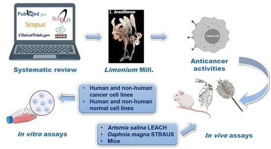
4. Materials and Methods

4.1. Study Design

This systematic review was performed following the recommendations of the Preferred Reporting Items for Systematic Reviews and Meta-Analyses (PRISMA) statement (Supplementary Materials, Board S1), Cochrane Handbook for Systematic Reviews of Interventions, and The Joana Briggs Institute (JBI) [40,71,72]. All the steps (i.e., article screening, full-text reading, data extraction, and methodological quality assessment) were conducted by two reviewers, independently. A third reviewer was consulted in the case of discrepancies. This study was registered in the Open Science Framework (OSF) with the registration DOI 10.17605/OSF.IO/WHBNE.

4.2. Systematic Literature Search and Eligibility Criteria

A systematic search was performed based on the electronic databases PubMed/MEDLINE, Scopus, and Scielo with no time or language restrictions (updated on 24 June 2021). The full search strategy is available in the Supplementary Materials (Board S2). A manual search was also conducted for the reference list of the included articles, in Clinical Trials.gov, and the Brazilian catalog of thesis from the Coordenação de Aperfeiçoamento de Pessoal de Nível Superior (CAPES).
We included in vitro, in vivo, ex vivo, or in silico experimental studies that evaluated the anticancer or anti-proliferative activity of Limonium species (e.g., crude extract, fraction, subfraction, or isolated compounds). Studies evaluating other biological activities of Limonium species and with other study designs (e.g., phytochemistry, agronomic perspective, botany, salinity, or cultivation studies) and those published in non-Roman characters were excluded.

4.3. Data Extraction and Reporting Evaluation

A standardized form was used to collect data on the studies’ general characteristics (e.g., authors, publication date, country), bioassay type, methodological aspects, and main results. The adapted tool for in vitro assays and the Systematic Review Center for Laboratory Animal Experimentation (SYRCLE) tool were used to assess the studies’ risk of bias and methodological quality in the Supplementary Materials (Tables S1 and S2) [70,73].

5. Conclusions

The literature on the potential anticancer effects of Limonium species mostly refers to preliminary assessments of the cytotoxicity of different compounds obtained from crude extracts, fractions, subfractions, and isolates and their impact on the viability of a range of cancer cell lines. Limonium tetragonum was the most evaluated species, with promising in vitro anti-melanogenesis effects. Isolated compounds of the flavonoid class, such as apigenin of L. duriusculum, also demonstrated a favorable cytotoxic effect against colorectal cancer, as well as lignanamides of L. gmelinii against cervix and breast adenocarcinoma cell lines. However, the complete mechanism of action of all isolated compounds and their effect in in vivo models remain unclear for most species.
These findings reinforce the biological potential of Limonium spp. as a source for the discovery and development of new potential cytotoxic phytochemicals. However, better planning, experimental designs, and reporting of the results will make future studies more robust and provide better proof to demonstrate the mechanism of action of these compounds.

Supplementary Materials

The following supporting information can be downloaded at: https://www.mdpi.com/article/10.3390/ph16020293/s1, Board S1 PRISMA 2020 Checklist, Board S2: Search Strategies, Table S1: Evaluation of the risk of bias by the adapted SYRCLE’s tool for in vitro studies, Table S2: Evaluation of the risk of bias by SYRCLE’s tool for in vivo studies, Board S3: Studies excluded after full reading, Figure S1: Chemical structures of isolated compounds of Limonium species drawn using ChemDraw version 14.0.0.118, Table S3: Checklist for reporting data on plant material for pharmacognostic studies.

Author Contributions

N.C.G., R.I., N.C.d.O., C.V.N., D.C.d.M.A., A.C.C.S., F.S.T., F.F.-L., D.C. and J.C.P.d.M. contributed to the study conception and design. N.C.G., D.C., R.I. and N.C.d.O. were involved in the collection, analysis, and data interpretation. N.C.G. and D.C. wrote the original draft, and all authors critically reviewed the content of the manuscript. All authors have read and agreed to the published version of the manuscript.

Funding

This work was supported by the Conselho Nacional de Desenvolvimento Científico e Tecnológico (CNPq, DC grant #150413/2020-3; JCPM grant #312309/2018-0) and Coordenação de Aperfeiçoamento de Pessoal de Nível Superior (CAPES, NCG grant #88882.448895/2019-01).

Institutional Review Board Statement

Not applicable.

Informed Consent Statement

Not applicable.

Data Availability Statement

Data sharing not applicable.

Acknowledgments

Thanks to the Post-Graduate Program in Pharmaceutical Sciences and the Laboratory of Pharmaceutical Biology (Palafito) at the State University of Maringá, and the efforts of all authors to make this work possible. The authors acknowledge Lucie Conte Herrig for the revision of the English.

Conflicts of Interest

The authors declare no conflict of interest.

References

  1. Desai, A.G.; Qazi, G.N.; Ganju, R.K.; El-Tamer, M.; Singh, J.; Saxena, A.K.; Bedi, Y.S.; Taneja, S.C.; Bhat, H.K. Medicinal plants and cancer chemoprevention. Curr. Drug Metab. 2008, 9, 581–591. [Google Scholar] [CrossRef]
  2. Pratheeshkumar, P.; Sreekala, C.; Zhang, Z.; Budhraja, A.; Ding, S.; Son, Y.O.; Wang, X.; Hitron, A.; Hyun-Jung, K.; Wang, L.; et al. Cancer prevention with promising natural products: Mechanisms of action and molecular targets. Anti-Cancer Agents Med. Chem. 2012, 12, 1159–1184. [Google Scholar] [CrossRef] [PubMed]
  3. Wang, H.; Khor, T.O.; Shu, L.; Su, Z.-Y.; Fuentes, F.; Lee, J.-H.; Kong, A.-N.T. Plants vs. cancer: A review on natural phytochemicals in preventing and treating cancers and their druggability. Anti-Cancer Agents Med. Chem. 2012, 12, 1281–1305. [Google Scholar] [CrossRef]
  4. Tariq, A.; Sadia, S.; Pan, K.; Ullah, I.; Mussarat, S.; Sun, F.; Abiodun, O.O.; Batbaatar, A.; Li, Z.; Song, D.; et al. A systematic review on ethnomedicines of anti-cancer plants. Phytother. Res. 2017, 31, 173–344. [Google Scholar] [CrossRef]
  5. Rauf, A.; Imran, M.; Butt, M.S.; Nadeem, M.; Peters, D.G.; Mubarak, M.S. Resveratrol as an anti-cancer agent: A review. Crit. Rev. Food Sci. Nutr. 2018, 58, 1428–1447. [Google Scholar] [CrossRef]
  6. Khan, Y.H.; Uttra, A.M.; Qasim, S.; Mallhi, T.H.; Alotaibi, N.H.; Rasheed, M.; Alzarea, A.I.; Iqbal, M.S.; Alruwaili, N.K.; Khan, S.-U.-D.; et al. Potential role of phytochemicals against matrix metalloproteinase induced breast cancer; an explanatory review. Front. Chem. 2021, 8, 592152. [Google Scholar] [CrossRef] [PubMed]
  7. Sultana, S.; Munir, N.; Mahmood, Z.; Riaz, M.; Akram, M.; Rebezov, M.; Kuderinova, N.; Moldabayeva, Z.; Shariati, M.A.; Rauf, A.; et al. Molecular targets for the management of cancer using Curcuma longa Linn. phytoconstituents: A review. Biomed. Pharmacother. 2021, 135, 111078. [Google Scholar] [CrossRef]
  8. Khan, H. Medicinal plants in light of history: Recognized therapeutic modality. J. Evid. Based Complement. Altern. Med. 2014, 19, 216–219. [Google Scholar] [CrossRef]
  9. Ijaz, S.; Akhtar, N.; Khan, M.S.; Hameed, A.; Irfan, M.; Arshad, M.D.; Ali, S.; Asrar, M. Plant derived anticancer agents: A green approach towards skin cancers. Biomed. Pharmacother. 2018, 103, 1643–1651. [Google Scholar] [CrossRef] [PubMed]
  10. Newman, D.J.; Cragg, G.M. Natural products as sources of new drugs over the nearly four decades from 01/1981 to 09/2019. J. Nat. Prod. 2020, 83, 770–803. [Google Scholar] [CrossRef] [PubMed] [Green Version]
  11. Plants of the World Online (POWO). Plumbaginaceae Juss. Available online: https://powo.science.kew.org/taxon/urn:lsid:ipni.org:names:30000293-2#children (accessed on 21 March 2022).
  12. Plants of the World Online (POWO). Limonium Mill. Available online: https://powo.science.kew.org/taxon/urn:lsid:ipni.org:names:331722-2#children (accessed on 21 March 2022).
  13. Murray, A.P.; Rodriguez, S.; Frontera, M.A.; Tomas, M.A.; Mulet, M.C. Antioxidant metabolites from Limonium brasiliense (Boiss.) Kuntze. Z. Nat. C 2004, 59, 477–480. [Google Scholar] [CrossRef] [PubMed]
  14. Medini, F.; Fellah, H.; Ksouri, R.; Abdelly, C. Total phenolic, flavonoid and tannin contents and antioxidant and antimicrobial activities of organic extracts of shoots of the plant Limonium delicatulum. J. Taibah Univ. Sci. 2014, 8, 216–224. [Google Scholar] [CrossRef]
  15. Benmeddour, T.; Laouer, H.; Flamini, G.; Akkal, S. Chemical composition of essential oil of Limonium bonduellei. Chem. Nat. Compd. 2018, 54, 188–190. [Google Scholar] [CrossRef]
  16. Gadetskaya, A.V.; Tarawneh, A.H.; Zhusupova, G.E.; Gemejiyeva, N.G.; Cantrell, C.L.; Cutler, S.J.; Ross, S.A. Sulfated phenolic compounds from Limonium caspium: Isolation, structural elucidation, and biological evaluation. Fitoterapia 2015, 104, 80–85. [Google Scholar] [CrossRef]
  17. Geng, D.; Chi, X.; Dong, Q.; Hu, F. Antioxidants screening in Limonium aureum by optimized on-line HPLC-DPPH assay. Ind. Crops Prod. 2015, 67, 492–497. [Google Scholar] [CrossRef]
  18. Medini, F.; Bourgou, S.; Lalancette, K.; Snoussi, M.; Mkadmini, K.; Coté, I.; Abdelly, C.; Legault, J.; Ksouri, R. Phytochemical analysis, antioxidant, anti-inflammatory, and anticancer activities of the halophyte Limonium densiflorum extracts on human cell lines and murine macrophages. S. Afr. J. Bot. 2015, 99, 158–164. [Google Scholar] [CrossRef]
  19. Blainski, A.; Gionco, B.; Oliveira, A.G.; Andrade, G.; Scarminio, I.S.; Silva, B.D.; Lopes, N.P.; Mello, J.C.P. Antibacterial activity of Limonium brasiliense (Baicuru) against multidrugresistant bacteria using a statistical mixture design. J. Ethnopharmacol. 2017, 198, 313–323. [Google Scholar] [CrossRef]
  20. Caleare, A.O.; Hensel, A.; Mello, J.C.P.; Pinha, A.B.; Panizzon, G.P.; Lechtenberg, M.; Petereit, F.; Nakamura, C.V. Flavan-3-ols and proanthocyanidins from Limonium brasiliense inhibit the adhesion of Porphyromonas gingivalis to epithelial host cells by interaction with gingipains. Fitoterapia 2017, 118, 87–93. [Google Scholar] [CrossRef]
  21. Sereia, A.L.; Oliveira, M.T.; Baranoski, A.; Marques, L.L.M.; Ribeiro, F.M.; Isolani, R.G.; Medeiros, D.C.; Chierrito, D.; Lazarin-Bidóia, D.; Zielinski, F.; et al. In vitro evaluation of the protective effects of plant extracts against amyloid-beta peptide-induced toxicity in human neuroblastoma SH-SY5Y cells. PLoS ONE 2019, 14, e0212089. [Google Scholar] [CrossRef]
  22. Al-madhagi, W.M.; Hashim, N.M.; Ali, N.A.A.; Othman, R. Phytochemical screening, cytotoxic and antimicrobial activities of Limonium socotranum and Peperomia blanda extracts. Trop Biomed. 2019, 36, 11–21. [Google Scholar]
  23. Souid, A.; Bellani, L.; Gabriele, M.; Pucci, L.; Smaoui, A.; Abdelly, C.; Hamed, K.B.; Longo, V. Phytochemical and biological activities in Limonium species collected in different biotopes of Tunisia. Chem. Biodivers. 2019, 16, e1900216. [Google Scholar] [CrossRef] [PubMed]
  24. Tuasha, N.; Petros, B.; Asfaw, Z. Plants used as anticancer agents in the Ethiopian traditional medical practices: A systematic review. Evid. Based Complement. Altern. Med. 2018, 18, 6274021. [Google Scholar] [CrossRef]
  25. Lellau, T.F.; Liebezeit, G. Cytotoxic and antitumor activities of ethanolic extracts of salt marsh plants from the lower Saxonian Wadden Sea, Southern North Sea. Pharm. Biol. 2003, 41, 293–300. [Google Scholar] [CrossRef]
  26. Tang, X.-H.; Yan, L.-F.; Gao, J.; Yang, X.-L.; Xu, Y.-X.; Ge, H.-Y.; Yang, H.-D. Antitumor and immunomodulatory activity of polysaccharides from the root of Limonium sinense Kuntze. Int. J. Biol. Macromol. 2012, 51, 1134–1139. [Google Scholar] [CrossRef]
  27. Kong, N.-N.; Fang, S.-T.; Wang, J.-H.; Wang, Z.-H.; Xia, C.-H. Two new flavonoid glycosides from the halophyte Limonium franchetii. J. Asian Nat. Prod. Res. 2014, 16, 370–375. [Google Scholar] [CrossRef] [PubMed]
  28. Tang, X.-H.; Yu, F.; Liu, J.; Gao, J.; Yan, L.-F.; Dong, M.-M. Isolation and identification of anti-tumor polysaccharide LSP21 from Limonium sinense (Girard) Kuntze. Int. J. Biol. Macromol. 2014, 70, 138–142. [Google Scholar] [CrossRef]
  29. Bae, M.-J.; Karadeniz, F.; Lee, S.-G.; Seo, Y.; Kong, C.-S. Inhibition of MMP-2 and MMP-9 activities by Limonium tetragonum extract. Prev. Nutr. Food Sci. 2016, 21, 38–43. [Google Scholar] [CrossRef]
  30. Rodrigues, M.J.; Neves, V.; Martins, A.; Rauter, A.P.; Neng, N.R.; Nogueira, J.M.F.; Varela, J.; Barreira, L.; Custódio, L. In vitro antioxidant and anti-inflammatory properties of Limonium algarvense flowers’ infusions and decoctions: A comparison with green tea (Camellia sinensis). Food Chem. 2016, 200, 322–329. [Google Scholar] [CrossRef]
  31. Bae, M.J.; Karadeniz, F.; Oh, J.H.; Yu, G.H.; Jang, M.; Nam, K.; Seo, Y.; Kong, C. MMP-Inhibitory effects of flavonoid glycosides from edible medicinal halophyte Limonium tetragonum. Evid. Based Complement. Altern. Med. 2017, 2017, 6750274. [Google Scholar] [CrossRef]
  32. Chen, J.; Teng, J.; Ma, L.; Tong, H.; Ren, B.; Wang, L.; Li, W. Flavonoids isolated from the flowers of Limonium bicolor and their in vitro antitumor evaluation. Pharm. Mag. 2017, 13, 222–225. [Google Scholar] [CrossRef]
  33. Cordeiro, M.F. Avaliação das atividades imunomoduladora, antineoplásica e antibacteriana de rizomas de Limonium brasiliense, sementes de Paullinia cupana e cascas do caule de Trichilia catigua. Ph.D. Thesis, Universidade Federal de Pernambuco, Centro de Biociências, Programa de Pós-Graduação em Inovação Terapêutica, Recife, Brazil, 2017; pp. 1–142. [Google Scholar]
  34. Lee, S.-G.; Karadeniz, F.; Seo, Y.; Kong, C.-S. Anti-melanogenic effects of flavonoid glycosides from Limonium tetragonum (Thunb.) Bullock via inhibition of tyrosinase and tyrosinase-related proteins. Molecules 2017, 22, 1480. [Google Scholar] [CrossRef] [PubMed] [Green Version]
  35. Sahli, R.; Rivière, C.; Neut, C.; Bero, J.; Sahuc, M.-E.; Smaoui, A.; Beaufay, C.; Roumy, V.; Hennebelle, T.; Rouillé, Y.; et al. An ecological approach to discover new bioactive extracts and products: The case of extremophile plants. J. Pharm. Pharmacol. 2017, 69, 1041–1055. [Google Scholar] [CrossRef]
  36. Amrani, A.; Lahneche, A.M.; Benaissa, O.; Boubekri, N.; Demirtas, I.; Benayache, F.; Benayache, S.; Zama, D. In vitro antiproliferative and inhibition of oxidative DNA damage activities of n-butanol extract of Limonium bonduelli from Algeria. Braz. Arch. Biol. Technol. 2019, 62, e19170779. [Google Scholar] [CrossRef]
  37. Hamadou, M.H.; Kerkatou, M.; Gatto, P.; Pancher, M.; Bisio, A.; Inga, A.; Menad, A.; Benayache, S.; Benayache, F.; Ameddah, S. Apigenin rich Limonium duriusculum (de Girard) Kuntze promotes apoptosis in HCT116 cancer cells. Nat. Prod. Res. 2019, 35, 2910–2914. [Google Scholar] [CrossRef] [PubMed]
  38. Rodrigues, M.J.; Monteiro, I.; Castañeda-Loaiza, V.; Placines, C.; Oliveira, M.C.; Reis, C.; Caperta, A.D.; Soares, F.; Pousão-Ferreira, P.; Pereira, C.; et al. Growth performance, in vitro antioxidant properties and chemical composition of the halophyte Limonium algarvense Erben are strongly influenced by the irrigation salinity. Ind. Crops Products. 2020, 143, 111930. [Google Scholar] [CrossRef]
  39. Tuohongerbieke, A.; Li, J.; Sabir, G.; Xin, X.; Hu, M.; Duan, X.; Liu, L.; Tang, D.; Zhu, J.; Aisa, H.A. Lignanamides from the roots of Limonium gmelinii (Willd.) Kuntze and their anti-diabetic, cytotoxic and anti-inflammatory activities. Phytochemistry 2021, 184, 112648. [Google Scholar] [CrossRef] [PubMed]
  40. Page, M.J.; McKenzie, J.E.; Bossuyt, P.M.; Boutron, I.; Hoffmann, T.C.; Mulrow, C.D.; Shamseer, L.; Tetzlaff, J.M.; Moher, D. The PRISMA 2020 statement: An updated guideline for reporting systematic reviews. PLoS Med. 2021, 18, e1003583. [Google Scholar] [CrossRef]
  41. Hassan, M.A.; Estrelles, E.; Soriano, P.; López-Gresa, M.P.; Bellés, J.M.; Boscaiu, M.; Vicente, O. Unraveling salt tolerance mechanisms in halophytes: A comparative study on four mediterranean Limonium species with different geographic distribution patterns. Front. Plant Sci. 2017, 8, 1438. [Google Scholar] [CrossRef]
  42. González-Orenga, S.; Grigore, M.N.; Boscaiu, M.; Vicente, O. Constitutive and induced salt tolerance mechanisms and potencial uses of Limonium Mill. Species. Agronomy 2021, 11, 413. [Google Scholar] [CrossRef]
  43. Dholwani, K.K.; Saluja, A.K.; Gupta, A.R.; Shah, D.R. A review on plant-derived natural products and their analogs with anti-tumor activity. Indian J. Pharmacol. 2008, 40, 49–58. [Google Scholar] [CrossRef]
  44. Kuete, V.; Efferth, T. African flora has the potential to fight multidrug resistance of cancer. Biomed. Res. Int. 2015, 2015, 914813. [Google Scholar] [CrossRef]
  45. American Cancer Society (ACS). How Chemotherapy Drugs Work. Available online: https://www.cancer.org/treatment/treatments-and-side-effects/treatment-types/chemotherapy/how-chemotherapy-drugs-work.html (accessed on 21 March 2022).
  46. Jain, C.K.; Majumder, H.K.; Roychoudhury, S. Natural compounds as anticancer agents targeting DNA topoisomerases. Curr. Genomics. 2017, 18, 75–92. [Google Scholar] [CrossRef] [PubMed]
  47. Lichota, A.; Gwozdzinski, K. Anticancer activity of natural compounds from plant and marine environment. Int. J. Mol. Sci. 2018, 19, 3533. [Google Scholar] [CrossRef] [PubMed]
  48. Vardanyan, R.S.; Hruby, V.J. Synthesis of Essential Drugs, 1st ed.; Elsevier Science: Amsterdam, The Netherlands, 2006; pp. 389–418. [Google Scholar]
  49. Ina, K.; Kataoka, T.; Ando, T. The use of Lentinan for treating gastric cancer. Anticancer. Agents Med. Chem. 2013, 13, 681–688. [Google Scholar] [CrossRef]
  50. Masuda, M.; Itoh, K.; Murata, K.; Naruto, S.; Uwaya, A.; Isami, F.; Matsuda, H. Inhibitory effects of Morinda citrifolia extract and its constituents on melanogenesis in murine B16 melanoma cells. Biol. Pharm. Bull. 2012, 35, 78–83. [Google Scholar] [CrossRef]
  51. Mosmann, T. Rapid colorimetric assay for cellular growth and survival: Application to proliferation and cytotoxicity assays. J. Immunol. Methods. 1983, 65, 55–63. [Google Scholar] [CrossRef] [PubMed]
  52. McCauley, J.; Zivanovic, A.; Skropeta, D. Bioassays for anticancer activities. In Metabolomics Tools for Natural Product Discovery. Methods in Molecular Biology (Methods and Protocols); Roessner, U., Dias, D., Eds.; Humana Press: Totowa, NJ, USA, 2013; pp. 191–205. [Google Scholar]
  53. Cox, P.A. Pharmacology, Biodiversity. In Encyclopedia of Biodiversity, 2nd ed.; Levin, S.A., Ed.; Academic Press: Cambridge, MA, USA, 2013; pp. 703–715. [Google Scholar]
  54. Aslantürk, Ö.S. In vitro cytotoxicity and cell viability assays: Principles, advantages, and disadvantages. In Genotoxicity—A Predictable Risk to Our Actual World; Larramendy, M.L., Soloneski, S., Eds.; IntechOpen: London, UK, 2017; pp. 1–17. [Google Scholar] [CrossRef]
  55. Michael, A.S.; Thompson, C.G.; Abramovitz, M. Artemia salina as a test organism for bioassay. Science 1956, 123, 464. [Google Scholar] [CrossRef]
  56. Waart, J.; Van Aken, F.; Pouw, H. Detection of orally toxic microbial metabolites in foods with bioassay systems. Zent. Bakteriol. Orig. A 1972, 222, 96–114. [Google Scholar]
  57. Adema, D.M.M. Daphnia magna as a test animal in acute and chronic toxicity tests. Hydrobiologia 1978, 59, 125–134. [Google Scholar] [CrossRef]
  58. Canton, J.H.; Adema, D.M.M. Reproducibility of short-term and reproduction toxicity experiments with Daphnia magna and comparison of the sensitivity of Daphnia magna with Daphnia pulex and Daphnia cucullata in short-term experiments. Hydrobiologia 1978, 59, 135–140. [Google Scholar] [CrossRef]
  59. Sandbacka, M.; Christianson, I.; Isomaa, B. The acute toxicity of surfactants on fish cells, Daphnia magna and fish—A comparative study. Toxicol. Vitr. 2000, 14, 61–68. [Google Scholar] [CrossRef]
  60. Arcanjo, D.D.R.; Albuquerque, A.C.M.; Melo-Neto, B.; Santana, L.C.L.R.; Medeiros, M.G.F.; Citó, A.M.G.L. Bioactivity evaluation against Artemia salina Leach of medicinal plants used in Brazilian Northeastern folk medicine. Braz. J. Biol. 2012, 72, 505–509. [Google Scholar] [CrossRef]
  61. Visconti, R.; Grieco, D. New insights on oxidative stress in cancer. Curr. Opin. Drug Discov. Devel. 2009, 12, 240–245. [Google Scholar] [PubMed]
  62. Arulselvan, P.; Fard, M.T.; Tan, W.S.; Gothai, S.; Fakurazi, S.; Norhaizan, M.E.; Kumar, S.S. Role of antioxidants and natural products in inflammation. Oxidative Med. Cell. Longev. 2016, 2016, 5276130. [Google Scholar] [CrossRef]
  63. Mohamed, S.I.A.; Jantan, I.; Haque, M.A. Naturally occurring immunomodulators with antitumor activity: An insight on their mechanisms of action. Int. Immunopharmacol. 2017, 50, 291–304. [Google Scholar] [CrossRef]
  64. Nuzzo, G.; Senese, G.; Gallo, C.; Albiani, F.; Romano, L.; d’Ippolito, G.; Manzo, E.; Fontana, A. Antitumor potential of immunomodulatory natural products. Mar. Drugs. 2022, 20, 386. [Google Scholar] [CrossRef]
  65. Ruiz-Riaguas, A.; Zengin, G.; Sinan, K.I.; Salazar-Mendías, C.; Llorent-Martínez, E.J. Phenolic profile, antioxidant activity, and enzyme inhibitory properties of Limonium delicatulum (Girard) Kuntze and Limonium quesadense Erben. J. Chem. 2020, 2020, 1016208. [Google Scholar] [CrossRef]
  66. Reuter, S.; Gupta, S.C.; Chaturvedi, M.M.; Aggarwal, B.B. Oxidative stress, inflammation, and cancer: How are they linked? Free Radic. Biol. Med. 2010, 49, 1603–1616. [Google Scholar] [CrossRef] [PubMed]
  67. Hamed, K.B.; Chibani, F.; Abdelly, C.; Magne, C. Growth, sodium uptake and antioxidant responses of coastal plants differing in their ecological status under increasing salinity. Biologia 2014, 69, 193–201. [Google Scholar] [CrossRef]
  68. Bakhshi, S.; Abbaspour, H.; Saeidisar, S. Study of phytochemical changes, enzymatic and antioxidant activity of two halophyte plants: Salsola dendroides Pall and Limonium reniforme (Girard) Lincz in different seasons. Bulg. Chem. Communications. 2018, 50, 374–382. [Google Scholar]
  69. Jafari, S.; Saeidnia, S.; Abdollahi, M. Role of natural phenolic compounds in cancer chemoprevention via regulation of the cell cycle. Curr. Pharm. Biotechnol. 2014, 15, 409–421. [Google Scholar] [CrossRef] [PubMed]
  70. Chierrito, D.; Villas-Boas, C.B.; Tonin, F.S.; Fernandez-Llimos, F.; Sanches, A.C.C.; de Mello, J.C.P. Using cell cultures for the investigation of treatments for attention deficit hyperactivity disorder: A systematic review. Curr. Neuropharmacol. 2019, 17, 916–925. [Google Scholar] [CrossRef] [PubMed]
  71. Higgins, J.P.T.; Thomas, J.; Chandler, J.; Cumpston, M.; Li, T.; Page, M.J.; Welch, V.A. Cochrane Handbook for Systematic Reviews of Interventions Version 6.2. 2021. Available online: https://www.training.cochrane.org/handbook (accessed on 24 October 2021).
  72. Joanna Briggs Institute (JBI). Methodology for JBI Scoping Reviews Joanna Briggs Institute. Joanna Briggs Institute Reviewers’ Manual. 2015 ed./Supplement. Australia: Joanna Briggs Institute. 2015. Available online: https://nursing.lsuhsc.edu/jbi/docs/reviewersmanuals/scoping-.pdf (accessed on 24 October 2021).
  73. Hooijmans, C.R.; Rovers, M.M.; Vries, R.B.; Leenaars, M.; Ritskes-Hoitinga, M.; Langendam, M.W. SYRCLE’s risk of bias tool for animal studies. BMC Med. Res. Methodol. 2014, 14, 43. [Google Scholar] [CrossRef] [PubMed] [Green Version]
Figure 1. Flow diagram of the systematic review. Fonte: Adapted from Page et al. [40].
Pharmaceuticals 16 00293 g001
Figure 2. Main results of in vivo and in vitro assays of eligible studies included in the systematic review. Fonte: Adapted from Medini et al. [18], Al-madhagi et al. [22], Lellau et al. [25], Tang et al. [26,28], Kong et al. [27], Bae et al. [29,31], Chen et al. [32], Cordeiro [33], Lee et al. [34], Sahli et al. [35], Amrani et al. [36], Hamadou et al. [37], Rodrigues et al. [30,38], Tuohongerbieke et al. [39].
Pharmaceuticals 16 00293 g002
Figure 3. The possible cellular mechanism involved in the anti-cancer activity of Limonium species included in the systematic review. (A) Cell proliferation inhibition pathways mediated by L. duriusculum extract. (B) Inhibition of matrix metalloproteinases (MMP-2 and 9), Zn dependent endoproteases related to various complications in cancer, such as metastasis, and anti-melanogenic activity linked to inhibition of melanin biosynthesis mediated by bioactive compounds of L. tetragonum. (C) Immunomodulatory activity related to reduced of interleukin (IL) 6, 17A, and 22, and interferon-gamma (IFN-ϒ) mediated by L. brasiliense crude extracts and fractions, suggesting an anticancer effect of this species. Note: Adapted from Hamadou et al. [37]; Bae et al. [31]; Lee et al. [34]; Cordeiro [33].
Pharmaceuticals 16 00293 g003
Table 1. Main characteristics of all eligible in vivo and in vitro studies included in the systematic review.
Reference NumberCountryPlant SpeciesPart of Plant UsedCell LineBioassay/
Model Used
Compound TestedPositive ControlTime of Treatment (h)Other Biological Activities
In vivo
[25]GermanyL. vulgareNRNRArtemia salina Daphnia magnaEtOH extractHgCl2 solution (1%)6
24
48
NR
[26]ChinaL. sinenseRootsHepG2MiceCrude LSPCyclophosphamide
Lentinan with 5-fluoracil
5-Fluoracil
312Immunomodulatory effects
[30]PortugalL. algarvenseFlowersNRArtemia salinaAq extractNR48Antioxidant and anti-inflammatory activities
In vitro
[26]ChinaL. sinenseRootsHeLa
HepG2
K562
MTTCrude LSPNR24Immunomodulatory effects
[28]ChinaL. sinenseRootsHepG2MTTLSP11
LSP21
LSP31
5-Fluorouracil24NR
[27]ChinaL. franchetiiWholeBGC-823MTT12 isolated compoundsNRNRNR
C6
HepG2
Sulforhodamine B
[18]TunisiaL. densiflorumLeavesA549
DLD-1
WS1
Resazurin reduction testDCM extract
EtOH extract
MeOH extract
Hex extract
Etoposide48Antioxidant and anti-inflammatory activities
[29]KoreaL. tetragonumNRHT-1080MTTDCM fractionNR48Determination of enzymatic activities of MMPs, mRNA expression of MMPs and TIMPs via RT-PCR, and detection of immunoreactive proteins via Western blotting
[31]KoreaL. tetragonumNRHT-1080MTTDCM extract
(Hex fraction and 85% MeOH fraction)
Aq extract
(BuOH fraction and Aq fraction)
NR48Determination of enzymatic activities of MMPs, mRNA expression of MMPs and TIMPs via RT-PCR, and detection of immunoreactive proteins via Western blotting
[34]KoreaL. tetragonumNRB16-F10Spectrophotometric methodHex fraction
85% MeOH fraction
BuOH fraction
Aq fraction
Kojic acid0.5DOPA oxidase activity, cellular tyrosinase activity, melanin content, melanogenesis-related mRNA expression via RT-PCR, and detection of TRP via Western blotting
[30]PortugalL. algarvenseFlowersHepG2
N9
S17
RAW-264.7
MTTAq extractNR72Antioxidant and anti-inflammatory activities
[38]PortugalL. algarvenseFlowers
Leaves
Peduncles
HEK-293
HepG2
RAW-264.7
MTTEtOH extractNR72Antioxidant activity
[32]ChinaL. bicolorFlowersLoVo
MCF-7
U-2OS
MTT15 isolated compounds5-Fluorouracil48NR
[33]BrazilL. brasilienseRhizomeHepG2
HL-60
K562
MOLT-4
PANC-1
PBMC
SK-MEL-28
T-47D
Toledo
Vero
MTTCE
Aq fraction
EAF Subfractions (A-K)
Isolated compounds
(SA, SB, EGCG)
Amsacrine72Selectivity index, anti-migration and anti-clonogenic potential, and immunomodulatory activity
[35]FranceL. virgatumLeaves
Stems
J774
WI-38
MTTMeOH extractCamptothecin72Antiradical, antimicrobial, and antiviral activity
[22]YemenL. sokotranumFlowers
Leaves
Stem
HepG2
MCF-7
Sulforhodamine BPE extract
DCM extract
MeOH extract
Doxorubicin48Antibacterial and antifungal activity
[36]AlgeriaL. bonduelleiFlowers
Leaves
HeLa
HT-29
xCELLigence RTCABuOH extractNR48
72
DNA damage inhibition efficiency
[37]AlgeriaL. duriusculumFlowers
Leaves
HCT116Calcein-AM
Hoechst 33342
BuOH extract
Apigenin
NR48Measures of relative levels of p53, MDM2, p21, total and p-ERK proteins, and PARP cleavage via western blotting
[39]ChinaL. gmeliniiRootsA549
HeLa
MCF-7
MTTEtOAc extract
19 isolated compounds
Doxorubicin48Anti-diabetic and anti-inflammatory activities
Abbreviations: A549: human lung carcinoma; Aq: aqueous; B16-F10: melanoma (mouse); BGC-823: human gastric adenocarcinoma; BuOH: n-butanol; C6: brain glioma (rat); Calcein-AM: calcein-acetomethoxy; CE: crude extract; DCM: dicloromethane; DLD-1: human colorectal adenocarcinoma; EAF: ethyl-acetate fraction; EGCG: epigallocatequin-3-O-gallate; EtOAc: ethyl-acetate; EtOH: ethanol; HCT116: human colorectal carcinoma; HeLa: human cervix adenocarcinoma; HepG2: human hepatocellular carcinoma; Hex: n-hexane; HL-60: human acute promyelocytic leukemia; HT-29: human colorectal adenocarcinoma; HT-1080: human fibrosarcoma; HEK-293: human embryonic kidney (normal cell); J774: sarcoma (mice); K562: human chronic myelogenous leukemia; LoVo: human colorectal adenocarcinoma; LSP: Limonium sinense polysaccharide; MMPs: matrix metalloproteinases; MCF-7: human breast adenocarcinoma; MeOH: methanol; MOLT-4: human acute lymphoblastic leukemia; N9: microglia (mice normal cell); NR: not related; PANC-1: human pancreas epithelioid carcinoma; PBMCs: human primary peripheral blood mononuclear cells (normal cells); PE: petroleum ether; p-ERK: phosphorylated ERK; RAW-264.7: Abelson murine leukemia virus-induced tumor (mouse); S17: bone marrow (mouse normal cell); SA: samarangenin A; SB: samarangenin B; SK-MEL-28: human malignant melanoma; T-47D: human breast carcinoma; TIMPs: tissue inhibitor of metalloproteinases; Toledo: human diffuse large cell lymphoma (non-Hodgkin’s B cell); TRP: tyrosinase-related proteins; U-2OS: human osteosarcoma; Vero: kidney (monkey normal cell); xCELLigence RTCA: xCELLigence real-time cell analyzes; WI-38: human lung fibroblast (normal cell); WS1: human skin fibroblast (normal cell). Note: The studies were described in chronological order. The same species were described together.
Table 2. Phytochemistry of Limonium species included in the systematic review.
Reference NumberPlant SpeciesClass of MetaboliteCompoundsNumber of Isolated Compounds Tested In Vitro *
Primary metabolites
[28]L. sinensePolysaccharideLSP21 (glucose, galactose and mannose)
[38]L. algarvenseAmino acidN-acetyl-tryptophan
Fatty acidsOxo-tridecanoic acid sulphate
Trihydroxy-10-octadecenoic acid
Trihydroxy-10,15-octadecadienoic acid
PolysaccharideHex-3-en-olxylopyranosyl-(1-6)-glicopyranoside
Sucrose or isomeric structures
Secondary metabolites
[27]L. franchetiiFlavonoidsApigenin
Dihydrokaempferol
Kaempferol-3-O-α-L-rhamnopyranoside
Luteolin
Myricetin
Myricetin-3-O-(2″-O-galloyl)-α-L-rhamnopyranoside
Myricetin-3-O-(3″-O-galloyl)-α-L-rhamnopyranoside
Myricetin-3-O-α-L-rhamnopyranoside
Quercetin
Quercetin-3-O-(2″-O-tigloyl)-α-L-rhamnopyranoside
Quercetin-3-O-(3″-O-tigloyl)-α-L-rhamnopyranoside
Quercetin-3-O- α-L-rhamnopyranoside
(1)
(2)
(3)
(4)
(5)
(6)

(7)

(8)
(9)
(10)

(11)

(12)
[18]L. densiflorumFlavonoidsCatechin hydrate
Isorhamnetin
Myricetin
Phenolic acidsEllagic acid
Gallic acid
Sinapic acid
trans 3-hydroxycinnamic acid
[30]L. algarvenseFlavonoidApigenin
Phenolic acidsCaffeic acid
Coumaric acid
Ferulic acid
Gallic acid
p-Hydroxybenzoic acid
Salicylic acid
Syringic acid
[38]LigninPinoresinol sulphate
Flavonoids2′-C-methyl myricetin-3-O-rhamnoside-galloyl
4′-methyl eriodictyol-galloyl-rhamnose
Apigenin
Apigenin derivative
Apigenin-O-glucoside
Apigenin-O-glucuronide
Dihydrokaempferol
Epigallocatechin gallate
Eriodictyol
Eriodyctiol-O-glucoside
Isorhamnetin-3-O-rutinoside
Licoagroside B
Luteolin
Luteolin-7-O-glucoside
Luteolin-7-O-rhamnoside
Methyl licoagroside B
Myricetin
Myricetin-3-O-(2″-O-galloyl)-glucoside
Myricetin-3-O-acetyl-deoxyhexose
Myricetin-3-O-acetyl-hexoside
Myricetin-3-O-pentoside
Myricetin-ethyl acetoacetate-galloyl
Myricetin-galloyl-acetyl deoxyhexose
Myricetin-O-(galloyl)-deoxyhexose
Myricitin-3-O-glucoside
Myricitin-3-O-rhamnose
Myricitin-3-O-rutinoside
Naringenin
Naringenin derivative
Quercetin
Quercetin derivative
Quercetin-3-O-rhamnoside
Quercetin-hexoside derivative
Quercetin-O-galloy-glucoside
Quercetin-O-hexoside
Quercetin-tetramethyl ether- -dihydroxyethylfructopyranose
Rutin
Phenolic acidsFeruloyltyramine
Glucosyringic acid
Syringic acid
TanninsDigalloyl-hexoside
Galloylglucoside derivative
Galloyl-hexoside
Galloylhexoside derivative
PhenylpropanoidSinapyl alcohol sulphate
[31]L. tetragonumFlavonoidsMyricetin 3-galactoside
Quercetin 3-O-β-galactopyranoside
(13)
(14)
[34]
[32]L. bicolorFlavonoidsAcacetin
Eriodictyol
Hesperidin
Isorhamnetin
Kaempferol
Kaempferol-3-O-(6″-O-galloyl)-β-D-glucoside
Kaempferol-3-O-α-L-rhamnoside
Kaempferol-3-O-β-D-glucoside
Luteolin
Myricetin-3-O-α-L-rhamnoside
Quercetin
Quercetin-3-O-α-L-rhamnoside
Quercetin-3-O-β-D-galactoside
Quercetin-3-O-β-D-glucoside
Rutin
(15)
(16)
(17)
(18)
(19)
(20)
(21)
(22)
(4)
(23)
(9)
(24)
(25)
(26)
(27)
[33]L. brasilienseTanninsEpigallocatequin-3-O-gallate
Samarangenin A
Samarangenin B
(28)
(29)
(30)
[35]L. virgatumPhenolic amideN-trans-feruloyl tyramine
[37]L. duriusculumFlavonoidsApigenin
Apigenin 7-O-β-D-(6”-methylglucuronide)
(31)
(32)
[39]L. gmeliniiLignanamides(2,3-trans)-3-(3-hydroxy-5-methoxyphenyl)-N-(4-hydroxyphenethyl)-7-{(E)-3-[(4-hydroxyphenethyl)amino]-3-oxoprop-1-en-1-yl}-2,3-dihydrobenzo[b][1,4]dioxine-2-carboxamide
Limoniumin F
3,3′ -demethyl-heliotropamide
Limoniumin A
Limoniumin B
Limoniumin C
Limoniumin D
6-hydroxy-4-(4-hydroxy-3-methoxyphenyl)-2-(4-hydroxyphenethyl)-7-methoxy-1H-benzo(f)isoindole-1,3(2H)-dione
Cannabisin I
Limoniumin E
Limoniumin G
Limoniumin H
Limoniumin I
Cannabisin D
Cannabisin B
Cannabisin C
Cannabisin A
Cannabisin F
Thoreliamide B
(33)


(34)
(35)
(36)
(37)
(38)
(39)
(40)


(41)
(42)
(43)
(44)
(45)
(46)
(47)
(48)
(49)
(50)
(51)
Phenolic amideN-cis-feruloyl tyramine
N-trans-feruloyl tyramine
* Chemical structures of the isolated compounds investigated for their cytotoxicities, numbered in bold, were drawn in the Supplementary Materials (Figure S1).
Table 3. Results of inhibitory concentration of 50% (IC50) of cell proliferation based on eligible studies included in the systematic review.
Reference
Number
Cell LineIC50 (µg/mL)/Compound TestedSelectivity Index (SI)/
Compound Tested
[18]A54929 (DCM extract), >200 (EtOH extract), 110 (MeOH extract), >200 (Hex extract), 10 (Etoposide PC)NR
DLD-185 (DCM extract), >200 (EtOH extract), >200 (MeOH extract), >200 (Hex extract), 80 (Etoposide PC)
WS1>200 (DCM extract), >200 (EtOH extract), 140 (MeOH extract), >200 (Hex extract), 26 (Etoposide PC)
[33]HepG2>200 (CE), 67.97 (Aq fraction), 59.47 (EAF)0.48 (CE)
2.94 (AF)
1.27 (EAF)
HL-6061.69 (CE), 49.68 (Aq fraction), 17.26 (EAF)1.56 (CE)
4.02 (AF)
4.39 (EAF)
PBMC96.78 (CE), >200 (Aq fraction), 75.82 (EAF)NR
T-47D90.68 (CE), >200 (Aq fraction), 77.70 (EAF)1.08 (CE)
1.00 (AF)
0.48 (EAF)
HL-6053.27 (SFa), 35.48 (SFb), 44.28 (SFc), 41.63 (SFd), 43.62 (SFe), 8.21 (SFf), 7.35 (SFg), 45.58 (SFh), 55.60 (SFi), 54.06 (SFj), 53.32 (SFk), 1.0 (Amsacrine PC)NR
K56243.72 (SFa), 52.21 (SFb), 52.75 (SFc), 43.95 (SFd), 47.79 (SFe), 36.13 (SFf), 40.88 (SFg), 49.91 (SFh), 51.85 (SFi), 50.16 (SFj), 37.77 (SFk), 0.9 (Amsacrine PC)
MOLT-437.43 (SFa), 34.34 (SFb), 35.99 (SFc), 46.47 (SFd), 45.25 (SFe), 40.42 (SFf), 7.92 (SFg), 20.36 (SFh), 54.92 (SFi), 52.76 (SFj), 9.62 (SFk), NR (Amsacrine PC)
PANC-1>100 (SFa), >100 (SFb), 76.81 (SFc), 45.54 (SFd), >100 (SFe), 58.65 (SFf), >100 (SFg), >100 (SFh), >100 (SFi), >100 (SFj), >100 (SFk), >100(Amsacrine PC)
SK-MEL-28NA
Toledo57.46 (SFa), 57.02 (SFb), 61.29 (SFc), 54.09 (SFd), 55.29 (SFe), 55.29 (SFf), 54.32 (SFg), 57.36(SFh), 60.65 (SFi), 58.68 (SFj), 59.38 (SFk), 0.5 (amsacrine PC)
K56237.04 (28), 29.24 (29), 51.17 (30)2.69 (28)
3.41 (29)
1.95 (30)
Vero>100 (28), >100 (29), >100 (30)NR
[37]HCT1167.60 (BuOH extract), 25.74 * (31), NA (32)NR
[22]MCF-719.65 and 14.57 (PE extracts), 17.60 and 21.8 (DCM extracts), 8.70 and 17.18 (MeOH extracts), 3.39 (Doxorubicin PC)NR
HepG29.97 and 16.97 (PE extracts), 20.62 and 11.15 (DCM extracts), 13.90 and 24.86 (MeOH extracts), 7.38 (Doxorubicin PC)NR
[39]HeLa25.25 (EtOAc extract), NA ((2), (7) and (13)), 19.24 * (17), 12.85 * (18), 31.57 * (19), 0.23 * (Doxorubicin PC)NR
MCF-7NA (EtOAc extract), 20.08 (2), 21.58 (7), 43.28 (13), 28.85 (17), 14.14 (18), NA (19)
Abbreviations: A549: human lung carcinoma; Aq: aqueous; BuOH: n-butanol; CE: crude extract; DCM: dichloromethane; DLD-1: human colorectal adenocarcinoma; EAF: ethyl-acetate fraction; HCT116: human colorectal carcinoma; HepG2: human hepatocellular carcinoma; EtOAc: ethyl-acetate; EtOH: ethanol; Hex: n-hexane; HL-60: human acute promyelocytic leukemia; MCF-7: human breast carcinoma; MeOH: methanol; MOLT-4: human acute lymphoblastic leukemia; NA: not affected; NR: not related; PANC-1: human pancreas epithelioid carcinoma; PBMCs: human primary peripheral blood mononuclear cells (normal cells); positive control (PC); PE: petroleum ether; RAW-264.7: Abelson murine leukemia virus-induced tumor (mouse); SFs: subfractions; SK-MEL-28: human malignant melanoma; T-47D: human ductal carcinoma; Toledo: human diffuse large cell lymphoma (non-Hodgkin’s B cell); Vero: kidney (monkey normal cell); WS1: human skin fibroblast (normal cell). * IC50 in µM. Note: Tested compounds were described in the order of crude extracts, fractions, subfractions, isolated compounds, and positive controls.
Disclaimer/Publisher’s Note: The statements, opinions and data contained in all publications are solely those of the individual author(s) and contributor(s) and not of MDPI and/or the editor(s). MDPI and/or the editor(s) disclaim responsibility for any injury to people or property resulting from any ideas, methods, instructions or products referred to in the content.

