Stomata in southern African Icacinaceae

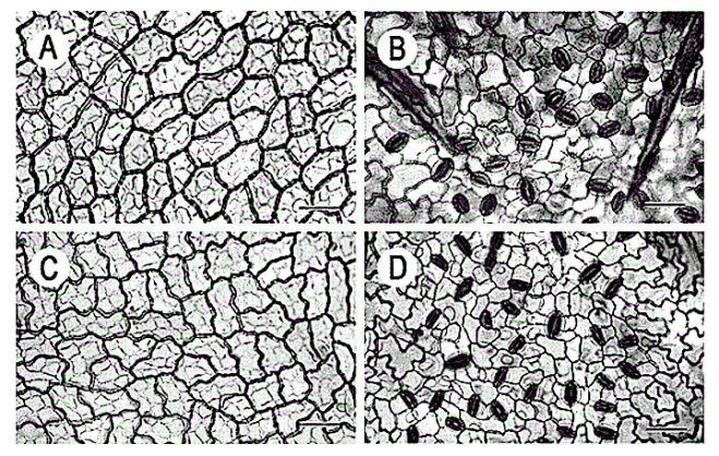
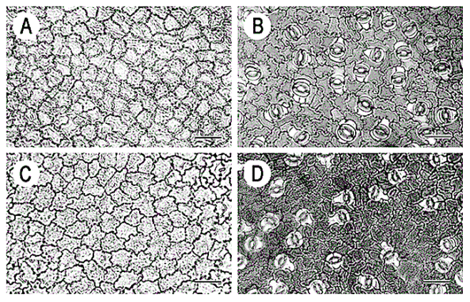
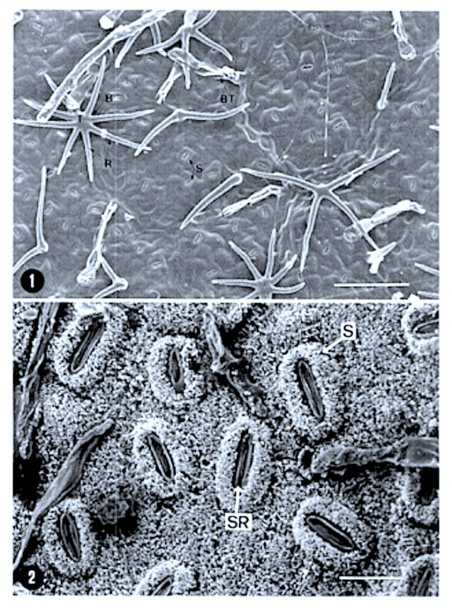
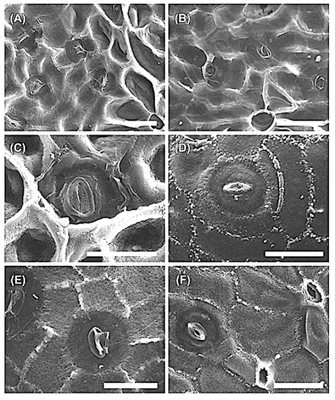
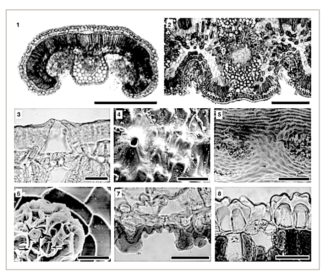
F. Cuticle preparation (abaxial) of A. dim/dia/a: scale bar = 10 )..lm .


Leaf anatomy of the southern African Icacinaceae and its taxonomic significance

by Potgieter M. J., van Wijk A. E. (1999)

M. J. Potgieter* and A. E. van Wijk1

University of LimpopoPolokwane, South Africa

Department of Botany, University of the North, Private Bag X1106, Sovenga, 0727 Republic of South Africa 1H.G.W J. Schweickerdt Herbarium, Department of Botany, University of Pretoria, 0002 Republic of South Africa

===

in S. Afr. J. Bot. 65(2): 53-62 –

https://ac.els-cdn.com/S0254629915309558/1-s2.0-S0254629915309558-main.pdf?_tid=f60e6b27-297f-4d31-9dee-0402cdc93c4a&acdnat=1525425559_8e6a06e676863296e8144758a39015ae

Screen Shot 2018-05-04 at 11.31.25
Figure 2 SEM micrographs of abaxial leaf surfaces. A. Stomata ofApodyles dimidiata. note cuticular striations. B. Stoma ofA. abbOllii. C. Stoma of A. geldenhuysii. Scale bars = 10 ~lIn . D, Trichome of A. dimidiara: scale bar = I 00 ~1l1 . E. Stoma of CasfllI/opsis iliciJofia. F. Stoma of C. tfni/olia, note peri stomal rim formed by cuticular striations. E & F scale bars = 10 Mm.

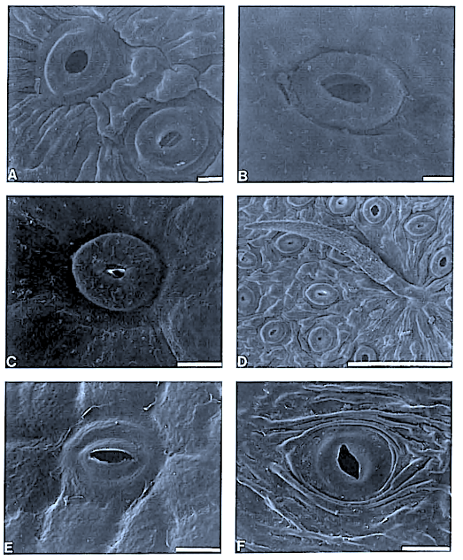
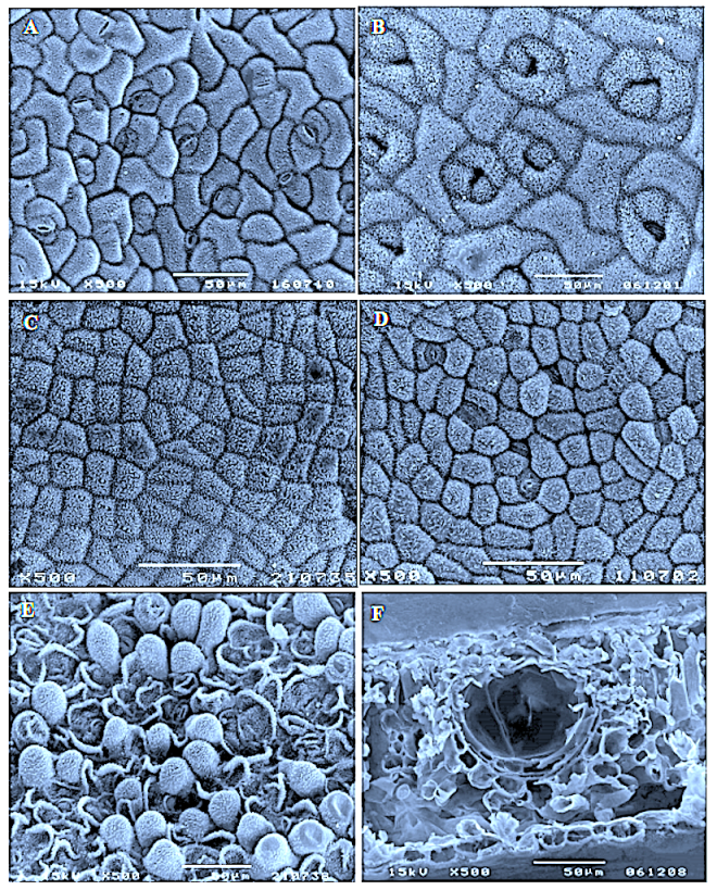
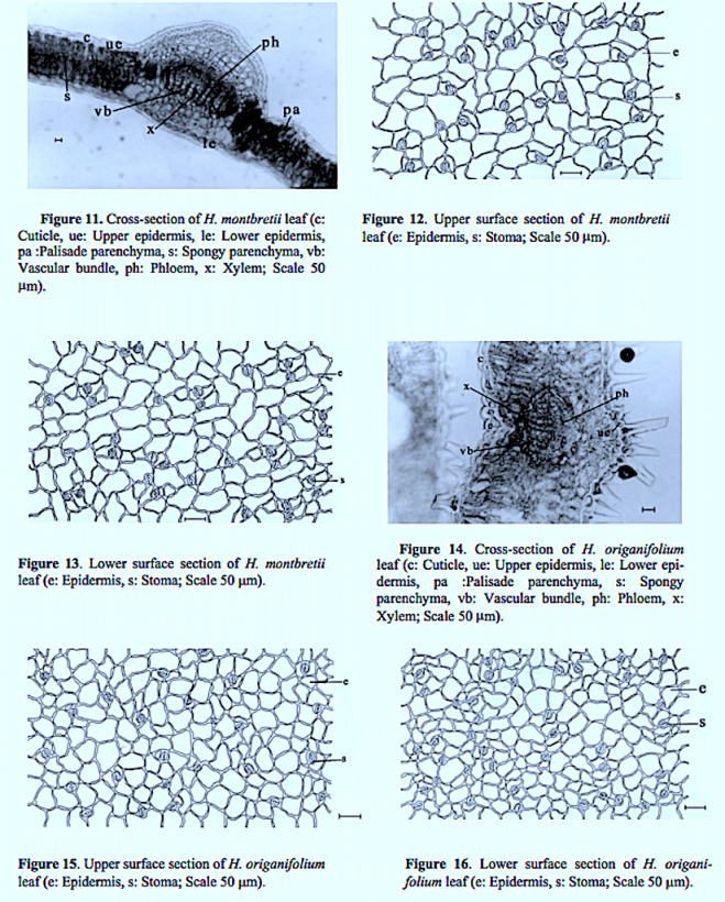
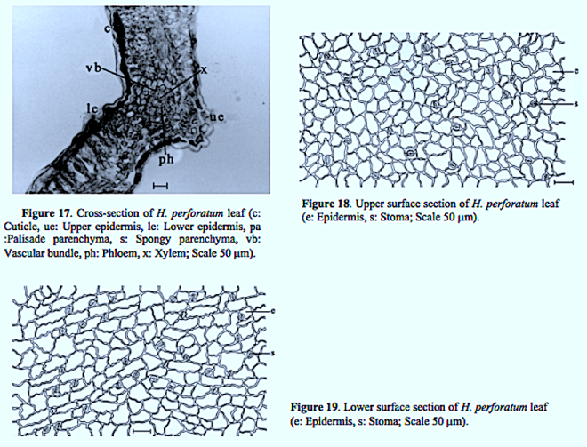
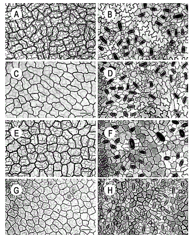
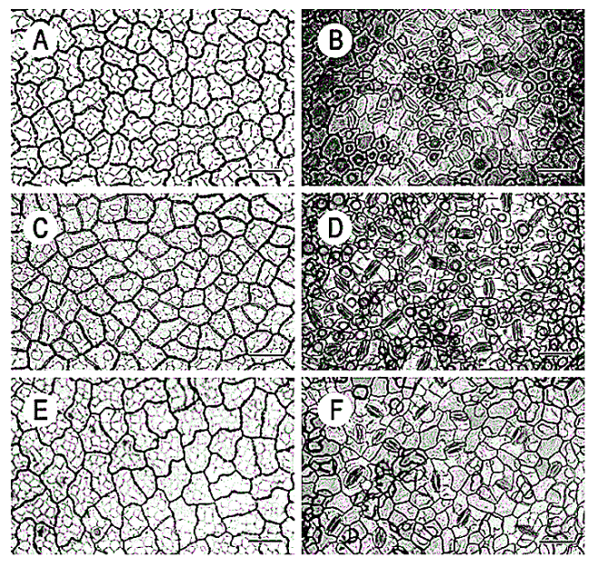
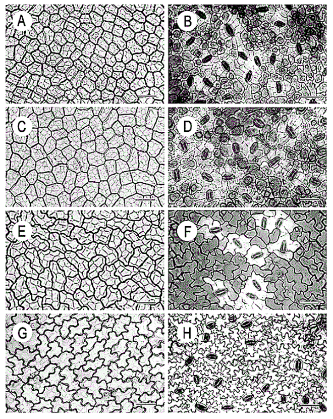
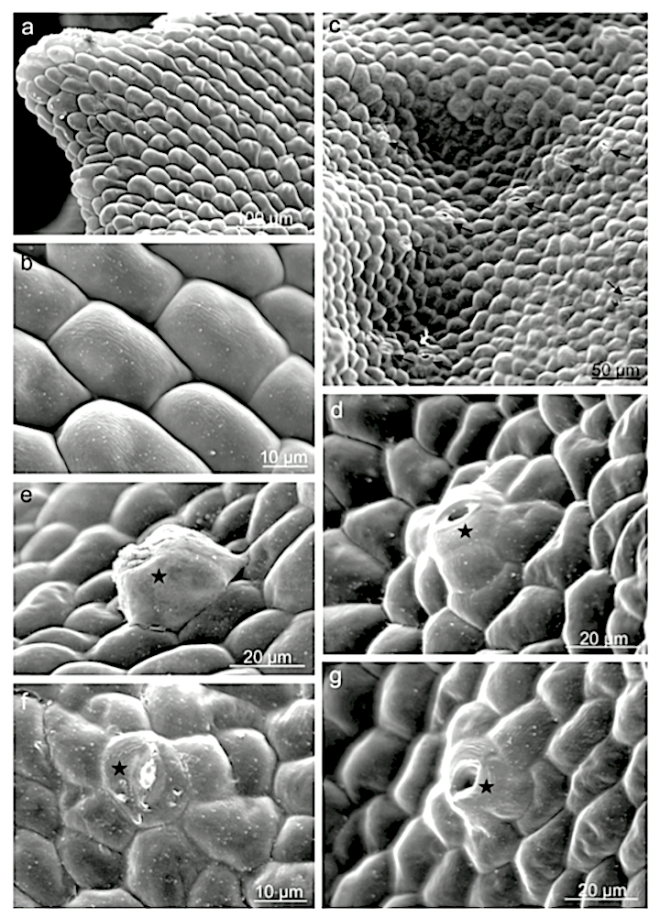
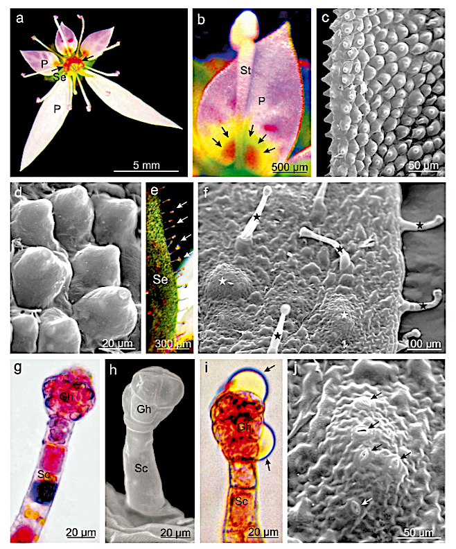
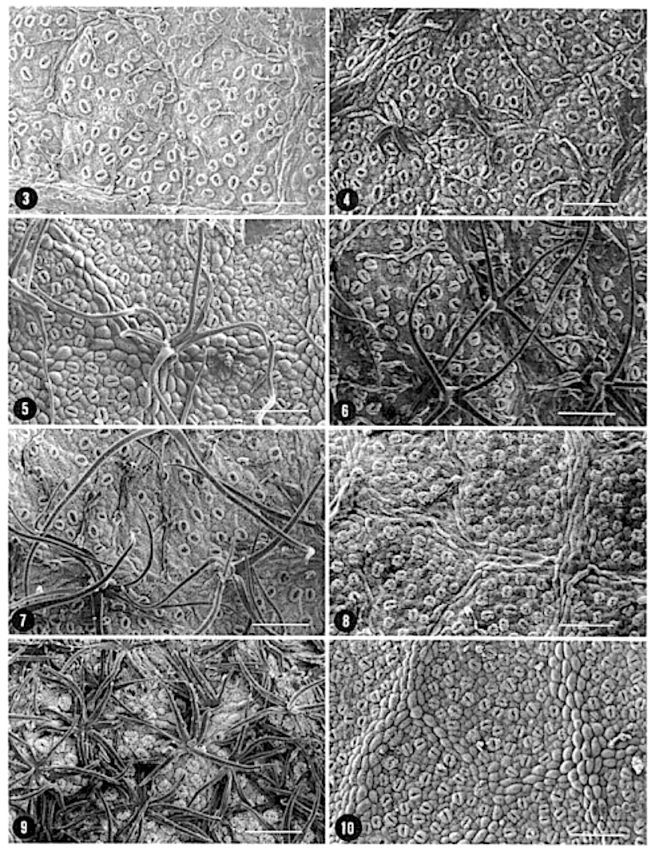
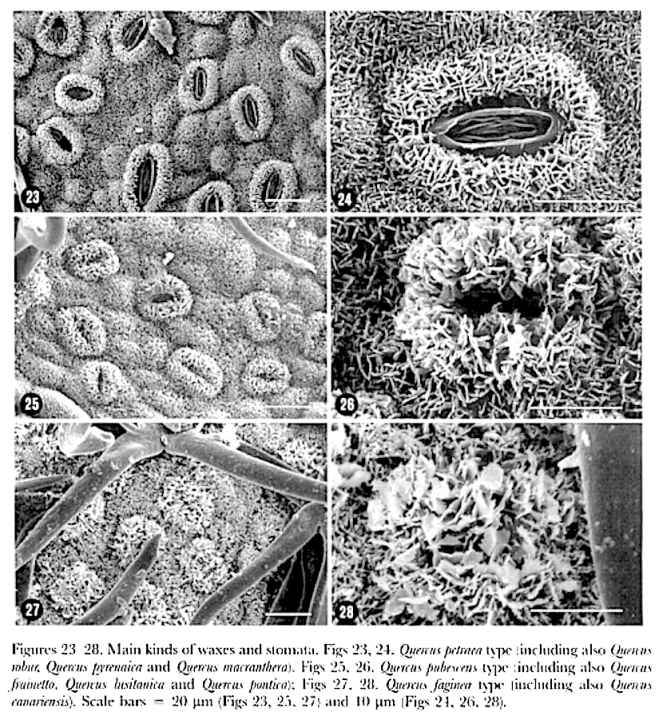
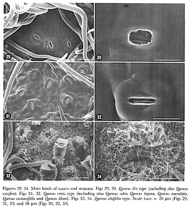
Leaf structure of the three southern African genera of Jcacinaceae was examined by light and scanmng electron microscopy. Diagnostic characters include the stoma and trichome type, and lamina characters, such as mucilage cells, pectic warts and ‘unidentified cell inclusions’,

In southern Africa both Cassinopsis Sand. and Pyrenacantha Hook. have cyclocytic stomata, as opposed to the anomocytic type of Apodytes E. Mey. Ex Arn. The presence of a stomatal ridge in A. dimidiata is a useful character in separating this species from the other two southern African members of this genus. A peristomal rim in Cassinopsis tinifolia Harv. and its absence in C. ilicifolia (Hochst.) Kuntze allow the separation of these two species.

Screen Shot 2018-05-04 at 11.34.20
Figure 4 SEM micrographs of abaxial kafsurfaces ofPyrenacantha. A, Stoma of P. grandijlora: scale bar = 1 () pm. S, Simple trichome in P grondijlora. note cuticular markings and bulging basal rim: scale bar = 100 /.un . C, Stoma of P kaurabassana. D. Trichome of P. kourabassana. E, Stoma of P. scandens. F. ‘Globular’ trichome of P scandens, Scale bars = 10 ).1111 .

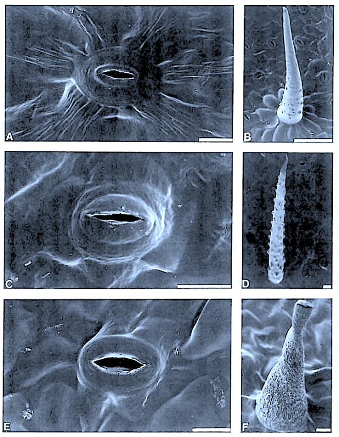
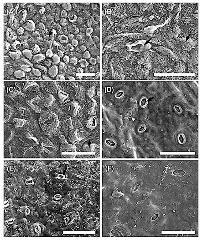
The indumentum of Pyrenacantha consists of simple (unmodified), ‘globular’ and ‘uncinate’ trichomes, whereas that of Apodytes and Cassinopsis consists of simple hairs.

Mucilage cells were found only in members of Apodytes. ln terce!lular, predominantly wart-like pectic protuberances are present in the mesophyll of mature leaf samples of A. ge/denhuysiiVan Wyk & Potgieter and Cassinopsis. Small,

Irregularly shaped , yellowish cell inclusions were found subepidermally to the abaxial epidermis in C. Ificifolia. Their chemical composition and function (if any) is still unknown.


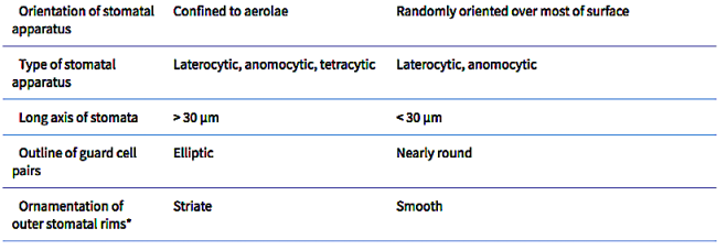
In southern Africa both Cassinopsis and Pyrenacantha have cyclocytic stomata, as opposed to the anomocytic type of Apodytes. According to Van Staveren and Baas (1973) anomocytic stomata are restricted to genera with a low level of specialization, thus representing a primitive condition. Cyclocytic stomata, according to the same authors, occur in genera with a wide range of specialization. Although this type of stomata can be found in genera with a low level of specialization, it is most frequent in highly specialized genera, indicating that this may represent an advanced condition.

Van Staveren and Baas (1 973) found the stomatal index for A. dimidiala to vary between 7 and 21. Research on Celastraceae and Winteraceae also indicates that the stomatal index may be extremely variable within a single species. This invalidates claims by authors in the past that the stomatal index is a diagnostic character at the species level. For this reason we have not considered the stomatal index in this study. In their survey of 26 genera of Maletian lcacinaceae, Van Staveren and Baas (1973) found peristomal rims only in the three genera Codiocarpus Howard, Platea Blume and Stemonurus Blume, indicating that this is a taxonomically useful character state in the Icacinaceae. The presence of a peristomal rim in C. i1iciiolia and its absence in C. tinifolia allows the separation of these two species.


The type and distribution of stomata in Hypericum

Screen Shot 2018-05-02 at 15.51.47
Figure 1. SEM micrographs of foliar epidermal surfaces: A – H. androsaemum abaxial epidermal cells, anomocytic and anisocytic stomata; B – H. calycinum abaxial epidermal cells and anisocytic stomata; C – H. canariense adaxial epidermal cells; D – H. grandifolium abaxial epidermal cells, anomocytic and brachyparacytic stomata (arrow); E – H. patulum abaxial epidermal papillate cells; F – H. calycinum leaf cross section with a cavity type internal secretory structure.


Leaf Phytognostic Characters of Six Species of Hypericum L. (Hypericaceae)

by Teixeira G., Monteiro A. (2017)

Generosa Teixeira1 , Ana Monteiro2

1 Center for Ecology, Evolution and Environmental Changes (CE3C), Faculdade de Farmácia, Universidade de Lisboa, Av. Prof. Gama Pinto, 1649-003, Lisboa, Portugals

2  LinkingLandscape, Environment, AgricultureandFood (LEAF), Instituto Superior de Agronomia, Universidade de Lisboa, Tapada da Ajuda, 1349-017 Lisboa, Portugal

===


in RA Journal of Applied Research ISSN (e): 2394-6709 – Vol.3(12): 1303-1314 –

https://www.repository.utl.pt/bitstream/10400.5/14490/1/REP-AnaMonteiro-hypericum7rajarv3I12p1303_1314_2017.pdf

Abstract:

Most of the studies on Hypericum genus deals with the phytochemical and pharmacological properties and the morphoanatomical data are scarce, although sometimes some species are poorly identified. In order to obtain differentiating phytognostic characters, the micromorphological, anatomical and histochemical study of mature leaves of six Hypericum species, H. androsaemum, H. calycinum, H. canariense, H. grandifolium, H. x inodorum and H. patulum, was carried out using different techniques of microscopy.

Qualitative characters of both epidermal surfaces were evaluated: cell shape, cell wall, epicuticular deposits and stomata type.

Only the last three characters showed some kind of differences. Five qualitative characters, stomata and gland indexes, total mesophyll thickness, palisade parenchyma and spongy parenchyma thickness, were subjected to analysis of variance on one factor ANOVA. Differences were found in the stomata and gland indexes as well as in the parenchyma cells arrangement and thickness.

Histochemical tests were performed to localize and identify chemical groups of metabolites and the differences found were semi-quantitative.

We conclude that the most important leaf phytognostic characters are: the epidermal cells shape, the type and distribution of stomata, the glands distribution and the mesophyll features.

Stomata in Hypericum

Screen Shot 2018-05-02 at 15.35.31


Morphological and anatomical investigations on some Hypericum L. species growing naturally in and around Eskisehir

by Potoglu Erkara, I., Tokur S. (2004)

İsmühan POTOĞLU ERKARA, Süleyman TOKUR,

Osmangazi Üniversitesi Fen Edebiyat Fakültesi Biyoloji Bölümü 26480 Eskişehir,

===

in Trakya Univ J Sci, 5(2): 97-105, 2004 ISSN 1302 647X DIC: 116IPREST510412040105 –

http://dergipark.gov.tr/download/article-file/214106

Screen Shot 2018-05-02 at 15.37.43

Abstract:

The purpose of the present study is to determine some morphological and anatomical characteristics of the species of Hypericum montbretii Spach., H. origanifolium Willd. and H. perforatum L. that show natural distribution in Eskişehir and its surrounding area. H. perforatum was determined to exist more abundantly in the research area in comparison with H. montbretii and H. origanifolium.

While H. montbretii and H. origanifolium have black glands, H. perforatum lack such glands.

Upon the examination of the root cross-rections, the pith was determined to be completely covered by xylem cells. As to stem cross-sections, it was observed that large parenchymatic cells were present in the pith of the young stems, while old stems were observed to have formed cavity in their pith. The species examined were determined to bear resemblance to one another in consideration of their leaf anatomy. Leaves are equifacial and amphistomatic and they have amaryllis type stomata. They are mesomorphic and there are schizo-lisigenous type secretion pockets in the leaves.

Compared to adjacent cells, stomata are anisocytic or diacytic. Stomata on the lower surface of the leaves show higher frequencies than those on the upper surface.

Stomata in Schizophragma and Pileostegia (Hydrangeaceae)

Characters of leaf epidermis of Schizophragma crassum var. ellipticum under light microscopy: adaxial on the left, abaxial on the right. Scale bars, 50 µm. [A, B, G. Forrest 29029 (Liu & Zhu, 2009, figs 15, 16)].


===

Screen Shot 2018-05-02 at 14.44.41
Characters of leaf epidermis under light microscopy: adaxial on the left, abaxial on the right. Scale bars, 50 µm. A–F, Schizophragma integrifolium var. integrifolium. G, H, S. integrifolium var. minus. [A, B, J. H. Xiong & Z. L. Zhou 91666, Chongqing; C, D, S. H. Chun 9604, Guangxi; E, F, X. Y. He 23711, Zhejiang; G, H, B. G. Li 96, Hunan (Liu & Zhu, 2009, figs 7, 8)].

Leaf epidermal characters and taxonomic revision of Schizophragma and Pileostegia (Hydrangeaceae)

by Liu W., Lin X.-Y. (2011)

Wei LIU,

Xiang-Yun ZHU,
Institute of Botany, Chinese Academy of Sciences, Beijing 100093, China
*

Corresponding author. E-mail: xiangyunzhu@ibcas.ac.cn

Search for other works by this author on:

XIANG-YUN ZHU

Institute of Botany, Chinese Academy of Sciences, Beijing 100093, China

===

in Botanical Journal of the Linnean Society 165(3): 285–314 – https://doi.org/10.1111/j.1095-8339.2010.01101.x

https://academic.oup.com/botlinnean/article/165/3/285/2418498

Screen Shot 2018-05-02 at 14.47.51
Characters of leaf epidermis of Schizophragma hypoglaucum under light microscopy: adaxial on the left, abaxial on the right. Scale bars, 50 µm. [A, B, J. H. Xiong & Z. L. Zhou 93885, Chongqing (Liu & Zhu, 2009, figs 5, 6); C, D, Sichuan Econ. Pl. Exped. 1030, Sichuan; E, F, Y. Tsiang 5550, Guizhou].

Abstract

This article provides leaf epidermal characters and a taxonomic revision of Schizophragma and Pileostegia (Hydrangeaceae).

Schizophragma and Pileostegia are treated as two separate genera on the basis of morphology and leaf epidermal characters. In Schizophragma, seven species and five varieties are recognized, and two new synonyms are proposed and one lectotype is designated.

Leaf epidermal characters are of great significance in taxonomic classification in Schizophragma. In Pileostegia, two species are recognized, and two new synonyms are proposed and two lectotypes are designated. Descriptions, maps, notes on ecology and phenology and keys to both genera are provided.

Screen Shot 2018-05-02 at 14.49.30
Characters of leaf epidermis under light microscopy: adaxial on the left, abaxial on the right. Scale bars, 50 µm. A, B, Schizophragma molle; C, D, S. fauriei. (A, B, M. H. Li & Y. Q. Kuang 909; C, D, Anonymous s.n.).


In Schizophragma, stomata are only present on the abaxial epidermis and confined to aerolae. Laterocytic, anomocytic and tetracytic types of stomatal apparatus are observed. Cuticular intrusions, known as ‘T’ pieces, are found between the ends of each guard cell in all the taxa of Schizophragma. The outline of guard cell pairs is elliptic in surface view, with length/width ratios of 1.3–1.5 : 1.

Screen Shot 2018-05-02 at 14.51.13
Characters of leaf epidermis under light microscopy: adaxial on the left, abaxial on the right. Scale bars, 50 µm. A, B, Schizophragma hydrangeoides var. hydrangeoides; C, D, S. hydrangeoides var. formosum; E, F, S. hydrangeoides var. concolor; G, H, S. hydrangeoides var. molle. (A, B, M. Furuse 45391; C, D, M. Furuse 15386; E, F, Y. Endo 3521; G, H, M. Furuse 10202).


Stomata are only present on the abaxial epidermis and are randomly oriented over most of the surface in Pileostegia. Laterocytic and anomocytic types of stomatal apparatus are observed, and ‘T’ pieces are also found between the ends of each guard cell in both species. The guard cell pairs usually have a nearly round outline in surface view, with length/width ratios of 1.1 : 1.

Screen Shot 2018-05-02 at 14.58.20
Characters of leaf epidermis under light microscopy: adaxial on the left, abaxial on the right. Scale bars, 50 µm. A, B, Pileostegia viburnoides; C, D, P. tomentella. (A, B, S. Y. Hu 11081; C, D, Y. Li 2001).
                                     Schizophragma       Pileostegia

Screen Shot 2018-05-02 at 15.00.30

Nectarostomata in Saxifraga

Screen Shot 2018-04-30 at 13.08.59
Fig. 3. Surface of the S. stolonifera nectaries with nectarostomata. a,b Epidermal cells of the nectary appendages. c The nectary cavities with numerous nectarostomata (arrows). d-g Visible nectarostomata (asterisks) located on the convexities (d,g), covered with secretions (e) or with secretion in stomata pores (f).


Micromorphology and anatomy of flowers and nectaries of Saxifraga stolonifera L. 

by Konarska A. (2014)

Agata Konarska, University of Life Sciences in Lublin, Poland

===

in Acta Agrobotaica 67(4): 3-12 – DOI: 10.5586/aa.2014.054 –

4712-9441-1-SM 3 (3).pdf

Screen Shot 2018-04-30 at 13.14.51
Fig. 1. Details of Saxifraga stolonifera flowers. a Flower with a yellow-orange nectary (arrows). b Visible yellow and contrasting spots (nectar guides) at the base of the petal (arrows). c Petal surface with papillae. d Papillae on the petal surface. e Fragment of the sepal with peltate glandular trichomes (arrows). f Surface of the abaxial side of the sepal with glandular trichomes (black asterisks) and depressions with stomata (white asterisks). g-i Glandular trichomes with anthocyanins in stalk cells (g), and with secretion drops (arrows) (i). j On the sepal visible a depression with stomata (arrows). P – petal; Se – sepal; St – stamen; Gh – glandular head; Sc – stalk cell.

Screen Shot 2018-04-30 at 13.00.05Screen Shot 2018-04-30 at 13.06.52


Stomata in Lithocarpus (Fagaceae)


Comparative morphology of leaf epidermis in the genus Lithocarpus and its implication in leaf epidermal feature evolution in Fagaceae

by Deng M., Li Q., Shuting Y., Liu Y.-c., Xu J. (2013)

Min_Deng4
Min Deng, Shanghai Institutes for Biological Sciences, Shanghai, China
Qiansheng_Li
Qiansheng Li, Shanghai Institute of Technology, Shanghai, China
Yang_Shuting
Yang Shuting, Chinese Academy of Sciences,  Beijing, China
Yan-chun_Liu
Yan-chun Liu, Chinese Academy of Sciences,  Beijing, China
Jin Xu
===

in Plant Systematics and Evolution 299(3) –

https://www.researchgate.net/publication/257447723_Comparative_morphology_of_leaf_epidermis_in_the_genus_Lithocarpus_and_its_implication_in_leaf_epidermal_feature_evolution_in_Fagaceae

Abstract
Leaf epidermal features are considered to be taxonomically important in Fagaceae. In this study, we examined and compared leaf epidermal features of 112 specimens, representing 105 species and one variety of Lithocarpus from China and adjacent areas and Notholithocarpus densiflorus.
As a result of the different interpretations of terms in previous studies, trichome terminology in Lithocarpus and its relatives was re-assessed aiming to reveal the trichome evolutionary patterns in Fagaceae. Twelve types of trichomes and five types of trichome bases were detected in Lithocarpus, among which the broad-based trichome (BBT) is newly reported.
Stomata in Lithocarpus are restricted to the cyclocytic type and their size range is 28.6 ± 8.2 μm × 26.5 ± 9.3 μm. The distribution of epidermal features in Lithocarpus revealed three distinct morphological groups: glabrous, BBT, and appressed parallel tufts (APT). The importance of epidermal features across Fagaceae for taxon delimitation is evaluated.
Species of Lithocarpus can be accurately identified by the presence of APT or flat epidermal cells combined with non-dark stained subsidiary cells and non-cutinized trichome bases only, or in addition, fasciculate trichome bases. The phylogenetic distribution of epidermal features and their evolutionary trends in Fagaceae is also discussed.

Stomata in European and Mediterranean oaks (Quercus)

Screen Shot 2018-04-24 at 16.32.30
Figurcs 1, 2. ‘l’crms uscd to tlescrilxd thc iiiaiii iiiicrornorpliolo~ical featurcs in thc aliaxial surface is markcd. I;ig. 1. Lcaf with flat Tvaxes (Qmczc rnri!): ‘I = (richome; B = hase; R = rays. or arms: BT = bulbous tricliomc; S = stomaia. Fig. 2. Leaf with scaly waxcs (Quu(vx~ robur). S = stomata; SR = stomata1 rim.


European and Mediterranean oaks (Quercus L.; Fagaceae): SEA4 characterization of the micromorphology of the abaxial leaf surface

by Bussotti F., Grossoni P. (1997)

Screen Shot 2018-04-24 at 16.35.48

===

in Bot. J. Linn. Soc. 124: 183-198 –

https://watermark.silverchair.com/j.1095-8339.1997.tb01789.x.pdf?

Screen Shot 2018-04-24 at 16.38.08

Screen Shot 2018-04-24 at 16.40.46

Screen Shot 2018-04-24 at 16.30.36

Screen Shot 2018-04-24 at 16.43.25

Screen Shot 2018-04-24 at 16.45.21

Stomata in Fagus

Screen Shot 2018-04-24 at 16.14.51
Figure 3. SEM micrographs of the inner surface of lower epidermis. (A) Fagus engleriana showing subsidiary cells of anomocytic type. (B) Fagus japonica showing subsidiary cells of anomocytic type. (C) Fagus longipetiolata showing subsidiary cells of anomocytic type. (D) Fagus sylvatica showing subsidiary cells of cyclocytic type. (E) Fagus hayatae showing subsidiary cells of cyclocytic type. (F) Fagus crenata showing subsidiary cells of cyclocytic type. (A–F) Scale bar = 20 μm. SEM = scanning electron microscopy.


Leaf cuticle micromorphology of Fagus L. (Fagaceae) species

by Cho S. H., Jeong K. S., Kim S.-H., Pak J.-H. (2014)

Seong Ho Cho, 12  Keum Seon Jeong, 1  Sun-Hye Kim, Jae-Hong Pak, 1

1 Department of Biology, College of Natural Sciences, Kyungpook National University, Daegu, Korea
2 Natural History Museum, Kyungpook National University, Gunwi, Korea

===

in Journal of Asia-Pacific Biodiversity 7(4): 378-387 –

https://www.sciencedirect.com/science/article/pii/S2287884X14000636

Screen Shot 2018-04-24 at 16.17.42
Figure 5. SEM micrographs of the outer surface of lower epidermis. (A) Fagus engleriana showing papillae (arrow), stomata, and well-developed waxes. (B) Fagus japonica showing trichome base (arrow) and papillae. (C) Fagus longipetiolata showing stomata and papillae (arrow). (D) Fagus crenata showing stomatal rims (arrow). (E) Fagus hayatae showing stomatal rims and weakly developed waxes. (F) Fagus sylvatica showing weakly developed waxes and stomatal rims. (A–F) Scale bar = 40 μm. SEM = scanning electron microscopy.

Abstract

Cuticle micromorphology of all eight species of Fagus and an outgroup were examined in the present study. The genus Trigonobalanus was selected as the outgroup. Thirteen characteristics of the inner surface and five of the outer surface of the cuticle were described. Some characteristics, such as the subsidiary cell shape, size of stomata, arrangement of subsidiary cells, shape of anticlinal and periclinal cell walls, texture of periclinal cell wall, development epicuticular wax, and presence of papillae, were considered important for infrageneric classification.

The topology was obtained from the analysis using two major lineages: (1) Fagus engleriana + Fagus japonica + Fagus longipetiolata and (2) Fagus sylvatica + Fagus crenata + Fagus lucida + Fagus hayatae + Fagus grandifolia. The first clade supported a bootstrap value of 98% and the second clade a bootstrap value of 97%. Based on the cuticle morphology, our results support the previous study, by revealing F. englerianaF. japonica, and F. longipetiolata with long peduncles in one group and the remaining extant species of short- to medium-length peduncles in another group.

In addition, molecular phylogenetic study of Fagus based on ribosomal DNA ITS and chloroplast DNA sequences data supports this assemblage.

This study shows that cuticle micromorphological characteristics provide useful and important information for analyzing the evolutionary aspects of Fagus.


Table 2. Characters used in the cladistic analysis of Fagus and its relatives.

1. Trichome of abaxial surface: stellate & solitary (0), solitary (1), solitary and conical (2), and peltate (3)
2. Epicuticular wax of adaxial surface: weakly developed (0), developed (1), and undeveloped (2)
3. Epicuticular wax of abaxial surface: well developed (0) and weakly developed (1)
4. Papillae of abaxial surface: well developed (0) and undeveloped (1)
5. Stomatal rim: unknown (0) and well developed (1)
6. Trichome surrounding cell shape: irregular circular (0), irregular polygonal (1), and circular (2)
7. Cell number of trichome base of abaxial surface: 6–7 (0), 7–8 (1), and 10–12 (2)
8. Anticlinal wall thickness of adaxial surface: thin (0) and thick (1)
9. Anticlinal cell wall pattern of adaxial surface: straight (0), undulate (1), and sinuous and straight (2)
10. Ornament on the adaxial inner surface: absent (0) and present (1)
11. Anticlinal wall thickness of abaxial surface: thin (0) and thick (1)
12. Anticlinal cell wall pattern of abaxial surface: straight & sinuous (0), undulate (1), and sinuous (2)
13. Periclinal wall texture of abaxial surface: rough (0) and smooth (1)
14. Stomatal apparatus shape: elliptical (0), circular (1), and elliptical and circular (2)
15. Subsidiary cell number: 4–6 (0) and 6–8 (1)
16. Subsidiary cell’s anticlinal wall shape: rectangular (0), irregular circular (1), lunate (2), and circular (3)
17. Subsidiary cell arrangement: anomocytic (0), and cyclocytic and anomocytic (1)
18. Stomata length: 15–20 μm (0), 10–15 μm (1), and 20–25 μm (2)

Stomata Diversification and Phylogenetics of Euphorbiaceae


Stomata Diversification and Phylogenenetic Analysis of 13 Species of Family Euphorbiaceae sensu lato

by Hidayat T., Kusdianti (2009)

Topik Hidayat, Kusdianti,
Department of Biology Education, Faculty of Mathematics and Natural Sciences Education, Indonesia University of Education (UPI), Bandung 40154.


===

in Biodiversitas 10(1): 19-22 – DOI 10.13057/biodiv/d100104 –

https://www.researchgate.net/publication/275516519_Stomata_Diversification_and_Phylogenenetic_Analysis_of_13_Species_of_Family_Euphorbiaceae_sensu_lato

Abstract
Investigation on diversity of stomata from 13 species of family Euphorbiaceae has been carried out.
Characters like type of stomata, position of stomata, presence/absence of ledge, and density both abaxial and adaxial leaf surface were examined. Stomata characters in the family were found quite diverse in this study.
Monophyletic nature of Euphorbiaceae has been proved in this study on the basis of phylogenetic analysis using parsimoni method.
Our data further suggested that the family can be classified into two major groups. However, surprisingly, genus Phyllanthus is non-monophyletic.

Sunken, covered, and encrypted stomata in relation to dry habitats

Screen Shot 2018-04-17 at 11.38.00
Stomatal protection in Proteaceae. Note that these structures all supplement the protection provided by the outer cuticular ledges present in most vascular plants. Figs. 1–3, 7–8 light micrographs; Figs. 4–6, scanning electron micrographs. 1.Transverse section of a Banksia ericifolia leaf showing the closely revolute margins. Note the dense hairs in the grooves created by the leaf margin.2.Transverse section of a Grevillea striata leaf showing two hair‐lined grooves that contain the stomata. Note the dense hairs in the left hand groove.3.Transverse section of a Hakea lissosperma leaf showing an individual crypt.4.External surface of a H. lissosperma leaf showing the aperture of an individual crypt.5.External, abaxial surface of a Banksia quercifolia leaf showing the hair‐filled apertures of two pits.6.Inner surface of the abaxial cuticle of a Banksia quercifolia leaf (prepared by maceration in chromium trioxide) showing the balloon‐like crypt. Two stomata are indicated with arrows. 7.Transverse section of a Telopea truncata leaf showing a papillose pit. 8.Transverse section of a Leucadendron pubescens leaf showing a papillose pit. Scale bars: Fig. 1 = 500 μm; Fig. 2, 5 = 200 μm; Figs. 3, 4, 6–8 = 50 μm.


The evolutionary relations of sunken, covered, and encrypted stomata to dry habitats in Proteaceae

by Jordan G. J., Weston P. H., Carpenter R. J., Dillon R. A., Brodribb T. J. (2008)

School of Plant Science, University of Tasmania, Private Bag 55, Hobart 7001, Australia

Gregory J. Jordan, Peter H. Weston, Raymond J. Carpenter, Rebecca A. Dillon, Timothy J. Brodribb

===

in Am. J. Bot. 95(5): 521-530 – https://doi.org/10.3732/ajb.2007333 –

https://onlinelibrary.wiley.com/doi/full/10.3732/ajb.2007333

Abstract

Sunken, covered, and encrypted stomata have been anecdotally linked with dry climates and reduced transpiration and therefore have been used to infer dry palaeoclimates from fossils.

This study assesses the evolutionary and ecological associations of such stomatal protection in a model system—the diverse southern hemisphere family Proteaceae. Analyses were based on the morphology of over 1400 Australian, South African, New Caledonian, New Zealand, and South American species, anatomy of over 300 of these species, and bioclimatic data from all 1109 Australian species.

Ancestral state reconstruction revealed that five or six evolutionary transitions explain over 98% of the dry climate species in the family, with a few other, minor invasions of dry climates.

Deep encryption, i.e., stomata in deep pits, in grooves, enclosed by tightly revolute margins or strongly overarched by cuticle, evolved at least 11 times in very dry environments.

Other forms of stomatal protection (sunken but not closely encrypted stomata, papillae, and layers of hairs covering the stomata) also evolved repeatedly, but had no systematic association with dry climates.

These data are evidence for a strong distinction in function, with deep encryption being an adaptation to aridity, whereas broad pits and covered stomata have more complex relations to climate.