Stomata in Heteromorpha (Apiaceae)

Photo credit: Google

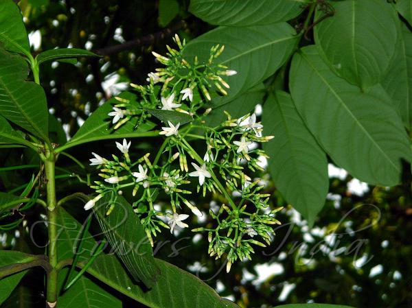
Heteromorpha arborescens var abyssinica, Parsley Tree

The taxonomic value of epidermal characters in the leaf of Heteromorpha and some related genera (Apiaceae). 

Winter P. J. D., Van Wyk B. E. (1994)

Department of Botany, Rand Afrikaans University, South Africa

citations
Pieter J. D. WINTER – Rand Afrikaans University, South Africa 
citations-1
Ben-Erik VAN WIJK – Rand Afrikaans University, South Africa

in Bothalia. 24. 187-194.- doi: 10.4102/abc.v24i2.770

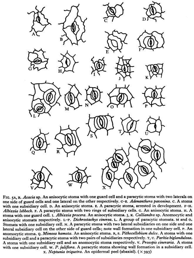
Heteromorpha (17 spp.), Polemannia (3), Polemanniopsis. SEM – hair types. Line drawing – hairs. Stomatal type, distribution and density. Epidermal cell number, size & outline differ with juvenile/adult & spp. – 

http://www.abcjournal.org/index.php/ABC/article/view/770/0

Abstract

All 17 species of Heteromorpha Cham. & Schltdl. (sensu Humbert 1956), all three species of Polemannia Eckl. & Zeyh. and the monotypic Polemanniopsis B.L. Burtt were investigated for leaf epidermal characters.

Stomatal type was anomocvtic. with an exception in only one Madagascar species, H. betsileensis.

The distribution and density of stomata (on both leaf surfaces) are diagnostic for some species.

The number, size and outline of normal epidermal cells are different in juvenile and adult leaves and these differences vary between species. Seven trichome types are recognized which, when combined with dispersion pattern, also serve to characterise the various species and forms.

Stomata in Caryophyllaceae


Foliar anatomy of the Caryophyllaceae family in Arasbaran, NW Iran.

by Zarinkamar F. (2001)

305440
Fatemeh Zarinkamar, School of Biological Sciences, Tarbiat Modares University

in Iranian J. Bot., Vol. 9 (1): 93-102. – 

http://en.journals.sid.ir/ViewPaper.aspx?ID=120545http://en.journals.sid.ir/ViewPaper.aspx?ID=120545

Abstract: 

The foliar anatomy of the Caryophyllaceae family from Arasbaran Protected Area including the following species were studied under light microscope:
Arenaria dianthoides, Arenaria gypsophiloides, Arenaria serpyllifolia, Cerastium glomeratum, Cerastium holosteoides, Cerastium szowitsii, Dianthus cretaceus, Dianthus crinitus, Gypsophila elegans, Herniaria hirsuta, Herniaria incana, Minuartia hirsuta, Mjnuartia acuminata, Minuartia lineata, Minuartia meyeri, Minuartia recurva, Silene alba, Silene rprechtii, Silene spergulifolia and Stellaria media.

The different anatomical characters on Caryophyllaceae family indicate flexibility of this family in various ecological circumstances.


Stomata in Chenopodiaceae

Photo credit : Google

Noaea mucronata (Forssk.) Asch. & Schweinf.

Foliar anatomy of Chenopodiaceae family and xerophytes adaptation

by Zarinkamar F. (2006)

305440
Fatemeh Zarinkamar, School of Biological Sciences, Tarbiat Modares University, Tehran


in Iran. J. Bot., 11: 169-175 –

http://ijb.areeo.ac.ir/article_102885_7388e65ca31e17a1280214595bd1ddc3.pdf

Abstract

Foliar anatomy of three species of Chenopodiaceae family, including Chenopodium album, Kochia prostrata and Noaea mucronata are studied.

Various anatomical characters such as stomatal densities, guard cell length for the adaxial and abaxial epidermis, type of stomata, and density of trichome on surface, cuticle thickness, mesophyll type and variety of crystal are compared.

Photosynthesis pathway is C4 in Kochia prostrata and Noaea mucronata. Foliar internal structure of species studied typically is characterized by xerophytes plants accompany by extensive central waterstorage tissue which explains Chenopodiaceae adaptation to arid and semiarid area.

Stomata in Sedum (Crassulaceae)

Photo credit: Google

Sedum spectabile

A leaf epidermis study on twelve species of Sedum in AnHui.

by Zheng Y., Gong J. (1999)

Zheng Yan,

Gong Jie

Department of Biology, Anhui Normal University, Wuhu, China


in Bulletin of Botanical Research , Vol. 19: 292- 297. –


http://bbr.nefu.edu.cn/EN/abstract/abstract2967.shtml

Abstract

The leaf epidermis structures of 12 species, which belong to Sedum from Anhui province in China, have been examined under LM and SEM.

The stomatal size, stomatal density and stomatal index were measured and counted under LM. Under SEM, it was described such as the shape of stomata, cuticular membrane, wax ornamentation, outer stomatal ledge (or rim) and trichome.

The results show that the type of stomatal apparatus in Sedum is the kind of typical anisocytic type. The characters, such as the type of stomatal apparatus, shape and pattern of anticlinal walls of epidermis cells, culticular membrane and wax ornamentation, are less important between species and can be used as secondary ones.

The features of stomatal size, distribution, density and trichome possess the classific value of species.

Stomatal development and stomatal differentiation


Ultrastructure of stomatal development in Arabidopsis (Brassicaceae) leaves.

by Zhao L., Sack F. D.  (1999)

Zhao L.

Fred Sack, Ohio State University

Sack
Fred Sack – Photo by: Elaine Simons Lane

in Amer. J. Bot., 86: 929-939. –

https://www.ncbi.nlm.nih.gov/pubmed/10406715

Abstract

Stomatal development was studied in wild-type Arabidopsis leaves using light and electron microscopy.

Development involves three successive types of stomatal precursor cells: meristemoid mother cells, meristemoids, and guard mother cells (GMCs). The first two types divide asymmetrically, whereas GMCs divide symmetrically.

Analysis of cell wall patterns indicates that meristemoids can divide asymmetrically a variable number of times. Before meristemoid division, the nucleus and a preprophase band of microtubules become located on one side of the cell, and the vacuole on the other.

Meristemoids are often triangular in shape and have evenly thickened walls. GMCs can be detected by their roughly oval shape, increased starch accumulation, and wall thickenings on opposite ends of the cells. Because these features are also found in developing stomata, stomatal differentiation begins in GMCs. The wall thickenings mark the division site in the GMC since they overlie a preprophase band of microtubules and occur where the cell plate fuses with the parent cell wall.

Stomatal differentiation in Arabidopsis resembles that of other genera with kidney-shaped guard cells. This identification of stages in stomatal development in wild-type Arabidopsis provides a foundation for the analysis of relevant genes and of mutants defective in stomatal patterning, cell specification, and differentiation.

Stomatal clusters in Cinnamon

Photo credit: Google

Cinnamomum camphora (Camphor Laurel)

Stomatal clustering in Cinnamomum camphora.

by Zhao X. Z., Yang Y. S., Shen Z. X., Zhang H., Wang G., Gan Y. (2006)

X. Zhao, Bureau of Linyi Environmental Protection, China

Y. YangZ. ShenH. ZhangG. WangY. Gan, Agro-Ecology Institute, Zhejiang University, Hangzhou, China

in South Africa. J. Bot. 72: 565-569.- DOI10.1016/j.sajb.2006.03.006 – 

https://www.infona.pl/resource/bwmeta1.element.elsevier-85683905-d4dd-3b26-88d9-70c0205b4fc9https://www.infona.pl/resource/bwmeta1.element.elsevier-85683905-d4dd-3b26-88d9-70c0205b4fc9

Abstract

Stomata play a crucial role in productivity and survival of land plants by regulating photosynthesis, respiration and transpiration through controlling exchanges of CO 2 , O 2 and water vapour between the interior of the leaf and the atmosphere.

Stomatal clusters (groups of two or more stomata that make direct contact) have been found only in a few plant species to data, while stomatal clustering has been found to be a very common phenomenon in Cinnamomum camphora in our observations.

The ontogeny of stomatal clusters, their distribution on leaves and their density under different soil moisture conditions were surveyed. The results showed that:

(1) the developmental pathway of stomatal clusters is similar to that of loss-of-function mutations in four lips (FLP) mutants that may result in additional divisions of guard mother cells (GMCs);

(2) stomatal clustering occurs along the veins, and the leaf bases have the most stomatal clusters; and

(3) there is a negative relationship between stomatal cluster density (SCD) and soil moisture (SM).

Therefore stomatal clusters are probably able to assist in water conservation.

Stomata in Ericaceae

Photo credit: Google

Cassiope_stellariana_15407.JPG

The taxonomic significance of certain anatomical variations on Ericaceae.THE ERICOIDEAE, CALLUNA AND CASSIOPE

by Watson L. (1964)

in J. Linn. Soc. (Bot.) 59: 111-125. – DOI: 10.1111/j.1469-8137.1964.tb07380.x – 

http://onlinelibrary.wiley.com/doi/10.1111/j.1469-8137.1964.tb07380.x/abstract

Summary

A review of the classical treatment of Ericaceae reveals how unconvincing are the usual definitions of the major groupings Ericoideae and Arbutoideae.

However, a survey of stomatal structure, stomatal distribution and pith structure supports the taxonomic soundness of the main body of the Ericoideae, which are further characterized by their distinctive habit. Calluna is exceptional among Ericoideae in pith structure and stomatal distribution, and differs from the others in various aspects of gross morphology.

There is a striking similarity between Calluna and Cassiope, a genus usually placed in the Arbutoideae; and there is little doubt that the two are closely related.

These conclusions lead to speculation about the origin and development of the Ericoideae in the minds of taxonomists, and about the mechanism by which closely similar genera become separated at the level of sub-family.

Giant stomata in Apocynaceae

Photo credit: Google

Tabernaemontana divaricata

Giant stomata in the family Apocynaceae

by Trivedi B. S., Upadhyay H. (1974)

in Current Science 43: 28, 29. –

https://eurekamag.com/research/000/102/000102832.php

batino
Alstonia macrophylla – http://www.flowersofindia.net/catalog/slides/Batino.jpg

Abstract

Giant stomata were observed in several species, including Alstonia macrophylla, Tabernaemontana divaricata and Strophanthus wightianus.

Conclusions on the significance of the leaf epidermis in Combretaceae


The significance of leaf epidermis in the taxonomy of the Combretaceae, Conclusions

by Stace C. A. (1980)

in Bot. J. Linn. Soc., (81) 327–339 – DOI: 10.1111/j.1095-8339.1980.tb01682.x – 

http://onlinelibrary.wiley.com/doi/10.1111/j.1095-8339.1980.tb01682.x/abstract

Abstract

The classification of the Combretaceae in relation to the anatomy of the leaf epidermis (especially to trichomes) is surveyed, with particular reference to Combretum. The new combination Calopyxis grandidieri (Drake) Capuron ex Stace is made and the new section Calopyxis sect. Petalantha Stace is described. A sectional review of Combretum is presented. The 69 sections are reduced to 45, and full bibliographic details, typification and synonymy are set out.


Stomata in Mimosa (dicots)

Scan 1Photo credit: Google

Mimosa pudica 

Epidermal structure and stomatal ontogeny in some Mimosaceae .

by Shah G. L., Parabia M. H., Kothari M. J.  (1972)

in Ann.Bot.,36:832-835. –

http://aob.oxfordjournals.org/content/36/4/823

Abstract

The structure of trichomes and stomata on leaflets of 21 species of the Mimosaceae are described.

Non-glandular trichomes in Mimosa pudica are of three types: unicellular, with a rounded thick-walled base and a terminal unicellular body, and multicellular. Capitate, clavate, or cylindric, 3–6-celled glandular hairs are observed on leaflets of Mimosa pudica only.

Leaflets are amphistomatic in all species except Adenanthera pavoninaCalliandra sp., Parkia biglandulosa, Pithecellobium dulce, and Samanea saman in which they are hypostomatic.

Only paracytic stomata are found in Leucaena leucocephala and Mimosa pudica. In the rest stomata are of more than one type. In spite of the diversity, the most frequent type in these species is paracytic. Anisocytic stomata, in all cases, are secondarily derived from paracytic ones by transverse or oblique wall formation in a subsidiary cell.

Similarly some stomata with one subsidiary cell are also secondary derivatives of the paracytic ones because of one of the subsidiary cells assuming the form of an epidermal cell.

The development has been traced in 14 species and that of paracytic stomata may be mesogenous or mesoperigenous, that of stomata with one subsidiary cell mesogenous but anomocytic stomata are ontogenetically perigenous. Occasionally a meristernoid is cut off from one of the subsidiary cells of a paracytic stoma. The organization of a stoma from such a meristemoid has been traced.

Scan

Scan 2

Scan 3

Scan 4