Introduction
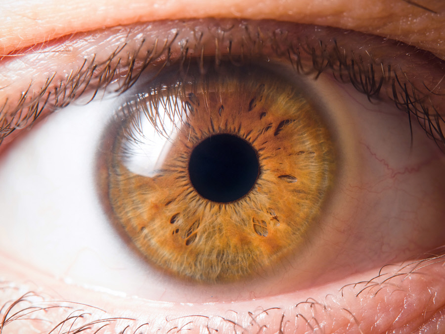
The iris is a vital part of the eye. It is the colored part of the eye which works by regulating how much light comes through the pupil.
Anatomy
The iris sits between the lens and cornea. More specifically, in front of the lens and ciliary body and behind the cornea. At the center of the iris is the circle-shaped pupil.
It is covered both in front and at the back with aqueous humor. The iris is made almost wholly of two smooth muscle fibers and connective tissue. These are the dilator muscle and sphincter muscle. These muscles perform the contrary actions of dilation and constriction. That is how they control the light that reaches the retina.
The iris contains color granules whose number determines an individual’s iris pigmentation. Blue eyes could be a result of very little pigment in the iris whereas black and brown eyes could be indications of increased pigment. The color of the iris may be associated with genetics, hair, skin type, etc.
Function
The iris controls the amount of light that enters the eye. The muscles in the iris cause the pupil to constrict (shrink) in bright light and dilate (widen) in a dark environment. This involuntary, brain-controlled action enables vision in both bright and dim surroundings. It also protects the eye from the effects of excessive light that falls on the eye. Both little or excess light can limit vision.
Associated symptoms & disorders
Some of the diseases associated with the iris include:
- Plateau iris is a condition that can lead to chronic angle closure glaucoma. It is the result of the narrowing of an anterior chamber angle or anatomic configuration of the iris.
- Ocular melanoma is cancer that grows in the iris. It starts from the middle of the three layers in the eye, namely the sclera, retina and uvea. It can cause vision distortion and a change in pupil size.
- Coloboma of the iris. It is a congenital condition in which a baby is born with a hole or defect in the iris. As a result, the pupil acquires an irregular shape. A coloboma can cause blurry vision, double vision, ghost images and decreased visual acuity.
- Aniridia. It occurs when a baby is born without a part or the whole of the iris. The absence of the pupil can lead to reduced photophobia (increased sensitivity to light) and visual acuity. The disease affects both eyes and can hamper the normal development of other structures in the eye.
- Iritis, an inflammation of the iris, also known as anterior uveitis. The iris forms part of the uveitis. The condition causes the iris to stick to the cornea or lens. This action prevents the fluid in the eye from flowing as it should. If it is not treated, iritis can lead to blindness. Secondary glaucoma can also result as a complication of iritis.
Diagnosis of associated disorders
Different tests can help diagnose various conditions. They may include:
- A physical examination.
- A visual acuity test using an eye chart and other standard tests.
- A complete medical history especially for congenital disorders like coloboma and aniridia.
- A biopsy or culture to reveal information about the tumor for cases like conjunctival melanoma.
- Slit-lamp examination after dilating the eye to see the inside of the eye using high magnification.
- Imaging tests such as magnetic resonance imaging especially if the professional suspects other problems.
- Fundus autofluorescence which makes use of a particular camera to capture the image. The camera causes damaged areas to reveal themselves as small points of light in a picture.
- Fluorescein angiography where the professional injects a vein with yellow dye. As the dye makes its way through the blood vessels, the professional takes images. Optical coherence tomography (OCT) may be used.
Treatment of associated disorders
Medical treatment options may include topical steroid eye drops for conditions like anterior uveitis. Dilating eye drops can also be used to relieve pain and prevent complications in iritis.
Scientists are still trying to discover a cure or reverse mechanism for coloboma. Currently, treatment options involve assisting the patient to make the most out of the vision available. The possibilities include glasses or contact lenses, drops to blur vision in the stronger eye and treating cataract if it's present. Other choices include preventing amblyopia from developing, using low-vision devices and so on.
Surgery for cataract removal can help with aniridia. If the cornea is affected in the case of aniridia, a corneal transplant (replacing a diseased cornea with a healthy one) can be done. A stem cell transplantation can aid in replacing some missing stem cells. Some individuals undergo cosmetic surgery to have an artificial iris. This procedure may lead to complications. Surgery is also the only option for plateau iris.
Artificial tears can assist in maintaining the health of the cornea.
Absorptive sunglasses can aid in symptoms of glare and photophobia. Colored contact lenses can be used for cosmetic reasons so that one appears to have an iris. The lenses also improve vision and can minimize glare.
Since glaucoma can be a complication of aniridia, it should be treated with drugs, laser or surgery.
For iris melanoma, cancer treatments can be considered. They include radiation, freezing therapy, chemotherapy and surgery.
