
by Piter Kehoma Boll
Here is a list of species described this month. It certainly does not include all described species. You can see the list of Journals used in the survey of new species here.
Bacteria
- 1 new cyanobacterium: Leptodesmis paradoxa;
- 22 new proteobacteria: Stenotrophobium rhamnosiphilum; Phenylobacterium soli; Acidihalobacter aeolianus, A. ferrooxydans; Pusillimonas thiosulfatoxidans; Brevundimonas lutea; Alterythrobacter maritimus; Photobacterium chitinilyticum; Roseovarius faecimaris; Azoarcus pumilus; Bosea psychrotolerans; Paracoccus subflavus; Marortus luteolus; Alteromonas sediminis; Dyella dinghuensis, D. choica; Halioglobus sediminis; Stagnimonas aquatica; Amphritea opalescens; Paraburkholderia dinghuensis; Pseudomonas kairouanensis, P. nabeulensis;
- 3 new actinobacteria: Marmoricola mangrovicus; Staphylospora marina; Streptomyces fodineus;
- 8 new firmicutes: Falsibacillus albus; Paraliobacillus zengyii; Planomicrobium iranicum; Suicoccus acidiformans; Lactobacillus suantsaii; Lactobacillus zhachilii; Filibacter tadaridae; Bacillus urbisdiaboli;
- 1 new thermotoga: Marinitoga lauensis;
Archaeans
- 1 new species: Methanofervidicoccus abyssi;
SARs
- 2 new ciliates: Australocirrus rubrus, Notohymena gangwonensis;
- 13 new apicomplexans: Karyolysus canariensis, Karyolysus galloti, Karyolysus stehlini, Karyolysus gomerensis, Karyolysus atlanticus, Karyolysus tinerfensis, Karyolysus makariogeckonis; Cryptosporidium proventriculi; Eimeria pantholopensis, Eimeria wudaoliangensis, Eimeria hodgsonii, Eimeria schalleri, Eimeria sui;
- 12 new diatoms: Actinella hermes-moreirae, Gomphonema mutunensis; Encyonopsis carraensis, Encyonopsis hibernica; Cymbella pavanaensis; Nitzschia pusilluhasta, Nitzschia australodesertorum; Tryblionella confusa, Tryblionella ornata; Clipeoparvus tibeticus; Ulnaria rhombus, Ulnaria wulingensis;
- 1 new foraminifer: Capsammina crassa;

Plants
- 1 new moss: Weissia wilsonii;
- 2 new lycopodiophytes: Isoetes shangrilaensis; Selaginella dianzhongensis;
- 2 new ferns: Christella kendujharensis; Woodsia kungiana;
- 76 new flowering plants: Dahlia mixtecana; Boswellia occulta; Calanthe himalaicum, Habenaria palpensis; Gundelia siirtica; Impatiens kamrupana; Impatiens damingensis; Rhododendron rimicola; Nepeta azadkouhensis; Fosterella atlantica, F. bodoquenensis, F. liliputiana; Rhabdosciadium hizanensei; Senna juchitanensis; Draba ancashensis; Curcuma tongii; Festuca greuteri; Arisaema melanostomum; Cousinia sharifii; Quintinia hyehenensis, Q. sessiliflora; Silene arsuzensis; Andropogon saxicola; Muscari fatmacereniae; Muscari tauricum; Aspidistra corniculata; Critoniopsis hermogenesii; Ponthieva boliviensis; Paepalanthus calvescens; Onobrychis garinensis; Chronopappus lanatus, Lychnophora grisea, L. haplopappa, Lychnophorella jacobinensis, Piptolepis riparia, Prestelia espeletoidea, Proteopsis hermogenesii; Hechtia gypsophila, H. minuta; Bellevalia sasonii; Aster brevicaulis; Echeveria xochipalensis; Campanula phitosiana; Myrciaria cambuca, M. una; Hedyotis dinganensis; Pteroceras dalaputtuwa; Myrcia auriculata; Cymbidium shidianense; Chresta artemisiifolia; Leptogonium hondae, L. quilombensis; Leucas sahyadriensis; Priogymnanthus colombianus; Notothlaspi viretum; Piptolepis rosmarinifolia; Hieracium wierzbickii; Moldenhawera congestiflora; Bonnaya milindii; Microchirita hairulii; Nothodissotis alenensis; Liparis napoensis; Indigofera monieriana, I. dumbeana; Sorbus lushanensis; Pibiria flava; Manglietia pubipedunculata; Eugenia petaloidea; Aristolochia lorenae; Trillium delicatum; Mimosa cerifera; Brongniartia bicornuta; Sida uniaristata; Bolanthus aziz-sancarii; Prestonia lauta, P. occultata; Hexasepalum nordestinum;


Excavates
- 3 new euglenozoans: Keelungia nitschei, Petalomonas acorensis, Ploeotia costaversata;
Fungi
- 24 new basidiomycetes: Tricholomopsis decora, T. sulphureoides; Ionosporus australis; Mycena castaneicola, M. hyalinostipitata, M. substylobates; Galzinia oberwinklerii; Pucciniastrum coronisporum, P. verruculosum; Neofomitella australiensis and N. guangxiensis; Hemistropharia subalbocrenulata; Strobilomyces minor; Gymnopilus minisporus; Milesina woodwardiana; Dextrinocystis calamicola, Subulicystidium acerosum, S.tropicum, Tubulicium bambusicola; Stephanospora mayana; Cantharelus densilamellatus, C. tomentosoides; Neoboletus antillanus; Rhizopogon laricinus;
- 43 new ascomycetes: Hydnobolites canaliculatus, Hydnobolites roseus, Hydnobolites shanxiensis, Hydnobolites yunnanensis; Melanocamarosporioides ugamica; Minimelanolocus nonramosus; Daldinia subvernicosa; Roussoella kunmingensis, R. yunnanensis; Colletotrichum yulongense; Phillipsia hydei; Caloplaca fluviatilis; Cyphellophora botryose, Cyphellophora guizhouensis; Aquimonospora tratensis; Alternaria glehniae; Pararhexoacrodictys catolensis, Pa. minima; Fuscidea multispora, Malmidea attenboroughii; Verruconis hainanensis, V. pseudotricladiata; Biatora alnetorum; Dendrostoma aurorae, D. castaneae, D. castaneicola, D. chinense, D. dispersum, D. parasiticum, D. qinlingense, D. quercus, D. shaanxiense, D. shandongense; Colletotrichum jishouense, C. tongrenense; Arthrinium balearicum, A. descalsii, A. esporlense, A. ibericum, A. italicum, A. piptatheri; Kurtzmaniella hittingeri; Wickerhamomyces menglaensis; Myxotrichum albicans;
- 2 new glomeromycetes: Sclerocarpum amazonicum, Diversispora sporocarpia;

Sponges
- 1 demosponge: Antho ridgwayi;
- 1 new hexactinellid: Rhizophyta yapensis;
Cnidarians
- 3 new anthozoans: Calcigorgia gigantea, C. pacifica, C. gracilis;
- 2 new myxosporeans: Myxobolus lienis, Myxobolus iecoris;
Flatworms
- 7 new trematodes: Endochortophagus protoporus; Pojmanskatrema balcanica; Masenia nkomatiensis; Provitellus chaometra, Provitellus infrequens, Provitellus infibrova, Ovipusillus geminus;
- 1 new polyclad: Pericelis tectivorum;
- 2 new planarians: Nerpa fistulata, Paucumara falcata;
- 1 new cestode: Raillietina saudiae;
Rotifers
Annelids
- 5 new polychaetes: Branchiosyllis belchiori, Branchiosyllis gonzaguinhai; Pectinaria nonatoi, Petta alissoni; Trophoniella radesiensis;
Mollusks
- 14 new gastropods: Rubyspira brasiliensis; Sulcospira guangxiensis, Sulcospira mashi; Phreatomascogos gregoi, Balconorbis sabinasense; Pseudamnicola sp., Bythinella sp. 1, Bythinella sp. 2, Valvata (Tropidina) sp.; Paladilhiopsis cattaroensis, Paladilhiopsis matejkoi, Bosnidilhia vitojaensis, Plagigeyeria feheri, Stygobium hercegnoviensis;
Nematodes
- 8 new species: Basiria khouzestanensis; Thalassironus koreanus; Coronostrongylus hasegawai, Dorcopsistrongylus supriyatnai; Paranticoma lepta; Longidorus polyae; Maldivea complexa; Dichelyne (Cucullanellus) bodiani;
Chelicerates
- 35 new spiders: Shaanxinus magniclypeus, S. shihchoensis, S. shoukaensis, S. hirticephalus, S. mingchihensis, S. makauyensis, S. lixiangae, S. curviductus, S. tsou, S. hehuanensis, S. seediq, S. meifengensis, S. atayal, S. tamdaoensis; Amphidraus araripe, A. boomerang, A. cornipalpis, A. draconitupan, A. manauara, A. sacrificatus, A. shenlong; Maratus aquilus, M. combustus, M. felinus; Mallinella bicanaliculata, M. calautica, M. laxa, M. obliqua; Harpactea popovi; Anisaspis camarita, Paratropis florezi, Stormtropis colima, S. muisca, S. paisa, S. parvum;
- 12 new mites: Neoaulobia cacatui, Lawrencipicobia calyptorhyncha, Lawrencipicobia sulphurea; Phyllotetranychus hadii; Trouessartia ampulicaudata, T. petrochelidon, T. bochkovi, T. cryptocaudata, T. progne; Halozercon tigerek, H. kazachok, H. capitaneus;
- 15 new pycnogonids: Nymphon rogersi, N. serpettiae, N. dentatum, N. gravidus, Tanystylum lamonti, T. tiara, Austrodecus bamberi, A. taylorae, Rhynchothorax swir, R. coralensis, Hedgpethia filamentus, H. shalei, Pycnogonum sentus, P. copleyi, P. cheni;
- 1 new pseudoscorpion: Lagynochthonius bailongtanensis;
- 3 new schizomids: Surazomus saturninoae; Troglocubazomus sp., Antillostenochrus sp.;

Myriapods
- 16 new millipedes: Zephronia laotica, Sphaerobelum bolavensis, S. phouloei, S. denticulatum, S. spinatum, S. lachneeis, S. peterjaegeri, S. nigrum, S. splendidum, S. laoticum, S. schwendingeri; Tasmanocambala greeni, T. tasmanica, T. taylori, Talomius weldensis; Antheromorpha nguyeni;
- 2 new centipedes: Lithobius (Ezembius) ternidentatus; Arrup akiyoshiensis;

Crustaceans
- 1 new copepod: Neoalbionella benzipirata;
- 2 new tanaids: Apseudomorpha drummi, Cryptapseudes mamua;
- 9 new decapods: Eumunida subsolanus, Heteroptychus galapagos, H. nautilus, U. compressus; Pachycheles coelhoi; Caridina kutchi; Cambarus franklini; Hippolyte chacei, H. nanhaiensis;
- 3 new isopods: Pectenoniscus liliae, Benthana xiquinhoi; Caecianiropsis goseongensis;
- 2 new syncarids: Indobathynella socrates; Fortescuenella serenitatis;
- 2 new amphipods: Dorotea papuana; Pseudaeginella freirei;
- 10 new cumaceans: Chalarostylis erebos, Chalarostylis nyx, Bathycuma sonneae, Leucon (Crymoleucn) fracturensis, Leucon (Crymoleucon) marinae, Leucon (Macrauloleucon) longiserratus, Leucon (Macrauloleucon) breviserratus, Atlantistylis paraborealis, Leptostylis abyssalis, Leptostylis danieli;
Hexapods
- 14 new orthopterans: Mimoscudderia longicaudata, Paraphyrrhicia leuca; Paterdecolyus magnimaculatus; Phlugiolopsis (Phlugiolopsis?) taiwanensis, Xiphidiopsis (Xiphidiopsis) trifoliata; Hilethera xinjiangensis; Trigonidium solis; Neophaloria dianxiensis; Conocephalus (Anisoptera) dorsalidentatus; Paranisitra flavofacia; Tettigoniidae sp. 1, 2, 3, 4;
- 79 new lepidopterans: Victrix akbet; Rhodostrophia reisseri, Rhodostrophia stueningi; Promalactis curvitaeniata, P. biprocessa, P. lvovskyi, P. subquadrata, P. tibetica, P. auremacularis, P. apicidigitata, P. campanulata, P. bicornea, P. spiniflagellata, P. grandilobata, P. arciformis, P. bornensis, P. cribrata, P. oliviformis; Panolis xundian; Ptilothyris subcucullata, P. crassiella, P. drepanodes, P. enormisella, P. hylodes, P. leifaarviki, P. pilosa, P. vokaensis; Chrysoclista karsholti; Neochloroglyphica perbella; Procinnus incanus. Micrallo macro; Mecothrix maputuana, Meganola shangaana; Siccia imana; Triscaedecia sulawesi, T. sarawaki, T. svetlanae, T. suva; Thubdora ealeaensis, T. ghesquierei, T. gladiator, T. kapangaensis, T. neobarbata, T. seydeli, Torodora amplignathosa, T. lichanosa, T. planusa, T. triloba; Timandra distorta, T. adunca, T. quadrata, T. accumulata, T. viminea, T. robusta, T. stueningi; Hoplosauris morenoi, Bulteriana phoenix, Warrenaria onca, Fueguina araucana, Aloba carolinae; Calisto gundlachi, Calisto siguanensis, Calisto disjunctus, Calisto sharkeyae,Calisto lastrai; Acrapex abyssinica, Acrapex dabaga, Acrapex jansei, Acrapex kifanya, Acrapex lusinga, Acrapex ngwenya, Acrapex njombea, Acrapex vetiveria, Acrapex zima; Sesapa strandiana; Longarista kareli; Breyeriana patagonica; Cyana smithi; Spininola longshengensis;
- 31 new hemipterans: Phytocoris (Compsocerocoris) darakiensis; Abbrosoga multispinosa; Plautia sakishimensis, P. himechabane, P. ishigaki; Orosius viraktamathi; Pseudolaryngodus spectabilis; Agnesiella (Draberiella) digita, A. (D.) furca, A. (D.) innota, A. (D.) sinuata; Ericerus farsicus; Zicca gloriosa; Hoplonannus australis; Caetana pulchra, Insolitana carinata; Pachymetopius falcatus; Ziczacella spinosa; Daveyoungana blockeri, Daveyoungana longibrachia, Daveyoungana pentacorni; Acusana adunca, Acusana spina, Acusana dilatata, Acusana longa; Undulivena thaiensis; Kuvera huoditangensis, Kuvera longwangshanensis; Myzus asterale, M. prunense, M. raphanense;
- 261 new coleopterans: Haptomerus maculosus; Diastatotropis blazeji;
Lepanus mckenziei, Lepanus sauroni, Lepanus gubara, Lepanus pecki, Lepanus podocarp, Lepanus pungalina, Lepanus lentil, Lepanus crenidens, Lepanus lingziae, Lepanus menendezae, Lepanus andersonorum, Lepanus tozerensis, Lepanus feehani, Lepanus guthrieae; Clavomicrus orousseti; Agrilus cicadelloides, A. draco, A. hergovitsi, A. hik, A. ika, A. jankae, A. jum, A. kon, A. mimicus, A. qom, A. titi, A. uxo, A. wos, A. xen, A. xia, A. xis, A. yoa, A. yuk, A. zao, A. zim; Agrilus cameronius,A. puncak,A. vendibilis; Methia similis; Oxytrechus juani, O. alexei; Lesbates chavesi; Cintaroa sikkimica; Hemiosus molanoi, H. quindiensis; Cercyon (s.str.) bellus, Cercyon (s.str.) biltoni; Megistophylla octobracchia; Neochila cassolawerneri; Paracupes mexicanus; Tychobythinus villasmundi; Megasybacodes brevitarsis; Omineus taiwanensis, O. chuangae; Neorthrius aduncus, Neorthrius aurantiacus, Neorthrius bipunctatus, Neorthrius bonasus, Neorthrius brunnorbis, Neorthrius buteocoloratus, Neorthrius cechovskyi, Neorthrius centromaculatus, Neorthrius chiangmaii, Neorthrius cornutus, Neorthrius crassopunctatus, Neorthrius ebenus, Neorthrius elegantulus, Neorthrius fortecruris, Neorthrius fulvus, Neorthrius fuscomaculosus, Neorthrius guttatus, Neorthrius longulus, Neorthrius majae, Neorthrius mariannae, Neorthrius molestus, Neorthrius schnitzeli, Neorthrius serratus, Neorthrius sexmaculatus, Neorthrius sigmoideus, Neorthrius tenuistriatus, Neorthrius tulipae, Neorthrius unicolor, Neorthrius uniformis, Neorthrius volsella, Neorthrius zebrinus; Faronus rica; Chandleriella yunnanica; Araucariocladus amfractus; Pentatrachyphloeus andersoni, P. baumi, P. brevithorax, P. bufo, P. endroedyi, P. exiguus, P. frici, P. grobbelaarae, P. hanzelkai, P. holubi, P. howdenae, P. hystrix, P. insignicornis, P. kalalovae, P. kuscheli, P. laevis, P. lajumensis, P. leleupi, P. lesothoensis, P. machulkai, P. marshalli, P. muellerae, P. musili, P. ntinini, P. oberprieleri, P. pavlicai, P. rudyardi, P. schoemani, P. soutpansbergensis, P. spinimanus, P. stingli, P. tenuicollis, P. tuberculatus, P. vavrai, P. vossi, P. vrazi, P. zikmundi; Beraba anae, Beraba angeli; Trigonopterus abnormis, T. adspersus, T. ambangensis, T. ampanensis, T. analis, T. arachnobas, T. armipes, T. artemis, T. asterix, T. barbipes, T. bonthainensis, T. carinirostris, T. castaneipennis, T. celebensis, T. cirripes, T. collaris, T. costatulus, T. curvipes, T. crenulatus, T. cricki, T. darwini, T. ejaculatorius, T. fuscipes, T. gracilipes, T. heberti, T. hirsutus, T. humilis, T. hypocrita, T. idefix, T. impressicollis, T. incendium, T. incognitus, T. indigenus, T. inhonestus, T. invalidus, T. jasminae, T. klabatensis, T. kolakensis, T. kotamobagensis, T. laevigatus, T. lampros, T. latipennis, T. lompobattangensis, T. luwukensis, T. mahawuensis, T. manadensis, T. mangkutanensis, T. matalibaruensis, T. mesai, T. minahassae, T. moatensis, T. modoindingensis, T. nanus, T. nitidulus, T. obelix, T. ovalipunctatus, T. ovatulus, T. pagaranganensis, T. palopensis, T. paracollaris, T. pauper, T. pendolensis, T. posoensis, T. prismae, T. procurtus, T. pseudallotopus, T. pseudanalis,, T. pseudovatulus, T. pseudovalipunctatus, T. pseudofulvicornis, T. pseudomanadensis, T. pseudosimulans, T. pumilus, T. rantepao, T. reticulatus, T. rhombiformis, T. rotundatus, T. rotundulus, T. rudis, T. rufipes, T. sampunensis, T. sampuragensis, T. satyrus, T. scabripes, T. scaphiformis, T. scitulus, T. selayarensis, T. serripes, T. seticnemis, T. silvicola, T. squalidulus, T. sulawesiensis, T. suturatus, T. tatorensis, T. tenuipes, T. tomohonensis, T. toraja, T. vicinus, T. viduus, T. volcanorum, T. wangiwangiensis, T. watsoni, and T. yoda; Lophorrhinides muellerae; Gymnephoria sp., Madiana sp.; Pachybrachis cubs, Pachybrachis ekstromorum, Pachybrachis troosevelti, Pachybrachis zellersorum; Anogdus championi, Colon (Myloechus) matthewsi; Photuris walldoxeyi; Tomarus laticaudus, T. maracaiboensis, T. moroni, T. paranaensis, T. pilcopataensis; Phaedon uinta; Phanaeus edmondsi; Amphicoma gandhara; Microogenius borealis, M. puna; Oxylymma soniae, O. surinamensis, O. birai, Paraeclipta albopilosa, P. vanderberghei, Odontocera ellanocarti, Giesberticus longiventris; Calochroa horii, C. fumikoae; - 51 new dipterans: Coelotrypes meremmiae, C. paralatilimbatus; Ablabesmyia (Ablabesmyia) lyrata, A. (Ablabesmyia) pectinata, A. (Ablabesmyia) huananensis, A. (Ablabesmyia) praegracilis, A. (Ablabesmyia) bifurca, A. (Karelia) daiensis; Olbiogaster halffteri, Olbiogaster nuxco, Olbiogaster puuc; Ablabesmyia akemiae, A. adamantis, A. monticola; Chaetonerius kotrbae, C. ebejeri, C. kirkspriggsi, C. madagasikara; Macrostomus montanus, M. rodriguezi, M. falcatus, M. trifidus, M. trilineatus; Hirtodrosophila rondonia, H. nungara, H. jukyry, H. crioula; Meoneura artoodetoo, Meoneura baechli, Meoneura gnomi, Meoneura joedaltoni, Meoneura mucki; Simulium mainitense, S. tukurongense; Hurleyella belizensis, Hurleyella salina; Polypedilum (Cerobregma) heberti; Agromyza arundinariae, A. indistincta, Calycomyza chrysopsidis, Cerodontha (Butomomyza) enigma, Cer. (Poemyza) arundinariella, Cer. (P.) saintandrewsensis, Liriomyza carphephori, L. polygalivora, L. triodanidis; Chvalaea annularis, C. ecuadoriensis, Chvalaea sinclairi; Stevenia gilasiani; Diamesa qiangi;
- 45 new hymenopterans: Orancistrocerus altus; Lomachaeta calamondin, L. eironeia, L. juanita, L. meloi, L. osita, L. warneri;
Clistopyga catarina, C. marcapatensis, C. polita; Campoletis katalinarum, Campoletis margaritae; Isolia bhima, I. bandoola; Rodrigama freidbergi; Pompiliodon katina; Adelius adeleae, A. bolivariensis, A. boliviensis, A. caatinga, A. canadensis, A. ecuadoriensis, A. excelsus, A. floridensis, A. gauldi, A. janzeni, A. monteiroi, A. morretesiensis, A. panamensis, A. pentagonalis, A. quiteriae, A. sancticaroli, Paradelius neotropicalis; Gilpinia infuscalae; Fagineura flactoserrula, F. xanthosoma; Recurvidris lekakuli; Calixomeria lasallei; Cryptopimpla aspeculosus, C. pentagonalis; Strumigenys hirsuta, S. lantaui, and S. nathistorisoc; Paralipsis brachycaudi, P. rugosa; - 9 new thysanopterans: Aeolothrips fukusimensis, A. setosus, Desmidothrips japonicus, Franklinothrips oblongus; Scirtothrips betulaceae, S. cryptomeriae, S. illicii, S. passiflorae, S. pini;
- 2 new ephemeropterans: Thraulodes catoles; Choroterpes (Euthraulus) kalladaensis;
- 6 new plecopterans: Anacroneuria bandido, A. tuberculata; Mesonemoura longiflagellata, M. tumida; Sphaeronemoura kunmingensis, S. campylura;
- 5 new trichopterans: Helicopsyche (Feropsyche) carajas; Atopsyche (Atopsyche) bravoi, A. (Atopsaura) beckeri, A. (Atopsaura) japi; Liophloeothrips arjanicus;
- 2 new collembolans: Schaefferia bermani; Cosberella mendozarum;
- 2 new psocodeans: Vinceopterus erythrocephali, Vinceopterus mindanensis;
Echinoderms
- 2 new holothuroids: Psolus tommasi, Psolus thandari;
Tunicates
- 4 new species: Pyura longispina, P. lopezlegentilae, P. carmanae, P. imesa;
Ray-finned fishes
- 1 new ophiidiform: Barathronus algrahami;
- 4 new cypriniforms: Barbatula zhangwuensis, Barbatula liaoyangensis, Barbatula emuensis, Barbatula linjiangensis;
- 2 new perciforms: Cerogobius petrophilus; Channa auroflammea;
- 2 new cupleiforms: Sardinella electra; Sardinella pacifica;
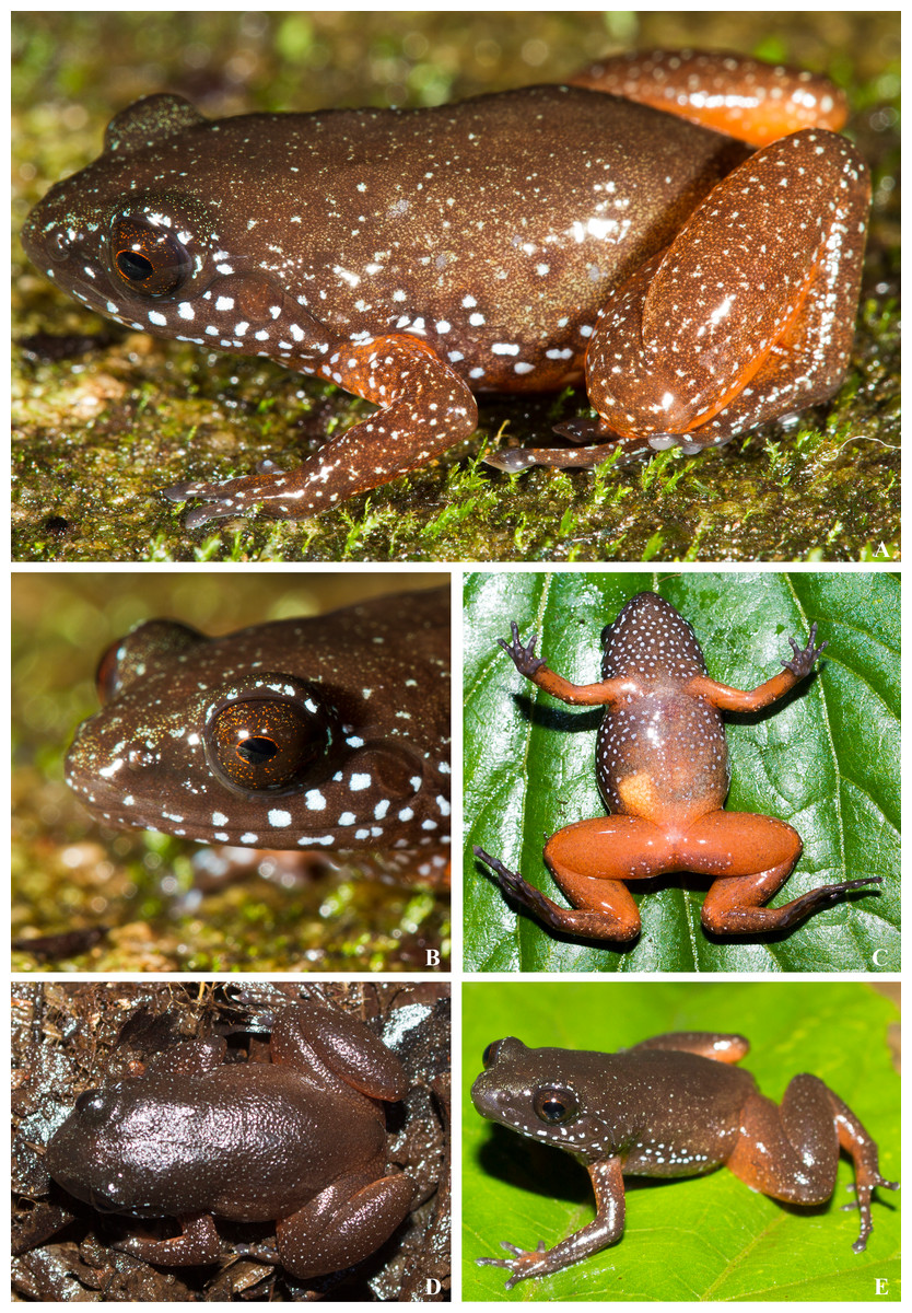
Amphibians
- 8 new anurans: Lynchius megacephalus; Pristimantis mallii; Astrobatrachus kurichiyana; Mini mum, Mini scule, Mini ature, Rhombophryne proportionalis, Anodonthyla eximia;
Reptiles and Birds
- 8 new squamates: Hemiphyllodactylus arakuensis, H. jnana, H. kolliensis; Salvadora gymnorhachis; Cnemaspis ingerorum; Cnemaspis agarwali; Bothrops mattogrossensis; Trachylepis raymondlaurenti;
- 1 new bird: Pycnonotus pseudosimplex;
– – –
* This work is licensed under a Creative Commons Attribution 4.0 International License.


